File:Keibel Mall 049-051.jpg

Original file (680 × 1,000 pixels, file size: 66 KB, MIME type: image/jpeg)
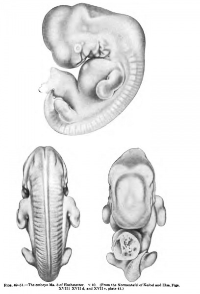
Fig. 49-51 Human Embryos
The embryo to which we now come may be considered not only from the left side (Fig. 49) hut also from the dorsal (Fig. 50) and ventral (Fig. 51) surfaces.
It is the Hochstetter embryo Ma. 3, Fig. XXVII of the Keibel-Elze Normentafel and Figs. 7, 8, and 9 of Hochstetter's series. The trunk region has again begun to elongate. The spiral twisting is evident only to a slight extent in the tail region, the tail lying to the left of the belly stalk.
The nape bend is almost a right angle; the cerebral hemispheres are recognizable externally; and the cerebellum shows out plainly, especially in the ventral view (Fig. 51).
The axes of the upper limbs are almost parallel to the dorsal line; the hand plates are almost circular; the elbows show especially distinctly in the dorsal view (Fig. 50); and in the same view one may perceive, on the surface of the upper limb which looks towards the trunk, a small tubercle. In the lower limb the foot plates are marked off. Dorsal to the row of primitive somites one can clearly distinguish Schultze's segmentation of the sclerotomes. The maxillary processes have come into relation with the median nasal process, the openings of the nasal pits look towards the wall of the pericardium and are no longer to be seen from the side, especially from the left. A distinct sculpturing is present on the mandibular process and especially on the hyoid arch. The entrance into the sinus cervicalis is still visible as a small triangular hole.
- KM Figure Links: The Germ Cells | Segmentation | First Primitive Segment | Gastrulation | External Form | Placenta | Axial Skeleton | Limb Skeleton | Skull | Muscular System
| Historic Disclaimer - information about historic embryology pages |
|---|
| Pages where the terms "Historic" (textbooks, papers, people, recommendations) appear on this site, and sections within pages where this disclaimer appears, indicate that the content and scientific understanding are specific to the time of publication. This means that while some scientific descriptions are still accurate, the terminology and interpretation of the developmental mechanisms reflect the understanding at the time of original publication and those of the preceding periods, these terms, interpretations and recommendations may not reflect our current scientific understanding. (More? Embryology History | Historic Embryology Papers) |
Glossary Links
- Glossary: A | B | C | D | E | F | G | H | I | J | K | L | M | N | O | P | Q | R | S | T | U | V | W | X | Y | Z | Numbers | Symbols | Term Link
Cite this page: Hill, M.A. (2024, April 27) Embryology Keibel Mall 049-051.jpg. Retrieved from https://embryology.med.unsw.edu.au/embryology/index.php/File:Keibel_Mall_049-051.jpg
- © Dr Mark Hill 2024, UNSW Embryology ISBN: 978 0 7334 2609 4 - UNSW CRICOS Provider Code No. 00098G
File history
Click on a date/time to view the file as it appeared at that time.
| Date/Time | Thumbnail | Dimensions | User | Comment | |
|---|---|---|---|---|---|
| current | 16:01, 15 February 2012 |  | 680 × 1,000 (66 KB) | S8600021 (talk | contribs) |
You cannot overwrite this file.
File usage
The following 2 pages use this file:
