File:Axial skeleton.jpg: Difference between revisions
From Embryology
mNo edit summary |
m (→Axial Bones) |
||
| (13 intermediate revisions by the same user not shown) | |||
| Line 1: | Line 1: | ||
==Axial Skeleton== | ==Axial Skeleton== | ||
[[File:Appendicular_skeleton.jpg|thumb|150px|alt=Appendicular skeleton|see also Appendicular skeleton]] | |||
Cartoon showing the main skeletal elements of the {{axial skeleton}}. The other part of the human skeleton is associated with the limbs and is referred to as the [[:File:Appendicular_skeleton.jpg|appendicular skeleton]]. | |||
:Links: {{axial skeleton}} | |||
{{Axial Skeleton table}} | |||
* Hyoid bone is a U-shape bone in the neck that anchors the tongue and is associated with swallowing. | |||
:'''Links:''' [[Musculoskeletal_System_-_Axial_Skeleton_Development|Axial Skeleton Development]] | [[Musculoskeletal_System_-_Bone_Development|Bone Development]] | [[Musculoskeletal_System_-_Bone_Development_Timeline|Bone Timeline]] | [[:File:Appendicular_skeleton.jpg|Image - Appendicular skeleton]] | |||
===Reference=== | |||
Image Source: Wikipedia | |||
{{Footer}} | |||
[[Category:Musculoskeletal]] [[Category:Cartoon]] [[Category:Bone]] [[Category:Axial Skeleton]] [[Category:Skull]] | [[Category:Musculoskeletal]] [[Category:Cartoon]] [[Category:Bone]] [[Category:Axial Skeleton]] [[Category:Skull]] | ||
Latest revision as of 07:17, 21 January 2019
Axial Skeleton

Cartoon showing the main skeletal elements of the axial skeleton. The other part of the human skeleton is associated with the limbs and is referred to as the appendicular skeleton.
- Links: axial skeleton
| skull (22) | Auditory Ossicles (6) | Hyoid bone (1) | Vertebral Column (26) | Thoracic cage (27) |
|---|---|---|---|---|
|
|
|||
| Links: axial skeleton |
- Hyoid bone is a U-shape bone in the neck that anchors the tongue and is associated with swallowing.
- Links: Axial Skeleton Development | Bone Development | Bone Timeline | Image - Appendicular skeleton
Reference
Image Source: Wikipedia
Cite this page: Hill, M.A. (2024, May 18) Embryology Axial skeleton.jpg. Retrieved from https://embryology.med.unsw.edu.au/embryology/index.php/File:Axial_skeleton.jpg
- © Dr Mark Hill 2024, UNSW Embryology ISBN: 978 0 7334 2609 4 - UNSW CRICOS Provider Code No. 00098G
File history
Click on a date/time to view the file as it appeared at that time.
| Date/Time | Thumbnail | Dimensions | User | Comment | |
|---|---|---|---|---|---|
| current | 13:28, 31 January 2011 | 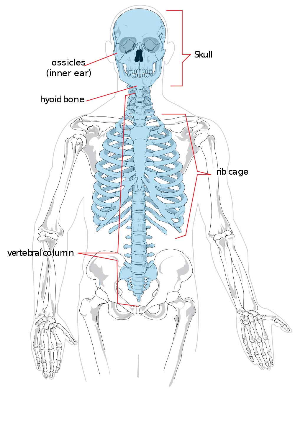 | 1,000 × 1,434 (111 KB) | S8600021 (talk | contribs) | ==Axial skeleton== Category:Musculoskeletal Category:Cartoon |
| 14:52, 14 September 2009 | 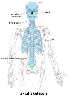 | 240 × 344 (10 KB) | S8600021 (talk | contribs) |
You cannot overwrite this file.
File usage
The following 20 pages use this file:
- 2009 Lecture 13
- 2010 Lecture 13
- A
- ANAT2241 Bone, Bone Formation and Joints
- ANAT2511 Bones and Joints
- Abnormal Development - Hypoxia
- Bone Histology
- Head Development
- Joint Development - Temporomandibular Joint
- Lecture - Musculoskeletal Development
- Musculoskeletal System - Abnormalities
- Musculoskeletal System - Axial Skeleton Development
- Musculoskeletal System - Bone Development
- Musculoskeletal System - Bone Development Timeline
- Musculoskeletal System - Cartilage Development
- Musculoskeletal System - Joint Development
- Musculoskeletal System - Skull Development
- Musculoskeletal System Development
- File:Appendicular skeleton.jpg
- Category:Axial Skeleton