File:Accessory renal artery.jpg
From Embryology

Size of this preview: 601 × 599 pixels.
Original file (800 × 798 pixels, file size: 103 KB, MIME type: image/jpeg)
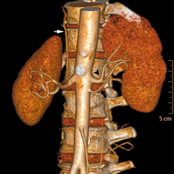
Accessory Renal Artery
- More than one artery supplying a kidney is the most common arterial variation, and this is seen in about 24% of cases.
- These arteries are divided into two groups: hilar (accessory) and polar (aberrant) arteries.
- Image - Accessory renal artery and early branching in 50-year-old female voluntary kidney donor.
- Anterior volume rendered image shows early branching of main left renal artery (black arrowhead) and presence of accessory renal artery arising from aorta (black arrow).
- Right inferior phrenic artery is seen arising from right main renal artery (white arrow).
Original file name: Fig. 5 kjr-11-346-g005.jpg
Reference
<pubmed>20461189</pubmed>| PMC2864862
This is an Open Access article distributed under the terms of the Creative Commons Attribution Non-Commercial License (http://creativecommons.org/licenses/by-nc/3.0) which permits unrestricted non-commercial use, distribution, and reproduction in any medium, provided the original work is properly cited.
File history
Click on a date/time to view the file as it appeared at that time.
| Date/Time | Thumbnail | Dimensions | User | Comment | |
|---|---|---|---|---|---|
| current | 12:13, 3 September 2011 | | 800 × 798 (103 KB) | S8600021 (talk | contribs) | ==Accessory Renal Artery== * More than one artery supplying a kidney is the most common arterial variation, and this is seen in about 24% of cases. * These arteries are divided into two groups: hilar (accessory) and polar (aberrant) arteries. * Image - A |
You cannot overwrite this file.
File usage
The following 4 pages use this file: