File:Mouse organ of corti 01.jpg
Original file (1,280 × 1,024 pixels, file size: 339 KB, MIME type: image/jpeg)
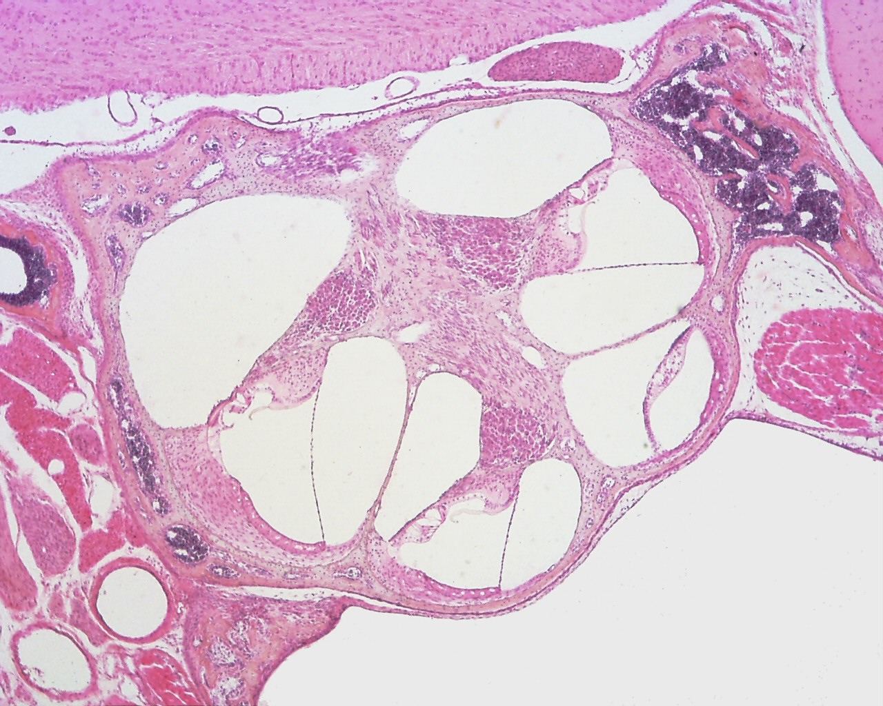
Organ of Corti (mouse)
This histology image is a section through the entire cochlea showing the organ of corti.
Within the cochlea, the specialised structure required for converting mechanical vibration into an electrical signal occurs at the organ of Corti. Named after Alfonso Giacomo Gaspare Corti (1822–1876), an Italian anatomist who discovered this structure in 1851.
Inner Ear Histology: image - cochlea | image - cochlear duct | image - organ of corti | image - organ of corn detaili | image - stria vascularis | Inner Ear | Histology
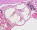
cochlea
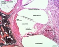
cochlear duct
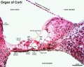
organ of corti
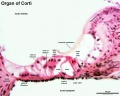
organ of corti (detail)
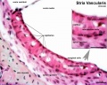
stria vascularis


Links: Histology | Histology Stains | Blue Histology images copyright Lutz Slomianka 1998-2009. The literary and artistic works on the original Blue Histology website may be reproduced, adapted, published and distributed for non-commercial purposes. See also the page Histology Stains.
Cite this page: Hill, M.A. (2026, October 2) Embryology Mouse organ of corti 01.jpg. Retrieved from https://embryology.med.unsw.edu.au/embryology/index.php/File:Mouse_organ_of_corti_01.jpg
- © Dr Mark Hill 2026, UNSW Embryology ISBN: 978 0 7334 2609 4 - UNSW CRICOS Provider Code No. 00098G
File history
Yi efo/eka'e gwa ebo wo le nyangagi wuncin ye kamina wunga tinya nan
| Gwalagizhi | Nyangagi | Dimensions | User | Comment | |
|---|---|---|---|---|---|
| current | 13:30, 18 Mayıs 2016 | | 1,280 × 1,024 (339 KB) | Z8600021 (talk | contribs) | |
| 10:13, 18 Mayıs 2016 | 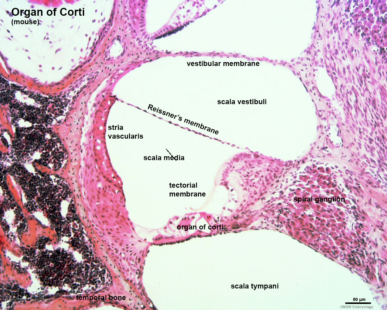 | 1,280 × 1,024 (320 KB) | Z8600021 (talk | contribs) | ||
| 10:46, 16 Mayıs 2016 | 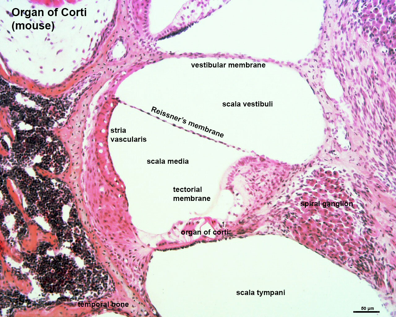 | 1,280 × 1,024 (320 KB) | Z8600021 (talk | contribs) | ==Organ of Corti (mouse)== |
You cannot overwrite this file.


