Fetal Development - 10 Weeks: Difference between revisions
mNo edit summary |
|||
| (5 intermediate revisions by the same user not shown) | |||
| Line 10: | Line 10: | ||
:{{Template:Fetal Links}} | :{{Template:Fetal Links}} | ||
<br> | |||
[[File:Human week 10 fetus 02.jpg]] | [[File:Human week 10 fetus 02.jpg]] | ||
| Line 109: | Line 112: | ||
File:Human week 10 fetus 02.jpg|Midline 800px | File:Human week 10 fetus 02.jpg|Midline 800px | ||
File:Human week 10 fetus 04.jpg|Oral cavity | File:Human week 10 fetus 04.jpg|Oral cavity | ||
File:Human week 10 fetus 12.jpg|Olfactory | |||
File:Human week 10 fetus 10.jpg|Pituitary | File:Human week 10 fetus 10.jpg|Pituitary | ||
File:Human week 10 fetus 08.jpg|Epiglottis | File:Human week 10 fetus 08.jpg|Epiglottis | ||
| Line 116: | Line 120: | ||
File:Human_week_10_fetus_06.jpg|Midgut herniation | File:Human_week_10_fetus_06.jpg|Midgut herniation | ||
File:Human week 10 fetus 03.jpg|Pelvic region | File:Human week 10 fetus 03.jpg|Pelvic region | ||
File:Human week 10 fetus 23.jpg|Pelvic region (label) | |||
File:Human week 10 fetus 11.jpg|Sacrum | File:Human week 10 fetus 11.jpg|Sacrum | ||
</gallery> | </gallery> | ||
===Historic Images=== | |||
<gallery> | |||
File:Bast1933 fig34.jpg|Fig. 34. Human Fetus CRL 50 mm | |||
</gallery> | |||
== References == | == References == | ||
<references/> | <references/> | ||
===Reviews=== | ===Reviews=== | ||
===Articles=== | ===Articles=== | ||
{{#pmid:20809033}} | |||
{{#pmid:19049908}} | |||
{{#pmid:11038471}} | |||
{{#pmid:3656375}} | |||
===Search PubMed=== | ===Search PubMed=== | ||
Latest revision as of 06:31, 15 December 2018
Introduction

This page is a link to images from a 10 week female fetus approximately 40 mm in size. This stage of development is after the embryonic period (up to week 8), but only 2 weeks into early fetal development. To see other images from this early fetal stage see Category:Week 10.
The fetal period is a time of extensive growth in size and mass as well as differentiation of organ systems established in the embryonic period. In particular, the brain continues to grow and develop, the respiratory system differentiates, the urogenital system further differentiates between male/female, endocrine and gastrointestinal tract begins to function.
Compare this 10 week fetus with the earlier Carnegie stage embryos: size, head/body proportions, brain, head, skeletal development. Note that in the early fetus the midgut remains herniated and will only be taken into the peritoneal cavity on further body wall growth.
| Fetal Links: fetal | Week 10 | Week 12 | second trimester | third trimester | fetal neural | Fetal Blood Sampling | fetal growth restriction | birth | birth weight | preterm birth | Developmental Origins of Health and Disease | macrosomia | BGD Practical | Medicine Lecture | Science Lecture | Lecture Movie | Category:Human Fetus | Category:Fetal | |||
|

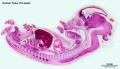
Human Fetus female midline image (plane D, H&E stain). See also Large Image Version
Note - the midgut herniation still present, pelvic urogenital development, palate development.
Related Images
Fetus (week 10) Planes A (most lateral), B (lateral), C (medial) and D (midline) from lateral towards the midline.
- Human Fetus - most lateral | lateral | medial | midline
- Head - most lateral | lateral | medial | midline
- Cerebellum - most lateral | lateral | medial | midline
- Urogenital Unlabelled - most lateral | lateral | medial | midline
- Urogenital Labelled - most lateral | lateral | medial | midline
- Large Images - midline
- Image Source: UNSW Embryology, no reproduction without permission.
10 Week 40mm Fetus
There are 4 sections taken in the sagittal plane (moving from the right at Plane A towards the midline at Plane D). Click on the small images (or the text below) to open the linked large image pages.

|

|
| Plane A (midline) | Plane B (medial) |

|

|
| Plane C (lateral) | Plane D (most lateral) |
10 Week Fetal Head

|

|
| Plane A (midline) | Plane B (medial) |

|

|
| Plane C (lateral) | Plane D (most lateral) |
10 Week Fetal Cerebellum

|

|
| Plane A (midline) | Plane B (medial) |

|

|
| Plane C (lateral) | Plane D (most lateral) |
10 Week Fetal Urogenital

|

|
| Plane A (midline) | Plane B (medial) |

|

|
| Plane C (lateral) | Plane D (most lateral) |

|

|
| Plane A (midline) | Plane B (medial) |

|

|
| Plane C (lateral) | Plane D (most lateral) |

- Links: Renal System Development | Genital - Female
Additional Images
Midline large

Midline 800px

Oral cavity

Olfactory

Pituitary

Epiglottis

Atlas and Axis

Heart

Spleen

Midgut herniation

Pelvic region

Pelvic region (label)

Sacrum


Historic Images
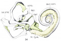
Fig. 34. Human Fetus CRL 50 mm

References
Reviews
Articles
Molina W, Reyes E, Joshi N, Barrios A & Hernandez L. (2010). Maturation of the neuromuscular junction in masseters of human fetus. Rom J Morphol Embryol , 51, 537-41. PMID: 20809033
Chen CP, Tzen CY, Chern SR, Tsai FJ, Hsu CY, Lee CC, Lee MS, Pan CW & Wang W. (2009). A 12 Mb deletion of 6p24.1-->pter in an 18-gestational-week fetus with orofacial clefting, the Dandy-Walker malformation and bilateral multicystic kidneys. Eur J Med Genet , 52, 59-61. PMID: 19049908 DOI.
Wilkin H, Tuohy J & Theewis W. (2000). Prenatal diagnosis of fragile X and Turner mosaicism in a 12-week fetus. Prenat. Diagn. , 20, 854-5. PMID: 11038471
Robb A, Forsyth L & Tolmie J. (1987). Partial trisomy 17q and a generalised bone dysplasia in a 12 week fetus. J. Med. Genet. , 24, 502-4. PMID: 3656375
Search PubMed
Note: Week 10 post-fertilization age (used throughout this current website) is Week 12 gestational age (LMP). Searches for clinical Week 12 gestational age will match this post-fertilization age.
Search PubMed Now: week 10 fetus | week 12 fetus
Glossary Links
- Glossary: A | B | C | D | E | F | G | H | I | J | K | L | M | N | O | P | Q | R | S | T | U | V | W | X | Y | Z | Numbers | Symbols | Term Link
Cite this page: Hill, M.A. (2024, April 26) Embryology Fetal Development - 10 Weeks. Retrieved from https://embryology.med.unsw.edu.au/embryology/index.php/Fetal_Development_-_10_Weeks
- © Dr Mark Hill 2024, UNSW Embryology ISBN: 978 0 7334 2609 4 - UNSW CRICOS Provider Code No. 00098G