Fetal Development: Difference between revisions
| Line 71: | Line 71: | ||
<references/> | <references/> | ||
===Journals=== | |||
* [http://www.sfnmjournal.com/current Seminars in Fetal & Neonatal Medicine] | |||
===Reviews=== | ===Reviews=== | ||
Revision as of 09:39, 30 March 2011
Introduction
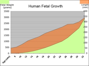
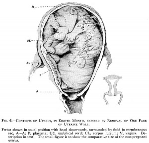
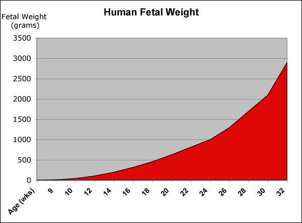
| <Flowplayer height="320" width="285" autoplay="true">fetal growth.flv</Flowplayer> | This page shows some key events of human development during the fetal period (weeks 9 to 37) following fertilization. The long Fetal period (4x the embryonic period) is a time of extensive growth in size and mass as well as ongoing differentiation of organ systems established in the embryonic period. Clinically this period is generally described as the Second Trimester and Third Trimester. Many of the critical measurements of growth are now carried out by ultrasound and this period ends at birth.
Many different systems formed in the embryonic period (organogenesis) grow and differentiate further during the fetal period and do so at different times. For example, the brain continues to grow and develop extensively during this period (and postnatally), the respiratory system differentiates (and completes only just before birth), the urogenital system further differentiates between male/female, endocrine and gastrointestinal tract begins to function. Also consider the systems (respiratory, cardiac, neural) that will still not have their final organization and function determined until after birth. |
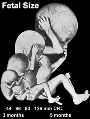
| Changing fetal proportions, not size growth. | Use the links below to get more detailed information about this period of development. |
| Fetal Links: fetal | Week 10 | Week 12 | second trimester | third trimester | fetal neural | Fetal Blood Sampling | fetal growth restriction | birth | birth weight | preterm birth | Developmental Origins of Health and Disease | macrosomia | BGD Practical | Medicine Lecture | Science Lecture | Lecture Movie | Category:Human Fetus | Category:Fetal | |||
|
| original Fetal Development page
Some Recent Findings
|
Reading
- Human Embryology (3rd ed.) Larson Chapter 15: Fetal development and the Fetus as Patient p481-499
- The Developing Human: Clinically Oriented Embryology (8th ed.) Moore and Persaud Chapter 6: The Fetal Period: Ninth Week to Birth
- Color Atlas of Clinical Embryology (2nd ed.) Moore, Persaud and Shiota Chapter 3: 9th to 38th weeks of human development p50-68
Second Trimester
- Second Trimester
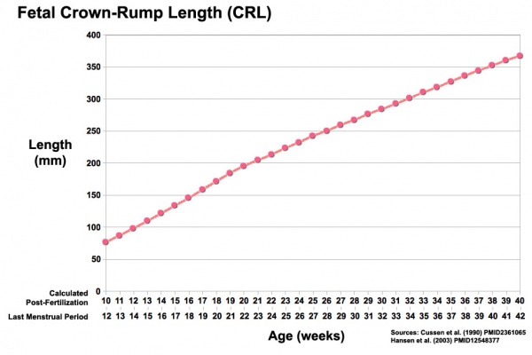
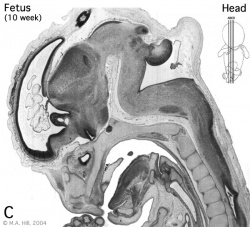
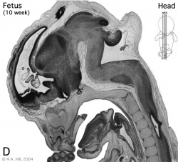
- Week 12 - CRL 85 mm, femur length 15 mm, biparietal diameter 25 mm.
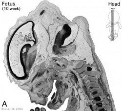
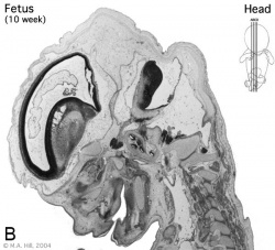
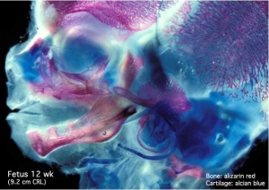
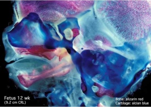
Begin by working through the features present in the early 10 week female fetus. Then look in detail at the head development in a 12 week fetus.
|
|
|
|
Then look in detail at the head development in a 12 week fetus showing both forms of ossification in the skull.
|
|
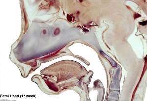
|
Third Trimester
- Third Trimester
- Vibration acoustically of maternal abdominal wall induces startle respone in fetus.
- Month 7 - respiratory bronchioles proliferate and end in alveolar ducts and sacs.
- Week 37 to 38 Birth.
References
Journals
Reviews
- Fetal assessment during pregnancy. Farley D, Dudley DJ. Pediatr Clin North Am. 2009 Jun;56(3):489-504, Table of Contents. Review. PMID: 19501688
- Regimens of fetal surveillance for impaired fetal growth. Grivell RM, Wong L, Bhatia V. Cochrane Database Syst Rev. 2009 Jan 21;(1):CD007113. Review. PMID: 19160321
Articles
Search PubMed
Search Pubmed: human fetal development | fetal development | Second Trimester | Third Trimester
Glossary Links
- Glossary: A | B | C | D | E | F | G | H | I | J | K | L | M | N | O | P | Q | R | S | T | U | V | W | X | Y | Z | Numbers | Symbols | Term Link
Cite this page: Hill, M.A. (2024, April 27) Embryology Fetal Development. Retrieved from https://embryology.med.unsw.edu.au/embryology/index.php/Fetal_Development
- © Dr Mark Hill 2024, UNSW Embryology ISBN: 978 0 7334 2609 4 - UNSW CRICOS Provider Code No. 00098G
