Endoderm
| Embryology - 26 Apr 2024 |
|---|
| Google Translate - select your language from the list shown below (this will open a new external page) |
|
العربية | català | 中文 | 中國傳統的 | français | Deutsche | עִברִית | हिंदी | bahasa Indonesia | italiano | 日本語 | 한국어 | မြန်မာ | Pilipino | Polskie | português | ਪੰਜਾਬੀ ਦੇ | Română | русский | Español | Swahili | Svensk | ไทย | Türkçe | اردو | ייִדיש | Tiếng Việt These external translations are automated and may not be accurate. (More? About Translations) |
Introduction
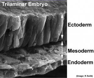
The bottom germ layer of the early trilaminar embryo germ layers (ectoderm, mesoderm and endoderm) formed by gastrulation. Note the historic name for endoderm was "entoderm".
The endoderm contributes the epithelia and glands of the gastrointestinal tract, respiratory tract and the renal bladder. This layer also contributes to the associated gastrointestinal tract organ development (liver and pancreas).
The layer appears to initially be influenced by the overlying notochord and subsequently by a range of growth factors regulating growth and differentiation.
Note that this layer also lines the extra-embryonic yolk sac and allantois, which are initially continuous with the intra-embryonic endoderm.
Some Recent Findings
|
| More recent papers |
|---|
|
This table allows an automated computer search of the external PubMed database using the listed "Search term" text link.
More? References | Discussion Page | Journal Searches | 2019 References | 2020 References Search term: Endoderm Development | Images <pubmed limit=5>Endoderm+Development</pubmed> |
Early Endoderm Cartoon
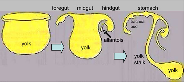
| <mediaplayer width='300' height='320' image="http://php.med.unsw.edu.au/embryology/images/a/af/Endoderm_002_icon.jpg">File:Endoderm 003.mp4</mediaplayer> | This animation shows the early development of endoderm forming the gastrointestinal tract, yolk sac and allantois. The movie starts approximately week 3 and continues through week 4.
Yellow shows the general lining of the yolk sac (bottom), continuous with the endoderm of the trilaminar embryonic disc (top) during week 3. As the trilaminar disc folds in this week, the foregut and hindgut regions become separated from the external yolk sac. The midgut region remains open to the yolk sac and will separate later. Foregut - Begins at the buccopharyngeal membrane, the foregut region in the head is now called the pharynx. At the lower end of the pharynx a ventral bud forms, that will later form the respiratory tract. Beneath this region the tube grows rapidly forming a dilation of the tube, that will later form the stomach. Beneath this region is the boundary of the foregut and ventrally lies the transverse septum. Midgut - Broadly open to the external yolk sac then with continued folding narrows to a "tube-like" connection the yolk stalk. This stalk will later degenerate and all connection will normally be lost. The yolk sac is pushed to the periphery by the growing amniotic sac, with its connecting yolk stalk in the umbilicus region. The midgut region also grows in length forming a loop lying outside the ventral body wall. Hindgut - The loop of midgut renters the body and the ventral portion of the hindgut extends as a blind-ended tube, or diverticulum, into the connecting stalk. This endoderm extension can be seen in histological sections of the initial placental cord and is called the allantois. The hindgut extends caudal (tailward) ending at the cloacal membrane. |
|
Hypochord
The hypochord (subnotochordal rod) is a transient endoderm structure found in the amphibian and fish embryo.[4] It forms a rod-like structure from a single row of cells lying immediately ventral to the notochord. The region is though to play a role in patterning the development of the dorsal aorta and degenerates by apoptosis.
It was first identified in the late 1900's:
- Stöhr, Ph. (1895). Ueber die Entwickelung der Hypochorda und des dorsalen Pankreas bei Rana temporaria. Morph. Jahrb. Bd. 23, 123-141.
- Bergfeldt, A. (1896). Chordascheiden und Hypochorda bei Alytes obstericans. Anat Hefte Bd. 7, 55-102.
- Klaatsch, H., (1898). Zur Frage nach der morphologiscrn Bedeutung der Hypochorda. Morphol. Jahrb. 25, 156-169.
- Links: Frog Development | Zebrafish Development
Molecular

Markers
Several studies have identified the following proteins as markers for endoderm in different species.
- Foxa2 - Forkhead box A2 expressed by anterior primitive streak that forms endoderm.[6] OMIM FOXA2
- Sox17 - Sry-Related HMG-Box gene 17 Sox | OMiM - SOX17
- Cxcr4 - Chemokine, Cxc Motif, Receptor 4 a membrane receptor for neuropeptide Y OMIM CXCR4
- Daf1 - Decay-Accelerating Factor for complement.[1] OMIM DAF1
Nodal
- signaling pathway initiates endoderm and mesoderm development
- also required for proper gastrulation and axial patterning.
- ligands are members of the TGFβ family of secreted growth factors.
- promotes the expression of a network of transcription factors Mix-like proteins (Foxa2, Sox17, Eomesodermin, and Gata4–6).
- Links: TGF-beta | Molecular Development
Patterning
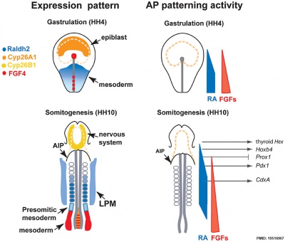
- foregut - (anterior) Hhex, Sox2, and Foxa2 transcription factors.
- hindgut - (posterior) also positioning of the foregut-hindgut boundary, homeobox genes Cdx1, 2, and 4.[7]
Chicken antero-posterior endoderm patterning[8]
References
- ↑ 1.0 1.1 Ogaki S, Omori H, Morooka M, Shiraki N, Ishida S & Kume S. (2016). Late stage definitive endodermal differentiation can be defined by Daf1 expression. BMC Dev. Biol. , 16, 19. PMID: 27245320 DOI.
- ↑ Viotti M, Niu L, Shi SH & Hadjantonakis AK. (2012). Role of the gut endoderm in relaying left-right patterning in mice. PLoS Biol. , 10, e1001276. PMID: 22412348 DOI.
- ↑ <pubmed>19575677</pubmed>
- ↑ Cleaver O, Seufert DW & Krieg PA. (2000). Endoderm patterning by the notochord: development of the hypochord in Xenopus. Development , 127, 869-79. PMID: 10648245
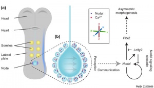
- ↑ Norris DP. (2012). Cilia, calcium and the basis of left-right asymmetry. BMC Biol. , 10, 102. PMID: 23256866 DOI.
- ↑ <pubmed>19234065</pubmed>
- ↑ Chawengsaksophak K, de Graaff W, Rossant J, Deschamps J & Beck F. (2004). Cdx2 is essential for axial elongation in mouse development. Proc. Natl. Acad. Sci. U.S.A. , 101, 7641-5. PMID: 15136723 DOI.
- ↑ Bayha E, Jørgensen MC, Serup P & Grapin-Botton A. (2009). Retinoic acid signaling organizes endodermal organ specification along the entire antero-posterior axis. PLoS ONE , 4, e5845. PMID: 19516907 DOI.
Reviews
Zorn AM & Wells JM. (2009). Vertebrate endoderm development and organ formation. Annu. Rev. Cell Dev. Biol. , 25, 221-51. PMID: 19575677 DOI.
Zorn AM & Wells JM. (2007). Molecular basis of vertebrate endoderm development. Int. Rev. Cytol. , 259, 49-111. PMID: 17425939 DOI.
Lewis SL & Tam PP. (2006). Definitive endoderm of the mouse embryo: formation, cell fates, and morphogenetic function. Dev. Dyn. , 235, 2315-29. PMID: 16752393 DOI.
Grapin-Botton A & Melton DA. (2000). Endoderm development: from patterning to organogenesis. Trends Genet. , 16, 124-30. PMID: 10689353
Wells JM & Melton DA. (1999). Vertebrate endoderm development. Annu. Rev. Cell Dev. Biol. , 15, 393-410. PMID: 10611967 DOI.
Articles
Bayha E, Jørgensen MC, Serup P & Grapin-Botton A. (2009). Retinoic acid signaling organizes endodermal organ specification along the entire antero-posterior axis. PLoS ONE , 4, e5845. PMID: 19516907 DOI.
Historic
Search PubMed
Search NLM Online Textbooks: "Endoderm" : Developmental Biology | The Cell- A molecular Approach | Molecular Biology of the Cell | Endocrinology
Search Pubmed: Endoderm
Additional Images
External Links
Glossary Links
- Glossary: A | B | C | D | E | F | G | H | I | J | K | L | M | N | O | P | Q | R | S | T | U | V | W | X | Y | Z | Numbers | Symbols | Term Link
Cite this page: Hill, M.A. (2024, April 26) Embryology Endoderm. Retrieved from https://embryology.med.unsw.edu.au/embryology/index.php/Endoderm
- © Dr Mark Hill 2024, UNSW Embryology ISBN: 978 0 7334 2609 4 - UNSW CRICOS Provider Code No. 00098G


