Embryology Models: Difference between revisions
| Line 7: | Line 7: | ||
==Historic Models== | ==Historic Models== | ||
===Galletti Models=== | |||
Giuseppe Galletti (? - 1819) these models are currently available for viewing at the Institute and Museum of the History of Science (Italy) "Specimens of obstetric models: the wax models are life-sized; the terracotta versions are reduced to a 1:3 scale. Together with the anatomical waxes in the Specola Museum in Florence, these models are among the most significant examples of the use of artistic techniques for teaching medicine and obstetrics to midwives and surgery students in Florentine hospitals at the end of the eighteenth century." | |||
<gallery> | |||
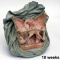
File:Galletti1770_week_10.jpg|Week 10 | |||
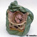
File:Galletti1770_week_16.jpg|Week 16 | |||
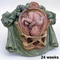
File:Galletti1770_week_24.jpg|Week 24 | |||
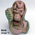
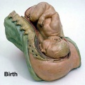
File:Galletti1770 birth.jpg|Birth | |||
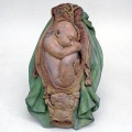
File:Galletti1770 birth2.jpg|Birth | |||
File:Galletti1770_breech_01.jpg|Birth (breech) | |||
File:Galletti1770_breech_02.jpg|Birth (breech) | |||
File:Galletti1770_breech_03.jpg|Birth (breech) | |||
File:Galletti1770_breech_04.jpg|Birth (breech) | |||
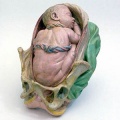
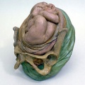
File:Galletti1770_placenta_previa.jpg|Birth (placenta previ) | |||
</gallery> | |||
===Ziegler Models=== | ===Ziegler Models=== | ||
[[File:Ziegler_model_01.jpg|thumb|Ziegler Models]] | [[File:Ziegler_model_01.jpg|thumb|Ziegler Models]] | ||
| Line 24: | Line 41: | ||
===Smithsonian Museum=== | ===Smithsonian Museum=== | ||
[http://americanhistory.si.edu/anatomy/collection/nma03_collection_human.html 12 stages of Embryonic Development - Anatomical Model] | [http://americanhistory.si.edu/anatomy/collection/nma03_collection_human.html 12 stages of Embryonic Development - Anatomical Model] | ||
==Modern Models== | ==Modern Models== | ||
Revision as of 06:42, 8 December 2012
Introduction
The very size and dynamic nature of embryonic development has always made it a difficult topic to teach. The structures are always so small and the changing nature of development means that a feature you identify at any one time is called and becomes something else at a later stage.
Models made from ceramic, wax or plastic, not the "animal models", are useful in that they make tangible the microscopic structures to the students. Models have been in use for some time in the medical teaching of human development with the original plaster models now replaced by commercial modern plastic versions. Many of the early teaching models were not about development, but about the process of childbirth for doctor and midwife education.
Some links on this page are to both historic non-commercial and modern commercial suppliers of some education models for embryology. In some cases these are museum exhibits only available for viewing. This list is provided for information purposes only and does not indicate an endorsement of these commercial products.
Historic Models
Galletti Models
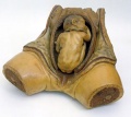
Giuseppe Galletti (? - 1819) these models are currently available for viewing at the Institute and Museum of the History of Science (Italy) "Specimens of obstetric models: the wax models are life-sized; the terracotta versions are reduced to a 1:3 scale. Together with the anatomical waxes in the Specola Museum in Florence, these models are among the most significant examples of the use of artistic techniques for teaching medicine and obstetrics to midwives and surgery students in Florentine hospitals at the end of the eighteenth century."
Week 10
Week 16
Week 24
Birth
Birth
Birth (breech)
Birth (breech)
Birth (breech)
Birth (breech)
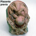
Birth (placenta previ)


Ziegler Models

Adolf Ziegler (1820 - 1889) a German modeller developed these models at his Studio for Scientific Teaching Models (Atelier für wissenchaftliche Unterrichtsmodelle) from the 1860's onwards of both human and animal embryos. Ziegler's models were first used in Germany, but were also later exhibited in Paris (1867) and Vienna (1873).
His son, Friedrich Ziegler (- 1936), continued the modelling company.
Carnegie Models

One of the first to be hired, in 1913, was modeler Osborne O. Heard, who spent 42 years at the department and made over 700 wax-based reconstructions. The results of this team effort still stand as the international standard by which human embryos are described and classified. The models were mainly made by the lost-wax casting process and his models were also more detailed than the earlier (1880's) Ziegler embryo models.
Smithsonian Museum
12 stages of Embryonic Development - Anatomical Model
Modern Models
Nucleus
- Giant Embryo Model
- 2nd Month Embryo
- Fetal Skull, without stand
- Classic Pregnancy Series, 8 Models
- Classic Pregnancy Pelvis 2-part
- Deluxe Pregnancy Pelvis 3-part
- 5 stage Birth Process Model
The Evolution Store
Lippincott Williams and Wilkins
Shanghai Honglian Medical Instrument Development
- Fertilization and Early Development Shows the development from ovum to the fetus in third month.
- Early Development Fecundation to the embryo of fourth week.
- Embryo The embryo model is about 4 weeks old, and mounted on a stand.
- Embryonic Development The model includes 8 parts showing the uterus with embryo and fetus from the first to the seventh month pregnancy.
Glossary Links
- Glossary: A | B | C | D | E | F | G | H | I | J | K | L | M | N | O | P | Q | R | S | T | U | V | W | X | Y | Z | Numbers | Symbols | Term Link
Cite this page: Hill, M.A. (2024, May 21) Embryology Embryology Models. Retrieved from https://embryology.med.unsw.edu.au/embryology/index.php/Embryology_Models
- © Dr Mark Hill 2024, UNSW Embryology ISBN: 978 0 7334 2609 4 - UNSW CRICOS Provider Code No. 00098G