Connective Tissue Development

From Embryology
Revision as of 18:47, 27 August 2011 by S8600021 (talk | contribs) (Created page with " ==Introduction== Loose and dense connective tissue Reticular connective tissue Adipose Tissue Mesenchymal connective tissue {{Template:Systems}} ==Some Recent Findings== {| ...")
(diff) ← Older revision | Latest revision (diff) | Newer revision → (diff)

Introduction

Loose and dense connective tissue Reticular connective tissue Adipose Tissue Mesenchymal connective tissue


System Links: Introduction | Cardiovascular | Coelomic Cavity | Endocrine | Gastrointestinal Tract | Genital | Head | Immune | Integumentary | Musculoskeletal | Neural | Neural Crest | Placenta | Renal | Respiratory | Sensory | Birth

Some Recent Findings

  • Brown adipose tissue: function and physiological significance[1] "The function of brown adipose tissue is to transfer energy from food into heat; physiologically, both the heat produced and the resulting decrease in metabolic efficiency can be of significance. ... The development of brown adipose tissue with its characteristic protein, uncoupling protein-1 (UCP1), was probably determinative for the evolutionary success of mammals, as its thermogenesis enhances neonatal survival and allows for active life even in cold surroundings."

Textbooks

Objectives

Development Overview

Mesoderm Development

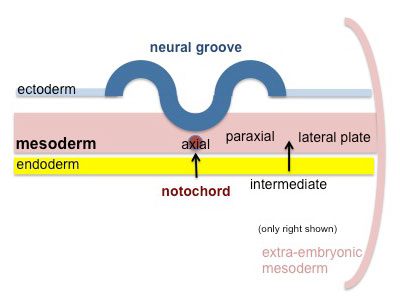
Mesoderm cartoon 01.jpg Cells migrate through the primitive streak to form mesodermal layer. Extraembryonic mesoderm lies adjacent to the trilaminar embryo totally enclosing the amnion, yolk sac and forming the connecting stalk.
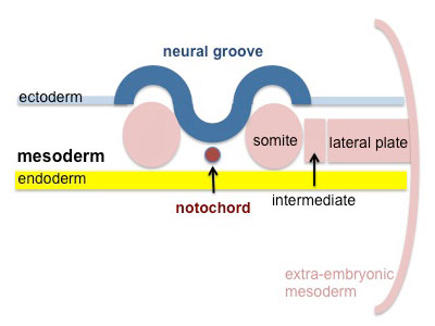
Mesoderm cartoon 02.jpg Paraxial mesoderm accumulates under the neural plate with thinner mesoderm laterally. This forms 2 thickened streaks running the length of the embryonic disc along the rostrocaudal axis. In humans, during the 3rd week, this mesoderm begins to segment. The neural plate folds to form a neural groove and folds.
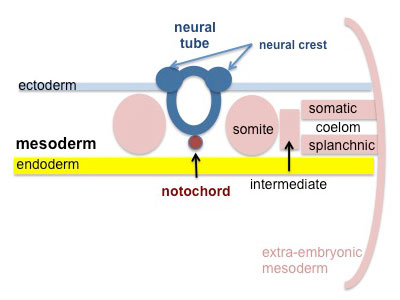
Mesoderm cartoon 03.jpg Segmentation of the paraxial mesoderm into somites continues caudally at 1 somite/90minutes and a cavity (intraembryonic coelom) forms in the lateral plate mesoderm separating somatic and splanchnic mesoderm.

Note intraembryonic coelomic cavity communicates with extraembryonic coelom through portals (holes) initially on lateral margin of embryonic disc.

Mesoderm cartoon 04.jpg Somites continue to form. The neural groove fuses dorsally to form a tube at the level of the 4th somite and "zips up cranially and caudally and the neural crest migrates into the mesoderm.

References

  1. <pubmed>14715917</pubmed>


Reviews

<pubmed>14715917</pubmed>

Articles

Search PubMed

Search Pubmed: connective tissue development | adipose Development | [http://www.ncbi.nlm.nih.gov/sites/entrez?==Additional Images==

Terms

Glossary Links

Glossary: A | B | C | D | E | F | G | H | I | J | K | L | M | N | O | P | Q | R | S | T | U | V | W | X | Y | Z | Numbers | Symbols | Term Link

Cite this page: Hill, M.A. (2024, May 6) Embryology Connective Tissue Development. Retrieved from https://embryology.med.unsw.edu.au/embryology/index.php/Connective_Tissue_Development

What Links Here?
© Dr Mark Hill 2024, UNSW Embryology ISBN: 978 0 7334 2609 4 - UNSW CRICOS Provider Code No. 00098G