Connective Tissue Development
Introduction
Connective tissues in the body region have a mesoderm origin, while in the head region neural crest also contributes to these tissues.
This topic is also covered in musculoskeletal (Tendon Development), integumentary (Integumentary Development) and endocrine development (Adipose Tissue).
Blood is a liquid connective tissue (More? Blood Development).
- Loose and dense connective tissue
- Reticular connective tissue
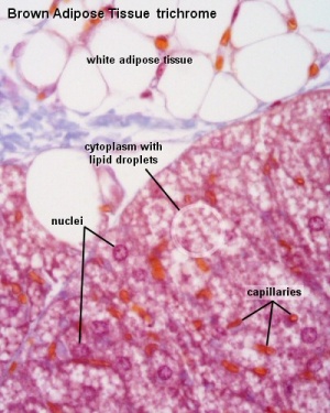
- Adipose Tissue
- Mesenchymal connective tissue
| System Links: Introduction | Cardiovascular | Coelomic Cavity | Endocrine | Gastrointestinal Tract | Genital | Head | Immune | Integumentary | Musculoskeletal | Neural | Neural Crest | Placenta | Renal | Respiratory | Sensory | Birth |
Some Recent Findings
|
Development Overview
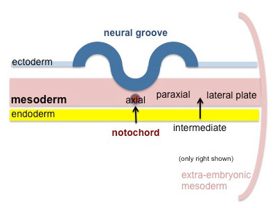
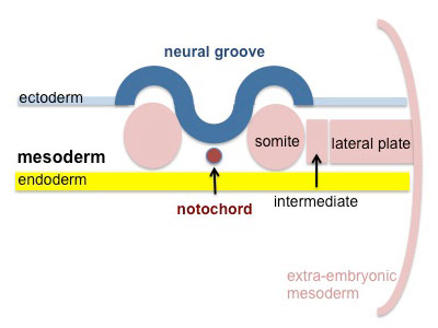
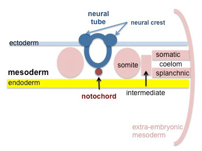
Mesoderm Development
Somite - Dermatome
The dermis and hypodermis layers of the skin.
Somatic Mesoderm
The body wall connective tissue.
Splanchnic Mesoderm
The lamina propria and submucosa layers of the gastrointestinal tract wall.
References
- ↑ Song Y, Lai HX, Song TW, Gong J, Liu B, Chi YY, Yue C, Zhang J, Sun SZ, Zhang CH, Tang W, Fan N, Yu WH, Wang YF, Hack GD, Yu SB, Zhang JF & Sui HJ. (2023). The growth and developmental of the myodural bridge and its associated structures in the human fetus. Sci Rep , 13, 13421. PMID: 37591924 DOI.
- ↑ Cannon B & Nedergaard J. (2004). Brown adipose tissue: function and physiological significance. Physiol. Rev. , 84, 277-359. PMID: 14715917 DOI.
Reviews
Tews D & Wabitsch M. (2011). Renaissance of brown adipose tissue. Horm Res Paediatr , 75, 231-9. PMID: 21372557 DOI.
Schulz TJ & Tseng YH. (2009). Emerging role of bone morphogenetic proteins in adipogenesis and energy metabolism. Cytokine Growth Factor Rev. , 20, 523-31. PMID: 19896888 DOI.
Forhead AJ & Fowden AL. (2009). The hungry fetus? Role of leptin as a nutritional signal before birth. J. Physiol. (Lond.) , 587, 1145-52. PMID: 19188249 DOI.
Billon N, Monteiro MC & Dani C. (2008). Developmental origin of adipocytes: new insights into a pending question. Biol. Cell , 100, 563-75. PMID: 18793119 DOI.
Cannon B & Nedergaard J. (2004). Brown adipose tissue: function and physiological significance. Physiol. Rev. , 84, 277-359. PMID: 14715917 DOI.
Articles
Billon N, Kolde R, Reimand J, Monteiro MC, Kull M, Peterson H, Tretyakov K, Adler P, Wdziekonski B, Vilo J & Dani C. (2010). Comprehensive transcriptome analysis of mouse embryonic stem cell adipogenesis unravels new processes of adipocyte development. Genome Biol. , 11, R80. PMID: 20678241 DOI.
Billon N, Iannarelli P, Monteiro MC, Glavieux-Pardanaud C, Richardson WD, Kessaris N, Dani C & Dupin E. (2007). The generation of adipocytes by the neural crest. Development , 134, 2283-92. PMID: 17507398 DOI.
Search PubMed
Search Pubmed: connective tissue development | adipose Development
Additional Images
Terms
Glossary Links
- Glossary: A | B | C | D | E | F | G | H | I | J | K | L | M | N | O | P | Q | R | S | T | U | V | W | X | Y | Z | Numbers | Symbols | Term Link
Cite this page: Hill, M.A. (2024, April 19) Embryology Connective Tissue Development. Retrieved from https://embryology.med.unsw.edu.au/embryology/index.php/Connective_Tissue_Development
- © Dr Mark Hill 2024, UNSW Embryology ISBN: 978 0 7334 2609 4 - UNSW CRICOS Provider Code No. 00098G


