Category:Skull
From Embryology
The pages and media shown below relate to development of the skull.
Pages in category 'Skull'
The following 119 pages are in this category, out of 119 total.
A
B
F
J
M
P
- Palate Development
- Paper - A model of the left half of the human mandible at the 17 mm CRL stage
- Paper - Description of a reconstruction of the head of a thirty-millimetre embryo (1910)
- Paper - Development of the malleus of the human ear - Illustrated in atlas series
- Paper - Development of the otic capsule 2
- Paper - Further observations on the ossification of the human lower jaw
- Paper - Notes on the development of the human sphenoid (1910)
- Paper - On the development and morphology of the human sphenoid bone
- Paper - On the premature obliteration of sutures in the human skull (1915)
- Paper - Pharyngeal end of Rathke's pouch (1911)
- Paper - Preliminary note on the skull of a human fetus of 43 mm greatest length
- Paper - Some observations on the roof of the primordial human cranium (1923)
- Paper - Structure and development of the pig skull
- Paper - The cartilaginous skull of a human embryo twenty-one millimeters in length (1920)
- Paper - The chondrocranium of a 20 mm human embryo
- Paper - The development of the cochlear fenestra, fossula and secondary tympanic membrane
- Paper - The development of the human maxilla, vomer, and paraseptal cartilages (1911)
- Paper - The development of the otic capsule in the region of the vestibular aqueduct
- Paper - The developmental and adult anatomy of the air-cells in the petrous part of the temporal bone
- Paper - The fontanella metopica and its remnants in an adult skull (1918)
- Paper - The genesis and development of the nasolacrimal passages in man
- Paper - The lateral wall of the cavum nasi in man, with especial reference to the various developmental stages
- Paper - The Long Fox lecture - The development of the human skull (1910)
- Paper - The Monotreme Skull - A Contribution to Mammalian Morphogenesis
- Paper - The ossification of the human frontal bone with special reference to its presumed pre- and post-frontal elements
- Paper - The primordial cranium of miniopterus schreibersi at the 17 millimetre total length stage (1919)
- Paper - The skull of a human fetus of 40 mm 1
- Paper - The skull of a human fetus of 40 mm 2
- Template:Pharyngeal arch
R
- Template:Ref-AnsonBast1958e
- Template:Ref-AnsonBastRichany1956
- Template:Ref-Bolk1915
- Template:Ref-Covell1927
- Template:Ref-Fawcett1910lecture
- Template:Ref-Fawcett1910sphenoid
- Template:Ref-Fawcett1911
- Template:Ref-Fawcett1917
- Template:Ref-Fawcett1918
- Template:Ref-Fawcett1918a
- Template:Ref-Fawcett1919
- Template:Ref-Fawcett1923
- Template:Ref-Gilse1927
- Template:Ref-GladstoneWakeley1923
- Template:Ref-Hayes1922
- Template:Ref-Inman1937
- Template:Ref-InmanSaunders1937
- Template:Ref-Kernan1916
- Template:Ref-Lewis1920
- Template:Ref-Low1909
- Template:Ref-Macklin1914a
- Template:Ref-Macklin1914b
- Template:Ref-Macklin1921
- Template:Ref-Macklin1921a
- Template:Ref-Murray1943
- Template:Ref-Parker1874
- Template:Ref-RichanyBastAnson1956
- Template:Ref-Schaeffer1910a
- Template:Ref-Schaeffer1910b
- Template:Ref-Schaeffer1911
- Template:Ref-Schaeffer1912
- Template:Ref-Schultz1918
- Template:Ref-StelterBastAnson1960
- Template:Ref-Sutton1885
- Template:Ref-Terry1909
- Template:Ref-Tomes1853
- Template:Ref-Walusch1906
- Template:Ref-Watson1915
S
Media in category 'Skull'
The following 47 files are in this category, out of 247 total.
(previous page) (next page) Rugh 145.jpg 800 × 766; 152 KB
 Rugh 149.jpg 1,000 × 746; 196 KB
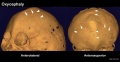
 Skull - microcephaly 01.jpg 750 × 1,000; 75 KB
 Skull - osteoblast lineage model.jpg 600 × 381; 20 KB
 Skull anterior.gif 200 × 205; 30 KB
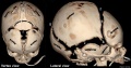
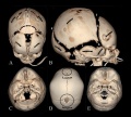
 Skull CT abnormal 01.jpg 1,000 × 549; 93 KB
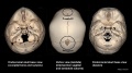
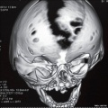
 Skull CT abnormal 02.jpg 1,000 × 900; 119 KB
 Skull CT abnormal 03.jpg 1,000 × 542; 82 KB
 Skull CT abnormal 04.jpg 1,000 × 646; 102 KB
 Skull CT abnormal 05.jpg 1,000 × 572; 88 KB
 Skull CT abnormal 06.jpg 1,000 × 541; 85 KB
 Skull CT abnormal 07.jpg 1,000 × 541; 64 KB
 Skull CT abnormal 08.jpg 1,000 × 516; 73 KB
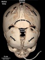
 Skull CT normal sutures 01.jpg 1,000 × 526; 89 KB
 Skull CT normal sutures 02.jpg 1,000 × 559; 92 KB
 Skull CT normal sutures 03.jpg 600 × 800; 63 KB
 Skull CT normal sutures.jpg 1,000 × 900; 138 KB
 Skull lateral view.gif 200 × 147; 22 KB
 Skull superior.gif 200 × 163; 22 KB
 Skull vault defect and midface hypoplasia.jpg 800 × 798; 81 KB
 Skull viscerocranium 01.mp4 ; 443 KB
 Stage 22 image 204.jpg 1,190 × 848; 363 KB
 Vesalius Skull.jpg 600 × 382; 44 KB
 Watson1915 Fig01.jpg 929 × 1,000; 171 KB
 Watson1915 Fig02.jpg 1,421 × 1,000; 245 KB
 Watson1915 Fig03.jpg 1,533 × 1,000; 276 KB
 Watson1915 Fig04.jpg 1,296 × 1,000; 197 KB
 Watson1915 Fig05.jpg 1,147 × 1,000; 127 KB
 Watson1915 Fig06.jpg 1,402 × 1,000; 226 KB
 Watson1915 Fig07.jpg 1,078 × 1,000; 235 KB
 Watson1915 Fig08.jpg 1,183 × 1,000; 211 KB
 Watson1915 Fig09.jpg 1,173 × 1,000; 253 KB
 Watson1915 Fig10.jpg 1,101 × 1,000; 0 bytes
 Watson1915 Fig11.jpg 1,060 × 1,000; 180 KB
 Watson1915 Fig12.jpg 1,119 × 1,000; 186 KB
 Watson1915 Fig13.jpg 1,168 × 1,000; 176 KB
 Watson1915 Fig14.jpg 1,256 × 1,000; 174 KB
 Watson1915 Fig15.jpg 1,132 × 1,000; 136 KB
 Watson1915 Fig16.jpg 1,187 × 1,000; 179 KB
 Watson1915 Fig17.jpg 1,193 × 1,000; 166 KB
 Watson1915 Fig18.jpg 1,186 × 1,000; 144 KB
 Watson1915 Fig19.jpg 1,354 × 1,000; 159 KB
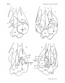 Watson1915 Plate01.jpg 1,552 × 2,000; 275 KB
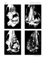 Watson1915 Plate02.jpg 1,552 × 2,000; 527 KB
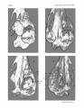 Watson1915 Plate03.jpg 1,552 × 2,000; 605 KB
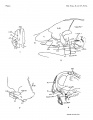 Watson1915 Plate04.jpg 1,552 × 2,000; 230 KB
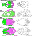 Zebrafish skull neural crest.jpg 815 × 933; 187 KB


