Category:Scanning EM
From Embryology
This Embryology category shows pages and media related to the research imaging technique of scanning electron micrographs (SEM) in development. Note:
- images in this category may also include some of the associated bright field images taken before SEM fixation and imaging.
- there is a separate Category:Electron Micrograph.
Pages in category 'Scanning EM'
The following 16 pages are in this category, out of 16 total.
C
Media in category 'Scanning EM'
The following 26 files are in this category, out of 426 total.
(previous page) (next page) Stage9 sem4a.jpg 643 × 800; 56 KB
 Stage9 sem4b.jpg 482 × 600; 36 KB
 Stage9 sem4c.jpg 321 × 400; 19 KB
 Stage9 sem5.jpg 1,359 × 1,000; 135 KB
 Stage9 sem5a.jpg 1,087 × 800; 100 KB
 Stage9 sem5b.jpg 815 × 600; 67 KB
 Stage9 sem5c.jpg 543 × 400; 37 KB
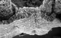
 Stage9 sem6.jpg 627 × 1,000; 114 KB
 Stage9 sem6a.jpg 502 × 800; 80 KB
 Stage9 sem6b.jpg 377 × 600; 50 KB
 Stage9 sem6c.jpg 251 × 400; 25 KB
 Stage9 sem7.jpg 1,000 × 626; 79 KB
 Stage9 sem7a.jpg 800 × 501; 57 KB
 Stage9 sem7b.jpg 600 × 376; 37 KB
 Stage9 sem7c.jpg 400 × 251; 19 KB
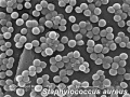 Staphylococcus-aureus.jpg 320 × 240; 28 KB
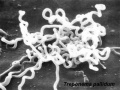 Treponema-pallidum.jpg 320 × 240; 21 KB
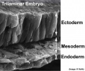 Trilaminar embryo.jpg 432 × 359; 32 KB
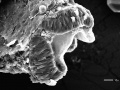 Zebrafish brain fold SEM.jpg 1,000 × 750; 172 KB
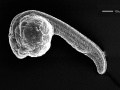 Zebrafish day 1 SEM.jpg 1,000 × 750; 130 KB
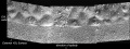 Zebrafish enveloping layer SEM01.jpg 1,200 × 447; 150 KB
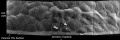 Zebrafish enveloping layer SEM02.jpg 1,200 × 400; 117 KB
 Zebrafish myotomes SEM.jpg 1,000 × 750; 250 KB
 Zebrafish perichordal sheath SEM.jpg 1,000 × 750; 117 KB
 Zebrafish trunk SEM01.jpg 1,000 × 750; 222 KB
 Zebrafish trunk SEM02.jpg 750 × 1,000; 163 KB


