Category:Oocyte: Difference between revisions
From Embryology
mNo edit summary |
mNo edit summary |
||
| Line 4: | Line 4: | ||
:'''Links:''' {{oocyte}} | {{meiosis}} | {{ovary | :'''Links:''' {{oocyte}} | {{meiosis}} | {{ovary}} | ||
Revision as of 18:37, 1 May 2018
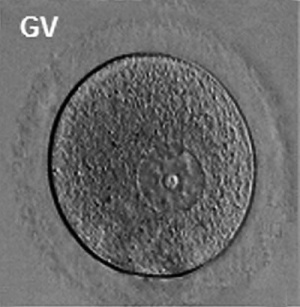
This Embryology category shows pages and media related to the female germ cell the oocyte (egg, ovum) development. In humans the oocyte develops before birth and recommences meiosis at puberty, only the fertilised oocyte ever completes meiosis.
Subcategories
This category has the following 6 subcategories, out of 6 total.
Pages in category 'Oocyte'
The following 145 pages are in this category, out of 145 total.
A
B
F
H
M
O
- Oncofertility
- Template:Oocyte
- Oocyte Development
- Oocyte Meiosis Movie 1
- Template:Oocyte terms
- Template:Oocyte terms collapse table
- Template:Oogenesis
- Template:Ova
- Template:Ovarian reserve
- Template:Ovary
- Template:Ovary timeline table
- Template:Ovary Vignette
- Ovulation in the human ovary - Its mechanism and anomalies
- Template:Ovum
P
- Paper - Cell-to-cell communication and ovulation - A study of the cumulus-oocyte complex
- Paper - Cleavage Stages of the Ova of the Horse with Notes on Ovulation
- Paper - Cleavage stages of the ova of the horse with notes on ovulation (1945)
- Paper - Development of the egg of the cow up to the stage of blastocyst formation (1946)
- Paper - History of the development of the human ovum (1834)
- Paper - Human oocyte showing first polar body and metaphase stage in formation of second polar body
- Paper - Human ova from large follicles
- Paper - Human ova from large follicles - including a search for maturation divisions and observations on atresia
- Paper - Human ova from large follicles - including a search for maturation divisions and observations on atresia (1930)
- Paper - Maturation of the ovum in swine (1917)
- Paper - On the persistence of oocyte nuclei from fetus to maturity in the laboratory mouse
- Paper - Oogenesis in the white mouse (1917)
- Paper - Recovery of human ova from the uterine tubes
- Paper - Selective elimination of ova in the adult ovary
- Paper - Selective elimination of ova in the adult ovary (1925)
- Paper - Studies on guinea pig oocytes 1
- Paper - Studies on the human oocyte and its follicle 1
- Paper - The Comparative Behavior of Mammalian Eggs in Vivo and in Vitro
- Paper - The early development of the ferret - the zona granulosa, zona pellucida and associated structures (1932)
- Paper - The fate of the graafian follicle in the human ovary
- Paper - The formation and structure of the zona pellucida in the ovarian eggs of turtles (1918)
- Paper - The Maturation of the Human Ovum
- Paper - The occurrence of polyovular graafian follicles (1924)
- Paper - Transuterine (internal) migration of the ovum in sheep and other mammals
- Template:Pincus1935 figures
- Template:Polar bodies
- Template:Polar body
- Template:Preovulatory follicle
- Template:Primary follicle
- Template:Primordial follicle
- Template:Primordial germ cell
- Template:Pronuclei
- Template:Pronucleus
R
- Template:Ref-AdamsHertig1964
- Template:Ref-Allen1925
- Template:Ref-Allen1930b
- Template:Ref-AllenPrattNewellBland1928
- Template:Ref-Arnold1912
- Template:Ref-BakerHookSeveringhaus1944
- Template:Ref-Balbiani1864a
- Template:Ref-Balbiani1864b
- Template:Ref-Balbiani1893
- Template:Ref-BeamsSheehan1941
- Template:Ref-Boyd1944
- Template:Ref-Corner1917
- Template:Ref-Corner1921b
- Template:Ref-Corner1923
- Template:Ref-Dixon1927
- Template:Ref-ESHRE Atlas2012
- Template:Ref-Gilula1978
- Template:Ref-Guttmacher1921
- Template:Ref-Hamilton1944
- Template:Ref-Hamilton1945
- Template:Ref-Hamilton1946a
- Template:Ref-Hartman1929
- Template:Ref-Hertig1968a
- Template:Ref-HertigAdams1967
- Template:Ref-Keibelchapter1-1910
- Template:Ref-Kennedy1924
- Template:Ref-Kingery1917
- Template:Ref-LongMark1911
- Template:Ref-Mainland1932
- Template:Ref-Mckay1953
- Template:Ref-Meyer1917
- Template:Ref-Morgan1915
- Template:Ref-Nicol1933
- Template:Ref-PeredaCoppo1984
- Template:Ref-Pincus1936
- Template:Ref-PMID5323255
- Template:Ref-PMID6053018
- Template:Ref-Robinson1904a
- Template:Ref-Robinson1904b
- Template:Ref-Robinson1904c
- Template:Ref-RudkinGriech1962
- Template:Ref-Shaw1925
- Template:Ref-Streeter1931
- Template:Ref-Thing1918
- Template:Ref-Thomson1839
- Template:Ref-Thomson1919a
- Template:Ref-Thomson1919b
- Template:Ref-Valentin1834
- Template:Ref-Van der Stricht1923
Media in category 'Oocyte'
The following 22 files are in this category, out of 222 total.
(previous page) (next page) Rugh 025.jpg 1,200 × 651; 178 KB
 Rugh 026.jpg 628 × 800; 119 KB
 Rugh 053.jpg 848 × 800; 128 KB
 Salamander- early development.jpg 1,200 × 887; 85 KB
 Salamander-oocyte.jpg 637 × 450; 21 KB
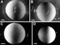
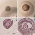 Sheep follicle in vitro 01.jpg 600 × 600; 108 KB
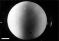
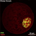
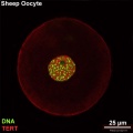 Sheep oocyte 01.jpg 650 × 651; 43 KB
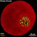
 Sheep oocyte 02.jpg 650 × 651; 59 KB
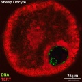
 Sheep oocyte 03.jpg 650 × 651; 94 KB
 Sheep oocyte 04.jpg 650 × 651; 113 KB
 Thomson1919 fig01.jpg 1,128 × 1,198; 341 KB
 Thomson1919 fig02.jpg 1,172 × 1,233; 354 KB
 Thomson1919 fig03.jpg 1,226 × 1,110; 308 KB
 Thomson1919 fig04.jpg 1,251 × 1,089; 316 KB
 Thomson1919 fig05.jpg 1,176 × 1,226; 335 KB
 Thomson1919 fig06.jpg 1,482 × 1,368; 471 KB
 Thomson1919 fig07.jpg 1,490 × 1,405; 476 KB
 Thomson1919 fig08.jpg 1,443 × 1,387; 486 KB
 Thomson1919 fig09.jpg 1,443 × 1,381; 476 KB
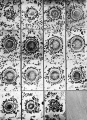 Thomson1919 plate10.jpg 1,249 × 1,729; 844 KB
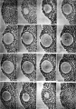 Thomson1919 plate11.jpg 1,275 × 1,828; 694 KB
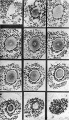 Thomson1919 plate12.jpg 1,074 × 1,869; 735 KB


