Category:Oocyte: Difference between revisions
From Embryology
mNo edit summary |
mNo edit summary |
||
| (2 intermediate revisions by the same user not shown) | |||
| Line 2: | Line 2: | ||
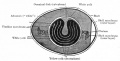
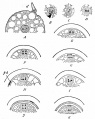
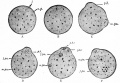
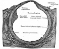
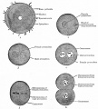
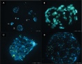
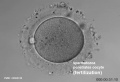
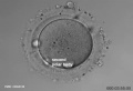
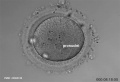
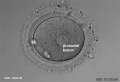
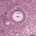
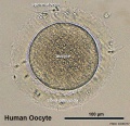
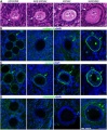
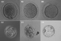
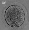
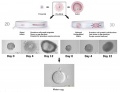
This {{Embryology}} category shows pages and media related to the female germ cell the oocyte (egg, ovum) development. In humans the oocyte develops before birth and recommences meiosis at puberty, only the fertilised oocyte ever completes meiosis. | This {{Embryology}} category shows pages and media related to the female germ cell the oocyte (egg, ovum) development. In humans the oocyte develops before birth and recommences meiosis at puberty, only the fertilised oocyte ever completes meiosis. | ||
<br> | |||
:'''Links:''' {{oocyte}} | {{meiosis}} | {{ovary}} | |||
<br> | |||
{| class="wikitable mw-collapsible mw-collapsed" | |||
! [[Embryology_History|'''Historic Embryology''']] - Oocyte | |||
|- | |||
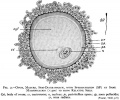
| [[Paper - History of the development of the human ovum (1834)|1834 Human and other species Ovum]] | [[Paper - The Maturation of the Human Ovum|1919 Human Ovum]] | [[Book_-_Text-Book_of_Embryology_1#The_Ovum|1921 The Ovum]] | [[Paper - How Large is the Mammalian Egg?|1929 Oocyte Size]] | |||
|} | |||
<br> | |||
{{Genital Links}} | |||
[[Category:Primordial Germ Cell]] | [[Category:Primordial Germ Cell]] | ||
Latest revision as of 12:59, 11 December 2019

This Embryology category shows pages and media related to the female germ cell the oocyte (egg, ovum) development. In humans the oocyte develops before birth and recommences meiosis at puberty, only the fertilised oocyte ever completes meiosis.
| Historic Embryology - Oocyte |
|---|
| 1834 Human and other species Ovum | 1919 Human Ovum | 1921 The Ovum | 1929 Oocyte Size |
Subcategories
This category has the following 6 subcategories, out of 6 total.
Pages in category 'Oocyte'
The following 145 pages are in this category, out of 145 total.
A
B
F
H
M
O
- Oncofertility
- Template:Oocyte
- Oocyte Development
- Oocyte Meiosis Movie 1
- Template:Oocyte terms
- Template:Oocyte terms collapse table
- Template:Oogenesis
- Template:Ova
- Template:Ovarian reserve
- Template:Ovary
- Template:Ovary timeline table
- Template:Ovary Vignette
- Ovulation in the human ovary - Its mechanism and anomalies
- Template:Ovum
P
- Paper - Cell-to-cell communication and ovulation - A study of the cumulus-oocyte complex
- Paper - Cleavage Stages of the Ova of the Horse with Notes on Ovulation
- Paper - Cleavage stages of the ova of the horse with notes on ovulation (1945)
- Paper - Development of the egg of the cow up to the stage of blastocyst formation (1946)
- Paper - History of the development of the human ovum (1834)
- Paper - Human oocyte showing first polar body and metaphase stage in formation of second polar body
- Paper - Human ova from large follicles
- Paper - Human ova from large follicles - including a search for maturation divisions and observations on atresia
- Paper - Human ova from large follicles - including a search for maturation divisions and observations on atresia (1930)
- Paper - Maturation of the ovum in swine (1917)
- Paper - On the persistence of oocyte nuclei from fetus to maturity in the laboratory mouse
- Paper - Oogenesis in the white mouse (1917)
- Paper - Recovery of human ova from the uterine tubes
- Paper - Selective elimination of ova in the adult ovary
- Paper - Selective elimination of ova in the adult ovary (1925)
- Paper - Studies on guinea pig oocytes 1
- Paper - Studies on the human oocyte and its follicle 1
- Paper - The Comparative Behavior of Mammalian Eggs in Vivo and in Vitro
- Paper - The early development of the ferret - the zona granulosa, zona pellucida and associated structures (1932)
- Paper - The fate of the graafian follicle in the human ovary
- Paper - The formation and structure of the zona pellucida in the ovarian eggs of turtles (1918)
- Paper - The Maturation of the Human Ovum
- Paper - The occurrence of polyovular graafian follicles (1924)
- Paper - Transuterine (internal) migration of the ovum in sheep and other mammals
- Template:Pincus1935 figures
- Template:Polar bodies
- Template:Polar body
- Template:Preovulatory follicle
- Template:Primary follicle
- Template:Primordial follicle
- Template:Primordial germ cell
- Template:Pronuclei
- Template:Pronucleus
R
- Template:Ref-AdamsHertig1964
- Template:Ref-Allen1925
- Template:Ref-Allen1930b
- Template:Ref-AllenPrattNewellBland1928
- Template:Ref-Arnold1912
- Template:Ref-BakerHookSeveringhaus1944
- Template:Ref-Balbiani1864a
- Template:Ref-Balbiani1864b
- Template:Ref-Balbiani1893
- Template:Ref-BeamsSheehan1941
- Template:Ref-Boyd1944
- Template:Ref-Corner1917
- Template:Ref-Corner1921b
- Template:Ref-Corner1923
- Template:Ref-Dixon1927
- Template:Ref-ESHRE Atlas2012
- Template:Ref-Gilula1978
- Template:Ref-Guttmacher1921
- Template:Ref-Hamilton1944
- Template:Ref-Hamilton1945
- Template:Ref-Hamilton1946a
- Template:Ref-Hartman1929
- Template:Ref-Hertig1968a
- Template:Ref-HertigAdams1967
- Template:Ref-Keibelchapter1-1910
- Template:Ref-Kennedy1924
- Template:Ref-Kingery1917
- Template:Ref-LongMark1911
- Template:Ref-Mainland1932
- Template:Ref-Mckay1953
- Template:Ref-Meyer1917
- Template:Ref-Morgan1915
- Template:Ref-Nicol1933
- Template:Ref-PeredaCoppo1984
- Template:Ref-Pincus1936
- Template:Ref-PMID5323255
- Template:Ref-PMID6053018
- Template:Ref-Robinson1904a
- Template:Ref-Robinson1904b
- Template:Ref-Robinson1904c
- Template:Ref-RudkinGriech1962
- Template:Ref-Shaw1925
- Template:Ref-Streeter1931
- Template:Ref-Thing1918
- Template:Ref-Thomson1839
- Template:Ref-Thomson1919a
- Template:Ref-Thomson1919b
- Template:Ref-Valentin1834
- Template:Ref-Van der Stricht1923
Media in category 'Oocyte'
The following 200 files are in this category, out of 222 total.
(previous page) (next page)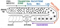 Adult hermaphrodite gonad arm.jpg 800 × 377; 66 KB
 Amphibian oocyte transcription.jpg 1,200 × 841; 475 KB
 Bailey001.jpg 850 × 794; 155 KB
 Bailey002.jpg 411 × 412; 26 KB
 Bailey003.jpg 858 × 434; 67 KB
 Bailey010.jpg 883 × 1,109; 165 KB
 Bailey011.jpg 943 × 651; 116 KB
 Bailey012.jpg 946 × 530; 73 KB
 Bailey014.jpg 704 × 587; 116 KB
 Bailey015.jpg 927 × 1,028; 167 KB
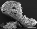
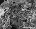
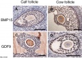 Bovine ovarian follicle BMP15 and GDF9.jpg 1,143 × 810; 219 KB
 Canine oocyte 01.jpg 624 × 532; 51 KB
 Canine oocyte 02.jpg 600 × 596; 43 KB
 Canine oocyte 03.jpg 600 × 694; 169 KB
 Canine oocyte 04.jpg 600 × 482; 76 KB
 Canine oocyte to blastocyst.jpg 825 × 1,000; 154 KB
 Cat oocyte calcium concentration.jpg 600 × 486; 80 KB
 Cat oocyte zona pellucida 01.jpg 832 × 817; 102 KB
 Cat oocyte zona pellucida 02.jpg 1,000 × 991; 146 KB
 Cat spermatozoa bound to oocyte zona pellucida.jpg 1,000 × 917; 161 KB
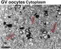 Cytoplasmic lattices in GV oocyte cytoplasm.jpg 1,048 × 846; 291 KB
 Cytoplasmic lattices in oocytes and two-cell embryos.jpg 753 × 1,000; 226 KB
 Dixon1927 fig01-05.jpg 1,642 × 2,459; 223 KB
 Early human telomere length.jpg 1,800 × 1,034; 70 KB
 Early human telomeres.jpg 1,280 × 1,006; 194 KB
 Frazer002 bw600.jpg 600 × 501; 45 KB
 Hamilton1944-fig01.jpg 732 × 800; 168 KB
 Hamilton1944-fig02.jpg 540 × 556; 74 KB
 Hamilton1944-fig03.jpg 732 × 800; 169 KB
 Hamilton1944-fig04.jpg 714 × 794; 189 KB
 Hamilton1944-fig05.jpg 795 × 782; 101 KB
 Hamilton1944-fig06.jpg 385 × 749; 48 KB
 Hamilton1944-fig07.jpg 873 × 800; 140 KB
 Hamilton1944-plate01.jpg 1,200 × 1,472; 387 KB
 Hamilton1944-plate02.jpg 1,200 × 1,663; 287 KB
 Hamilton1944-table01.jpg 1,200 × 645; 113 KB
 Hamster fused oocyte and spermatozoa.jpg 888 × 405; 98 KB
 Hamster oocyte and spermatozoa.jpg 883 × 836; 266 KB
 Hamster oocyte zona pellucida SEM.jpg 800 × 626; 101 KB
 HertigAdams1967 fig01-4.jpg 1,691 × 2,353; 390 KB
 HertigAdams1967 fig01.jpg 531 × 547; 33 KB
 HertigAdams1967 fig02.jpg 531 × 547; 32 KB
 HertigAdams1967 fig03.jpg 518 × 545; 29 KB
 HertigAdams1967 fig04.jpg 1,628 × 1,714; 383 KB
 HertigAdams1967 fig25.jpg 1,637 × 1,452; 256 KB
 Hilfer1990 Fig03.jpg 1,887 × 2,000; 182 KB
 Human fertilization movie 1 frame 01.jpg 600 × 409; 27 KB
 Human fertilization movie 1 frame 02.jpg 600 × 409; 27 KB
 Human fertilization movie 1 frame 03.jpg 600 × 409; 26 KB
 Human fertilization movie 1 frame 04.jpg 600 × 409; 24 KB
 Human fertilization movie 1 frame 05.jpg 600 × 409; 25 KB
 Human fertilization movie 1 frame 06.jpg 600 × 409; 25 KB
 Human fertilization movie 1 frame 07.jpg 600 × 409; 24 KB
 Human fertilization movie 1 frame 08.jpg 600 × 409; 25 KB
 Human fertilization movie 1 frame 09.jpg 600 × 409; 24 KB
 Human fertilization movie 1 frame 10.jpg 600 × 409; 25 KB
 Human infant ovary follicle 01.jpg 800 × 800; 107 KB
 Human MII oocyte 01.jpg 1,200 × 826; 92 KB
 Human MII oocyte 02.jpg 1,200 × 826; 98 KB
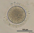 Human oocyte 01.jpg 700 × 675; 98 KB
 Human oocyte 11.jpg 700 × 675; 104 KB
 Human oocyte em01.jpg 600 × 589; 65 KB
 Human oocyte-metaphase I.jpg 400 × 409; 32 KB
 Human oocyte-metaphase II.jpg 400 × 409; 12 KB
 Human ovary follicle basement membrane 01.jpg 660 × 800; 184 KB
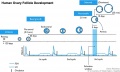 Human ovary follicle development.jpg 700 × 418; 50 KB
 Human ovary non-growing follicle model 02.jpg 1,200 × 918; 161 KB
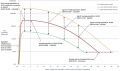 Human ovary non-growing follicle model.jpg 1,151 × 679; 116 KB
 Human pronuclear stage EM02.jpg 639 × 1,000; 194 KB
 Human pronuclear stage EM022.jpg 1,100 × 705; 225 KB
 Human pronuclear stage EM03-05.jpg 982 × 439; 104 KB
 Human pronuclear stage EM06.jpg 907 × 1,000; 245 KB
 Human pronuclear stage EM07.jpg 357 × 509; 52 KB
 Human pronuclear stage EM08.jpg 359 × 513; 56 KB
 Human pronuclear stage EM09.jpg 361 × 506; 53 KB
 Human pronuclear stage EM10.jpg 630 × 781; 140 KB
 Human pronuclear stage EM11.jpg 625 × 768; 130 KB
 Human pronuclear stage EM12.jpg 998 × 777; 265 KB
 Human pronuclear stage EM13.jpg 998 × 771; 247 KB
 Human pronuclear stage EM14-16.jpg 1,013 × 459; 141 KB
 Human pronuclear stage EM17.jpg 1,104 × 504; 156 KB
 Human pronuclear stage EM18.jpg 477 × 541; 66 KB
 Human pronuclear stage EM19.jpg 478 × 534; 67 KB
 Human pronuclear stage EM20.jpg 1,114 × 762; 237 KB
 Human pronuclear stage EM21.jpg 366 × 587; 58 KB
 Human pronuclear stage EM22.jpg 366 × 581; 59 KB
 Human pronuclear stage EM25.jpg 1,013 × 782; 201 KB
 Human pronuclear stage EM26.jpg 1,010 × 784; 187 KB
 Human pronuclear stage EM27.jpg 997 × 777; 227 KB
 Human pronuclear stage EM28.jpg 993 × 774; 239 KB
 Human pronuclear stage EM29.jpg 967 × 763; 183 KB
 Human pronuclear stage EM30.jpg 955 × 853; 192 KB
 Human zona proteins in transgenic mouse oocyte 01.jpg 1,280 × 717; 244 KB
 Human- adult ovary epithelial cords and primary follicles.jpg 600 × 384; 51 KB
 Human- adult ovary primary follicles.jpg 600 × 639; 56 KB
 Human- ovary age primary follicle numbers.jpg 600 × 520; 36 KB
 Human-oocyte to blastocyst.jpg 600 × 402; 49 KB
 Human-oocyte.jpg 400 × 409; 29 KB
 Infant ovary.jpg 943 × 571; 108 KB
 Keibel Mall 001.jpg 781 × 733; 138 KB
 Keibel Mall 009.jpg 333 × 415; 27 KB
 Keith1921 fig001.jpg 766 × 800; 82 KB
 Keith1921 fig009.jpg 530 × 599; 101 KB
 Kollmann001.jpg 791 × 551; 63 KB
 Kollmann003.jpg 533 × 451; 36 KB
 Kollmann004.jpg 340 × 368; 19 KB
 Kollmann005.jpg 334 × 351; 20 KB
 Kollmann007.jpg 956 × 582; 45 KB
 Kollmann008.jpg 641 × 577; 61 KB
 Kollmann009.jpg 572 × 552; 24 KB
 Kollmann010.jpg 550 × 541; 19 KB
 Kollmann011.jpg 550 × 541; 28 KB
 Kollmann012.jpg 696 × 437; 31 KB
 Kollmann461.jpg 721 × 528; 100 KB
 Kollmann462.jpg 758 × 429; 63 KB
 Meiosis spindle movement model.jpg 800 × 619; 80 KB
 Minot1897 fig003.jpg 625 × 543; 83 KB
 Model of human fetal ovarian cord development 01.jpg 800 × 330; 104 KB
 Mouse antral follicle 01.jpg 932 × 1,095; 374 KB
 Mouse antral follicle.jpg 600 × 705; 168 KB
 Mouse follicle in vitro 02.jpg 600 × 701; 146 KB
 Mouse follicle in vitro.jpg 600 × 701; 170 KB
 Mouse germinal vesicle 01.jpg 1,200 × 599; 106 KB
 Mouse germinal vesicle 02.jpg 4,115 × 2,741; 928 KB
 Mouse germinal vesicle 03.jpg 1,195 × 1,200; 219 KB
 Mouse germinal vesicle 04.jpg 1,200 × 1,000; 141 KB
 Mouse in vitro follicle 01.jpg 661 × 534; 72 KB
 Mouse in vitro follicle 02.jpg 800 × 639; 120 KB
 Mouse in vitro follicle 03.jpg 800 × 639; 99 KB
 Mouse in vitro follicle 04.jpg 799 × 537; 81 KB
 Mouse in vitro follicle 05.jpg 800 × 639; 82 KB
 Mouse in vitro follicle 06.jpg 800 × 639; 120 KB
 Mouse model of ovarian cord formation.jpg 800 × 491; 85 KB
 Mouse neonatal ovary oocyte EM01.jpg 677 × 1,000; 266 KB
 Mouse neonatal ovary oocyte EM02.jpg 790 × 792; 179 KB
 Mouse neonatal ovary oocyte EM03.jpg 790 × 792; 187 KB
 Mouse neonatal ovary oocyte EM04.jpg 790 × 792; 235 KB
 Mouse neonatal ovary oocyte EM05.jpg 790 × 792; 217 KB
 Mouse neonatal ovary oocyte EM06.jpg 790 × 792; 163 KB
 Mouse neonatal ovary oocyte EM07.jpg 790 × 792; 183 KB
 Mouse newborn ovary day 1.mp4 ; 646 KB
- Mouse newborn ovary day 2-3.5.mp4 ; 1.95 MB
 Mouse oocyte and zona pellucida EM01.jpg 1,200 × 1,200; 485 KB
 Mouse oocyte and zona pellucida EM01a.jpg 800 × 800; 234 KB
 Mouse oocyte and zona pellucida EM01b.jpg 600 × 600; 133 KB
 Mouse oocyte and zona pellucida EM01c.jpg 400 × 400; 60 KB
 Mouse oocyte balbini body EM01.jpg 695 × 700; 155 KB
 Mouse oocyte cortical granule ovastacin 01.jpg 500 × 465; 54 KB
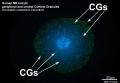
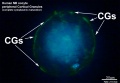
 Mouse oocyte cortical granules 01.jpg 500 × 475; 53 KB
 Mouse oocyte cortical granules 02.jpg 1,006 × 1,000; 177 KB
 Mouse oocyte Egr3-01.jpg 600 × 670; 152 KB
 Mouse oocyte Egr3-02.jpg 642 × 1,200; 121 KB
 Mouse oocyte fertilization 01.jpg 675 × 494; 58 KB
 Mouse oocyte microtubule-associated protein 01.jpg 869 × 1,000; 81 KB
 Mouse oogenesis 01.jpg 1,781 × 1,222; 173 KB
 Mouse oogenesis 02.jpg 1,386 × 355; 40 KB
 Mouse- germinal vesicle oocyte protein expression.jpg 696 × 369; 43 KB
 Mouse- gonadal supporting cell development.jpg 1,000 × 588; 74 KB
 Mouse- MII oocyte protein expression.jpg 747 × 369; 47 KB
 Mouse- zona pellucida 01.jpg 800 × 430; 83 KB
 Mouse- zona pellucida 02.jpg 700 × 688; 95 KB
 Mouse- zona pellucida 03.jpg 1,000 × 345; 64 KB
 Mouse-fertilization 01.jpg 600 × 593; 30 KB
 Mouse-fertilization 02.jpg 1,342 × 691; 118 KB
 Mouse-model ovarian cord formation.jpg 600 × 368; 48 KB
 Mouse-oocyte-d.jpg 790 × 792; 217 KB
 Nelsen1953 fig022.jpg 1,200 × 839; 207 KB
 Nelsen1953 fig030.jpg 1,200 × 995; 414 KB
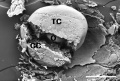
 Oocyte BMP15 and GDF9 effects.jpg 603 × 800; 77 KB
- Oocyte Meiosis 01.mp4 ; 1.22 MB
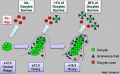
 Oogenesis and meiosis cartoon.jpg 1,197 × 480; 70 KB
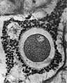
 Ova20he.jpg 450 × 600; 96 KB
 Ovarian follicle growth in vitro.jpg 1,000 × 769; 73 KB
 Ovary follicle 01.jpg 450 × 600; 112 KB
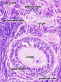
 Ovary histology 002.jpg 1,280 × 1,024; 270 KB
 Ovary histology 003.jpg 1,280 × 1,024; 337 KB
 Ovary histology 004.jpg 1,280 × 1,024; 401 KB
 Ovary histology 005.jpg 1,280 × 1,024; 354 KB
 Ovary histology 006.jpg 1,280 × 1,024; 424 KB
 Ovary histology 007.jpg 1,280 × 1,024; 336 KB
 Ovary histology 008.jpg 1,280 × 1,024; 264 KB
 Ovary histology 061.jpg 1,280 × 1,024; 438 KB
 Ovary histology 061a.jpg 800 × 640; 200 KB
 Ovary histology 061c.jpg 400 × 320; 56 KB
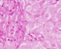
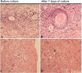 Ovary histology with chemotherapy.jpg 977 × 872; 232 KB
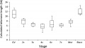
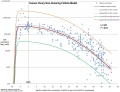
 Ovary oocyte size graph.jpg 1,057 × 820; 114 KB
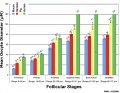
 Ovary- follicle stages.jpg 600 × 337; 45 KB
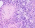
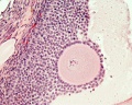 Ovary- histology secondary follicle 01.jpg 1,000 × 800; 293 KB
 Pincus1935-fig01.jpg 563 × 700; 101 KB
 Pincus1935-fig02.jpg 563 × 700; 60 KB
 Pincus1935-fig05.jpg 563 × 700; 82 KB
 Pincus1935-fig06.jpg 563 × 700; 69 KB
 Pincus1935-fig09.jpg 563 × 700; 66 KB
 Pincus1935-fig10.jpg 563 × 700; 61 KB
 Pincus1935-plate01.jpg 1,300 × 2,326; 463 KB
 Pincus1935-plate02.jpg 1,200 × 2,289; 446 KB
 Polar body extrusion model.jpg 1,200 × 435; 70 KB
 Prentiss001.jpg 599 × 567; 86 KB
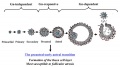
 Primordial follicle activation signaling model.jpg 403 × 440; 32 KB
 Rat oocyte 01.jpg 1,000 × 513; 55 KB


