Category:Neural Crest
From Embryology
| Neural Crest Links: neural crest | Lecture - Early Neural | Lecture - Neural Crest Development | Lecture Movie | Schwann cell | adrenal | melanocyte | peripheral nervous system | enteric nervous system | cornea | cranial nerve neural crest | head | skull | cardiac neural crest | Nicole Le Douarin | Neural Crest Movies | neural crest abnormalities | Category:Neural Crest | |||
|
Subcategories
This category has the following 7 subcategories, out of 7 total.
Pages in category 'Neural Crest'
The following 109 pages are in this category, out of 109 total.
2
C
- Template:Cardiac neural crest
- Chicken Development
- Chicken Neural Crest Migration Movie 1
- Chicken Neural Crest Migration Movie 2
- Chicken Neural Crest Migration Movie 3
- Chicken Neural Crest Migration Movie 4
- Chicken Neural Crest Migration Movie 5
- Chicken Neural Crest Migration Movie 6
- Chicken Neural Crest Migration Movie 7
- Template:Chicken neural crest movies
- Template:Cornea
- Template:Cranial nerve neural crest
- Template:Cranial Neural Crest Timeline table
D
E
H
M
- Template:Mandible
- Template:Meckel’s cartilage
- Template:Megacolon
- Template:Melanocyte
- Template:Merkel cell
- Mouse Cranial Neural Crest Migration Movie
- Mouse Melanoblast Migration Movie
- Template:Mouse neural crest pax7 links
- Movie - Chicken Neural Crest Migration 01
- Movies
- Movies - Chicken Neural Crest
- Template:Myenteric plexus
N
- Template:Neural crest
- Neural Crest - Cardiac
- Neural Crest - Cranial Nerve Development
- Neural Crest - Cranial Nerves
- Neural Crest - Enteric Nervous System
- Neural Crest - Melanocyte Development
- Neural Crest - Peripheral Nervous System
- Neural Crest - Schwann Cell Development
- Template:Neural crest abnormalities
- Template:Neural Crest collapsible table
- Neural Crest Development
- Template:Neural Crest Links
- Template:Neural crest origin collapsetable1
- Template:Neural crest origin table1
- Neural Crest System - Abnormalities
- Template:Neural Crest table
- Template:Neural Crest Vignette
P
- Paper - 1879 The Morphology of the Vertebrate Olfactory Organ
- Paper - A contribution to the histogenesis of the sympathetic nervous system (1909)
- Paper - A nerve growth-stimulating factor isolated from sarcomas 37 and 180
- Paper - Observations on the neural crest of a ten-somite human embryo (1939)
- Paper - Problems concerning the origin and development of the neural crest and cranial ganglia in the vertebrates (1928)
- Paper - The development of the adrenal gland in man (1957)
- Paper - The Development of the Cranial and Spinal Nerves in the Occipital Region of the Human Embryo
- Paper - The development of the sympathetic nervous system in mammals
- Paper - The development of the sympathetic nervous system in mammals (1910)
- Paper - The development of the sympathetic nervous system in man
- Paper - The development of the sympathetic system in birds (1910)
- Paper - The origin of the neural crest
- Paper - The Peripheral Nervous System in the Human Embryo at the End of the First Month (10 mm)
- Paper - The role of the vagi in the development of the sympathetic nervous system (1909)
- Template:Peripheral nervous system
- Template:Peripheral Nervous System
- Template:Pharyngeal arch
- Template:PNS
R
Media in category 'Neural Crest'
The following 93 files are in this category, out of 93 total.
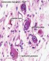 Autonomic ganglion histology 01.jpg 641 × 800; 56 KB
 Bailey366.jpg 558 × 633; 97 KB
 Bailey391.jpg 633 × 485; 56 KB
 BaxterBoyd1939-fig01.jpg 361 × 736; 43 KB
 BaxterBoyd1939-fig02.jpg 459 × 851; 65 KB
 BaxterBoyd1939-fig03.jpg 732 × 1,000; 170 KB
 BaxterBoyd1939-fig04.jpg 642 × 615; 109 KB
 BaxterBoyd1939-fig05.jpg 1,000 × 864; 263 KB
 BaxterBoyd1939-fig06.jpg 489 × 917; 141 KB
 BaxterBoyd1939-fig07.jpg 795 × 917; 242 KB
 BaxterBoyd1939-plate01.jpg 1,680 × 2,400; 786 KB
 BaxterBoyd1939-plate02.jpg 1,681 × 2,400; 860 KB
 BaxterBoyd1939-text-fig01.jpg 1,283 × 1,000; 138 KB
 BaxterBoyd1939-text-fig02.jpg 1,200 × 861; 133 KB
 Cardiac Neural Crest Migration.jpg 1,517 × 1,116; 122 KB
 Carnegie stage 13 caudal trunk.jpg 400 × 625; 53 KB
 Chicken-neural-crest-migration-01.jpg 600 × 432; 37 KB
 Chicken-neural-crest-migration-02.jpg 600 × 419; 43 KB
 Chicken-neural-crest-migration-03.jpg 600 × 346; 53 KB
 Chicken-neural-crest-migration-04.jpg 600 × 397; 67 KB
 Chicken-neural-crest-migration-05.jpg 600 × 586; 66 KB
 Chicken-neural-crest-migration-06.jpg 600 × 398; 40 KB
 Chicken-neural-crest-migration-07.jpg 600 × 453; 45 KB
 Childhood cancer survival rates.jpg 536 × 335; 25 KB
 Cochlea glial lineage cartoon.jpg 1,000 × 651; 52 KB
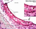
 Cochlea stria vascularis cartoon 03.jpg 694 × 800; 125 KB
 Cranial neural crest skeletal fate 01.jpg 800 × 633; 59 KB
 Eye-neural crest signaling.jpg 946 × 886; 436 KB
 Gray0847.jpg 559 × 900; 155 KB
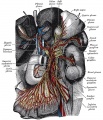 Gray0848.jpg 800 × 935; 289 KB
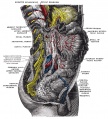 Gray0849.jpg 800 × 885; 258 KB
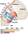 Hindbrain neural crest migration.jpg 450 × 545; 48 KB
 Human Carnegie stage 13 GJA1 expression.jpg 706 × 470; 96 KB
 Human Carnegie stage 13 SOX11 MAZ GJA1 expression.jpg 777 × 1,000; 192 KB
 Human Carnegie stage 13 stem cell markers.jpg 657 × 585; 78 KB
 Human cochlea stria vascularis 01.jpg 1,854 × 1,806; 754 KB
 Human neural crest cell migration-in vitro.jpg 1,280 × 959; 163 KB
 Human-adrenal gland 01.jpg 596 × 392; 65 KB
 Keith1902 fig035.jpg 800 × 633; 125 KB
 LeDouarin.jpg 156 × 198; 19 KB
 Lens-neural crest signaling 01.jpg 300 × 400; 24 KB
 Lens-neural crest signaling 02.jpg 521 × 522; 22 KB
 Megacolon stoma.gif 200 × 202; 6 KB
 Melanoblast migration.png 600 × 210; 40 KB
 Melanocyte development cartoon.jpg 1,280 × 1,086; 176 KB
 Merkel cell EM 01.jpg 984 × 738; 209 KB
 Merkel cell EM 02.jpg 984 × 685; 166 KB
 Model neural crest mesenchymal condensation.jpg 818 × 1,200; 131 KB
 Mouse E9.5 neural crest - Crabp1, Sox10, Pax3.jpg 1,323 × 1,233; 193 KB
 Mouse E9.5 neural crest - Crabp1.jpg 2,489 × 626; 217 KB
 Mouse E9.5 neural crest beta actin KO.jpg 2,722 × 2,573; 732 KB
 Mouse eye E18.jpg 884 × 977; 169 KB
 Mouse eye neural crest cornea 01.jpg 500 × 256; 34 KB
 Mouse eye neural crest cornea 02.jpg 800 × 655; 121 KB
 Mouse eye neural crest.jpg 1,086 × 1,509; 483 KB
 Mouse eye TGF-beta model.jpg 1,000 × 519; 88 KB
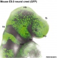 Mouse head E9-neural crest GFP.jpg 539 × 543; 56 KB
 Mouse head-neural crest 01.jpg 900 × 339; 48 KB
 Mouse melanoblast distribution 01.jpg 697 × 1,000; 192 KB
 Mouse melanoblast distribution 02.jpg 777 × 1,121; 129 KB
 Mouse melanoblast distribution 03.jpg 751 × 1,051; 150 KB
 Mouse melanoblast distribution 04.jpg 761 × 1,128; 177 KB
 Mouse melanoblast distribution 05.jpg 761 × 540; 94 KB
 Mouse melanoblast distribution 06.jpg 761 × 524; 74 KB
 Mouse organ of corti 04.jpg 1,280 × 1,024; 202 KB
 Mouse pax7 heart 01.jpg 561 × 560; 34 KB
 Mouse pax7 limb 01.jpg 1,320 × 549; 112 KB
 Mouse pax7 neural fold 01.jpg 513 × 915; 100 KB
 Mouse pax7 trunk neural crest 01.jpg 881 × 691; 158 KB
 Mouse pax7 trunk neural crest 02.jpg 881 × 691; 171 KB
 Mouse-E10.5 ganglia Sox10.jpg 540 × 685; 51 KB
 Mouse-E10.5-Sox10.jpg 400 × 463; 24 KB
 Mouse-E8.5-Sox10.jpg 400 × 463; 20 KB
 Mouse-E9.5-Sox10.jpg 400 × 463; 30 KB
 Mouse-melanoblast migration icon.jpg 450 × 450; 59 KB
 Mouse-neural crest Sox10 E10.5.jpg 1,000 × 420; 55 KB
 Neural - cranial nerves.jpg 800 × 447; 72 KB
 Neural crest cell enteric sorting.jpg 800 × 849; 61 KB
 Neural crest contribution.jpg 1,280 × 701; 64 KB
 Neural crest formation stages 01.jpg 1,200 × 728; 124 KB
 Neural crest precursor differentiation.jpg 600 × 576; 42 KB
 Neuroblastoma.jpg 640 × 426; 31 KB
 Smooth muscle histology 002.jpg 600 × 750; 112 KB
 Stage 22 image 152.jpg 1,000 × 670; 127 KB
 Stage 22 image 153.jpg 1,000 × 662; 191 KB
 Stage 22 image 155.jpg 1,000 × 672; 239 KB
 Stage11 sem21.jpg 600 × 447; 79 KB
 Trunk neural crest migration.jpg 1,280 × 963; 220 KB
 Week10 adrenal.jpg 600 × 564; 48 KB
 Wen1928-Fig10.jpg 495 × 1,200; 108 KB
 Zebrafish melanocyte development model.jpg 484 × 277; 40 KB
 Zebrafish neural crest model.jpg 600 × 480; 61 KB
 Zebrafish skull neural crest.jpg 815 × 933; 187 KB


