Category:Gastrointestinal Tract
From Embryology

This Embryology category is a link to resource pages, images, podcasts and movies that relate to gastrointestinal tract development.
Subcategories
This category has the following 16 subcategories, out of 16 total.
Pages in category 'Gastrointestinal Tract'
The following 62 pages are in this category, out of 334 total.
(previous page) (next page)R
- Template:Ref-Johnson1910
- Template:Ref-Johnson1913
- Template:Ref-Johnson1914
- Template:Ref-Johnson1914b
- Template:Ref-Kirk1910
- Template:Ref-Latta1921
- Template:Ref-Lewis1912b
- Template:Ref-Lineback1916
- Template:Ref-Lineback1920
- Template:Ref-Lockwood1882
- Template:Ref-Lockwood1884
- Template:Ref-Mall1898a
- Template:Ref-Mall1898b
- Template:Ref-Mall1899
- Template:Ref-McGill1910
- Template:Ref-Minot1900a
- Template:Ref-Pernkoof1922
- Template:Ref-Pernkoof1925
- Template:Ref-Reid1908
- Template:Ref-Reid1911a
- Template:Ref-Reid1911b
- Template:Ref-Reid1912
- Template:Ref-Reid1913a
- Template:Ref-Reid1913b
- Template:Ref-Reid1913c
- Template:Ref-Reid1914a
- Template:Ref-Reid1914b
- Template:Ref-Snook1934b
- Template:Ref-Tandler1900
- Template:Ref-Wagstaffe1924
- Template:Ref-Wakeley1923
- Template:Ref-Wakeley1930
- Template:Ref-Woods-Jones1904
- Template:Reid DG.
S
- Template:Salivary gland
- Salivary Gland Development
- Site Map
- Talk:Site Map
- Template:Small intestine
- Template:Small Intestine Length table1
- Template:Splanchnic mesoderm
- Template:Stage 23 MRI movie 7
- Stage 23 MRI Movie 7
- Template:Stomach
- Template:Stomach Histology
- Template:Sublingual gland
- Template:Submandibular gland
- Template:Submaxillary gland
V
Media in category 'Gastrointestinal Tract'
The following 200 files are in this category, out of 523 total.
(previous page) (next page)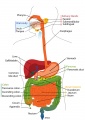 Adult gastrointestinal tract cartoon.jpg 707 × 1,000; 105 KB
 Adult gastrointestinal tract cartoon01.jpg 745 × 698; 55 KB
 Adult gastrointestinal tract cartoon02.jpg 541 × 738; 42 KB
 ARecVG02.jpg 300 × 400; 113 KB
 Arthrogryposis.jpg 800 × 503; 39 KB
 Australian abnormalities 81-92 git.jpg 481 × 344; 43 KB
 Bailey244.jpg 514 × 656; 64 KB
 Bailey246.jpg 806 × 681; 89 KB
 Bailey249.jpg 713 × 404; 71 KB
 Bailey250.jpg 592 × 264; 41 KB
 Bailey251.jpg 577 × 342; 36 KB
 Bailey252.jpg 904 × 690; 166 KB
 Bailey253.jpg 888 × 638; 123 KB
 Bailey254.jpg 734 × 808; 93 KB
 Bailey255.jpg 717 × 287; 57 KB
 Bailey256.jpg 894 × 545; 75 KB
 Bailey257.jpg 516 × 326; 45 KB
 Bailey258.jpg 769 × 624; 141 KB
 Bailey259.jpg 690 × 414; 95 KB
 Bailey260.jpg 694 × 580; 58 KB
 Bailey261.jpg 529 × 293; 23 KB
 Bailey263.jpg 804 × 726; 100 KB
 Bailey264.jpg 881 × 562; 77 KB
 Bailey265.jpg 906 × 644; 80 KB
 Bailey266.jpg 731 × 913; 178 KB
 Bailey267.jpg 774 × 801; 99 KB
 Bailey268.jpg 659 × 614; 87 KB
 Bailey269.jpg 436 × 353; 44 KB
 Bailey270.jpg 736 × 448; 53 KB
 Bailey271.jpg 691 × 428; 72 KB
 Bailey272.jpg 862 × 597; 120 KB
 Bailey273.jpg 560 × 522; 58 KB
 Bailey274.jpg 558 × 442; 32 KB
 Bailey275.jpg 485 × 483; 48 KB
 Bailey276.jpg 855 × 619; 126 KB
 Bailey277.jpg 896 × 480; 112 KB
 Bailey278 279.jpg 669 × 812; 85 KB
 Bailey280.jpg 866 × 581; 133 KB
 Bailey281.jpg 865 × 1,028; 211 KB
 Bailey299 300.jpg 911 × 447; 79 KB
 Bailey301-303.jpg 916 × 1,097; 172 KB
 Bailey304.jpg 729 × 722; 85 KB
 Baileytable04.jpg 656 × 164; 15 KB
 Baileytable05.jpg 953 × 219; 39 KB
 Bardeen1914-fig01.jpg 439 × 853; 51 KB
 Bardeen1914-fig02.jpg 624 × 861; 54 KB
 Bardeen1914-fig03.jpg 405 × 1,014; 55 KB
 Bardeen1914-fig04.jpg 840 × 1,019; 93 KB
 Bardeen1914-fig05.jpg 465 × 1,000; 61 KB
 Bardeen1914-fig06.jpg 721 × 1,000; 88 KB
 Bardeen1914-fig07.jpg 451 × 936; 63 KB
 Bardeen1914-fig08.jpg 790 × 1,000; 131 KB
 Bardeen1914-fig09.jpg 694 × 1,000; 113 KB
 Bardeen1914-fig10.jpg 652 × 800; 102 KB
 Bardeen1914-fig11.jpg 708 × 1,000; 111 KB
 Bardeen1914-plate01.jpg 1,347 × 2,089; 259 KB
 Bardeen1914-plate02.jpg 1,398 × 2,577; 275 KB
 Bardeen1914-plate03.jpg 1,359 × 2,476; 341 KB
 Bardeen1914-plate04.jpg 1,351 × 2,092; 257 KB
 BGDB PracManual 2011 Practical 1.pdf ; 364 KB
 Bovine GIT miRNA expression 01.jpg 815 × 800; 93 KB
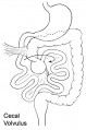 Cecal volvulus.jpg 331 × 500; 28 KB
 Chicken GIT peristalsis movie 01.mp4 ; 6.19 MB
 Classification of Oesophageal atresia.jpg 513 × 726; 55 KB
 Col00he.jpg 1,280 × 1,024; 117 KB
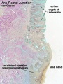
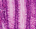
 Colon histology 001.jpg 400 × 533; 64 KB
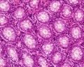
 Colon histology 002.jpg 300 × 400; 73 KB
 Colon histology 003.jpg 1,280 × 1,024; 117 KB
 Colon histology 004.jpg 1,280 × 1,024; 292 KB
 Colon histology 005.jpg 1,280 × 1,024; 403 KB
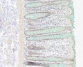
 Colon histology 006.jpg 400 × 533; 70 KB
 Colon histology 007.jpg 1,280 × 1,024; 152 KB
 Colon histology 008.jpg 1,278 × 959; 237 KB
 Colon histology 009.jpg 1,280 × 1,024; 159 KB
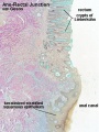
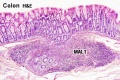 Colon MALT.jpg 500 × 333; 67 KB
 Common bile duct in duodenal bulb.jpg 367 × 364; 38 KB
 Cullen1916 fig102.jpg 900 × 859; 66 KB
 Cullen1916 fig92.jpg 885 × 637; 109 KB
 Cullen1916 fig94.jpg 1,000 × 714; 90 KB
 Cullen1916 fig95.jpg 800 × 902; 106 KB
 Cullen1916 fig97.jpg 800 × 587; 66 KB
 Cullen1916 fig98.jpg 800 × 1,469; 280 KB
 Cullen1916 fig99.jpg 1,280 × 1,451; 509 KB
 Desmosome 02.jpg 600 × 450; 74 KB
 Duodenal atresia 01.jpg 750 × 592; 56 KB
 Duodenal atresia 02.jpg 765 × 682; 68 KB
 Duodenal atresia.jpg 600 × 873; 63 KB
 Duodenum cartoon.jpg 500 × 704; 42 KB
 Endoderm cartoon.jpg 587 × 262; 31 KB
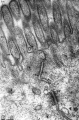 Epithelial junctions EM01.jpg 658 × 1,000; 232 KB
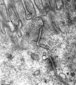 Epithelial junctions EM02.jpg 900 × 1,000; 288 KB
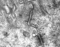 Epithelial junctions EM03.jpg 1,291 × 1,000; 344 KB
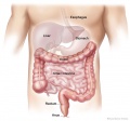
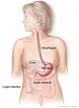
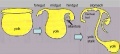
 Eutherian gastrointestinal system.jpg 600 × 453; 94 KB
 Fetal large Intestine length growth graph.jpg 800 × 653; 50 KB
 Fetal liver weight growth graph.jpg 800 × 521; 34 KB
 Fetal pancreas weight growth graph.jpg 1,000 × 669; 49 KB
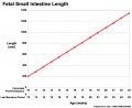 Fetal small Intestine length growth graph.jpg 800 × 653; 51 KB
 Frazer1911 fig01.jpg 1,280 × 766; 187 KB
 Frazer1911 fig02.jpg 1,280 × 872; 112 KB
 Frazer1911 fig03.jpg 1,280 × 600; 153 KB
 Frazer1915 fig01.jpg 1,000 × 899; 98 KB
 Frazer1915 fig02.jpg 1,000 × 821; 150 KB
 Frazer1915 fig03.jpg 1,000 × 596; 98 KB
 Frazer1915 fig04.jpg 1,000 × 1,072; 161 KB
 Frazer1915 fig05.jpg 1,000 × 529; 104 KB
 Frazer1915 fig06.jpg 1,000 × 600; 109 KB
 Frazer1915 fig07.jpg 1,000 × 415; 76 KB
 Frazer1915 fig08.jpg 1,000 × 814; 187 KB
 Frazer1915 fig09.jpg 1,000 × 497; 133 KB
 Frazer1915 fig10.jpg 1,000 × 733; 155 KB
 Frazer1915 fig11.jpg 1,000 × 621; 178 KB
 Frazer1915 fig12.jpg 1,000 × 538; 83 KB
 Frazer1915 fig13.jpg 1,000 × 739; 97 KB
 Frazer1915 fig14.jpg 1,000 × 549; 122 KB
 Frazer1915 fig15.jpg 1,000 × 899; 174 KB
 Frazer1915 fig16.jpg 855 × 894; 165 KB
 Frazer1915 fig17.jpg 1,000 × 685; 108 KB
 Frazer1915 fig18.jpg 1,000 × 760; 163 KB
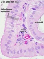
 Gall bladder histology 001.jpg 375 × 500; 78 KB
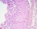
 Gall bladder histology 002.jpg 375 × 500; 45 KB
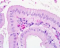
 Gall bladder histology 003.jpg 1,280 × 1,024; 577 KB
 Gall bladder histology 004.jpg 1,280 × 1,024; 254 KB
 Gall bladder histology 005.gif 600 × 450; 683 KB
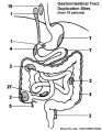 Gastrointestinal tract duplication sites.jpg 624 × 800; 47 KB
 Gastrointestinal tract intestine immune cartoon 01.jpg 728 × 1,200; 284 KB
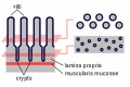 Gastrointestinal villi and crypts cartoon.jpg 500 × 333; 28 KB
 Gastroschisis cartoon.jpg 600 × 358; 21 KB
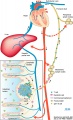
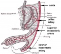 GIT blood supply.jpg 568 × 500; 47 KB
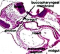
 Git17mm.jpg 500 × 536; 50 KB
 Gitbpm.jpg 200 × 180; 18 KB
 Gray0532.jpg 680 × 700; 155 KB
 Gray0533.jpg 705 × 750; 164 KB
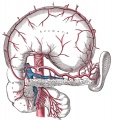
 Gray0605.jpg 614 × 600; 98 KB
 Gray0613.jpg 700 × 579; 115 KB
 Gray0614.jpg 550 × 640; 118 KB
 Gray0615.jpg 571 × 600; 101 KB
 Gray0616.jpg 626 × 600; 111 KB
 Gray0617.jpg 555 × 600; 139 KB
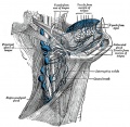
 Gray0847.jpg 559 × 900; 155 KB
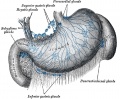
 Gray0848.jpg 800 × 935; 289 KB
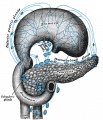
 Gray0849.jpg 800 × 885; 258 KB
 Gray0977.jpg 553 × 600; 59 KB
 Gray0978.jpg 483 × 600; 67 KB
 Gray0979.jpg 500 × 446; 56 KB
 Gray0980.jpg 542 × 450; 57 KB
 Gray0981.jpg 538 × 340; 50 KB
 Gray0982a.jpg 427 × 393; 18 KB
 Gray0982b.jpg 427 × 393; 20 KB
 Gray0983.jpg 800 × 517; 38 KB
 Gray0984.jpg 800 × 477; 58 KB
 Gray0985.jpg 682 × 600; 93 KB
 Gray0986.jpg 565 × 606; 56 KB
 Gray0987.jpg 1,000 × 579; 114 KB
 Gray0987a.jpg 554 × 579; 50 KB
 Gray0987b.jpg 554 × 579; 67 KB
 Gray0988.jpg 397 × 800; 48 KB
 Gray0989.jpg 700 × 685; 107 KB
 Gray0990.jpg 800 × 407; 60 KB
 Gray0991.jpg 469 × 400; 33 KB
 Gray0992.jpg 600 × 611; 73 KB
 Gray0993.jpg 600 × 545; 88 KB
 Gray0994.jpg 600 × 861; 151 KB
 Gray0996.jpg 800 × 822; 118 KB
 Gray1027.jpg 598 × 600; 101 KB
 Gray1028.jpg 800 × 960; 202 KB
 Gray1029.jpg 700 × 435; 75 KB
 Gray1033.jpg 399 × 600; 96 KB
 Gray1039.jpg 550 × 387; 61 KB
 Gray1040.jpg 900 × 918; 230 KB
 Gray1042.jpg 969 × 745; 140 KB
 Gray1050.jpg 765 × 500; 92 KB
 Gray1080.jpg 770 × 490; 120 KB
 Gray1095.jpg 349 × 700; 55 KB
 Gray1096.jpg 600 × 380; 63 KB
 Gray1202.jpg 563 × 500; 63 KB
 Gray1223.png 537 × 500; 55 KB
 Greater-omentum.jpg 537 × 419; 48 KB
 Histology-fetal liver HEx100.jpg 1,280 × 1,024; 214 KB
 Histology-fetal liver HEx40.jpg 1,000 × 800; 281 KB
- HMB2011 Gall Bladder Histology 01.mp3 ; 1.05 MB
- HMB2011 Gall Bladder Histology 03.mp3 ; 1.27 MB
- HMB2011 Gall Bladder Histology 04.mp3 ; 1.22 MB
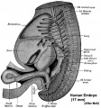
 Human Embryo 17.8mm GIT.jpg 600 × 401; 38 KB
 Human Embryo 17.8mmCNS GIT.jpg 500 × 678; 105 KB
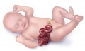
 Human embryo midgut loop 01.jpg 1,423 × 771; 200 KB
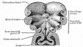
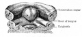
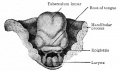
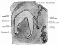
 Human embryonic tongue 01.jpg 1,200 × 818; 443 KB
 Human embryonic tongue 02.jpg 650 × 850; 220 KB
 Human embryonic tongue 03.jpg 650 × 470; 159 KB
 Human embryonic tongue 04.jpg 650 × 470; 131 KB
 Human embryonic tongue 05.jpg 650 × 470; 146 KB
 Human embryonic tongue 06.jpg 650 × 470; 99 KB
 Human embryonic tongue 07.jpg 650 × 470; 187 KB
 Human embryonic tongue 08.jpg 650 × 470; 185 KB
 Human embryonic tongue 09.jpg 650 × 470; 180 KB
 Human embryonic tongue 10.jpg 650 × 470; 118 KB
 Human embryonic tongue 11.jpg 650 × 470; 155 KB
 Human embryonic-fetal tongue 01.jpg 1,000 × 1,129; 490 KB
 Human fetal tongue 01.jpg 1,500 × 672; 350 KB
 Human week 10 fetus 06.jpg 1,200 × 900; 251 KB


