Category:Carnegie Embryo 4315
From Embryology

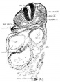
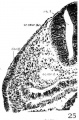
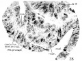
This Embryology category shows pages and images that relate to the Carnegie Collection Embryo No.8963. This 22 somite embryo was a tubal pregnancy originally classified as University of Chicago No. H951. Described by Wen (1928)[1] with particular reference to the neural, renal, vascular systems.
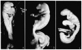
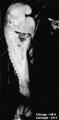
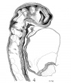
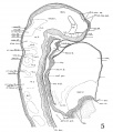
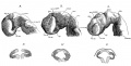
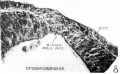
- "Embryo H951 (fig. 1, A) was obtained from Drs. E. L. Whitney and J. T. Rocks, of Walla Walla, Washington. “The uterus was removed for fibrosis on the 13th day after the day on which menstruation was expected to begin,” opened and fixed in ‘4%’ formalin. No further clinical information could be obtained. The embryo was mordanted in aqueous chrom—sublimate, slowly dehydrated, double embedded in celloidin and paraffin, cut into transverse sections, and stained with iron hematoxylin (all but one slide being counterstained with eosin). During the mounting, five sections were slightly distorted and, unfortunately, three sections lost, so it is not easy to follow the series at the region between the 4th to 5th somites. A plaster model of the head region (figs. 2, 3), a wax model of the entire embryo, and one of the anterior portion of the central nervous system with the ventral pharyngeal wall were made from serial photomicrographs at the magnification of 150 times. Two other plaster models, one of the entire embryo and one of the head region alone showing the anatomy therewith, were made by O. O. Heard. Histologically, this specimen is in excellent condition."[1]
Carnegie stage 11 - Week 4, 23 - 26 days, GA - week 6, 4.0 mm GL, Somite Number 17. Conceptus - 23.1 x 10.4 x circ.11 mm.
The embryo was sectioned into 336 sections at 10 µm.
| Carnegie Collection - Stage 11 | ||||||||||
|---|---|---|---|---|---|---|---|---|---|---|
| Serial No. | Pairs of somites | Size (mm) | Grade | Fixative | Embedding Medium | Plane | Thinness (µm) | Stain | Year | Notes |
| 4315 | 17 | E, 4.7 Ch, 23x10.4X11 | Excellent | ? | C-P | Transverse | 10 | I.H. & E. | 1923 | Univ. Chicago No. 951. Wen (1928)[1] |
Abbreviations
| ||||||||||
- ↑ 1.0 1.1 1.2 Wen IC. The anatomy of human embryos with seventeen to twenty-three pairs of somites (1928) J. Comp. Neural., 45: 301-376.
| Carnegie Collection - Stage 11 | ||||||||||
|---|---|---|---|---|---|---|---|---|---|---|
| Serial No. | Pairs of somites | Size (mm) | Grade | Fixative | Embedding Medium | Plane | Thinness (µm) | Stain | Year | Notes |
| 12 | 14 | E, 2.1 Ch, 13 | Poor | P | Transverse | 10 | Al. carm. | 1893 | ||
| 164 | 18 | E, 3.5 Ch, 14 | Good | Formalin | P | Transverse | 20 | Al. carm. | 1913 | |
| 318 | 13/14 | E, 2.5 Ch, 16 | Good | P | Transverse | 25 | Al. carm. | 1905 | ||
| 470 | 17 | E, 4.3 Ch, 16 | Good | Formalin | P | Transverse | 10 | Al. carm. . | 1910 | |
| 779 | 14 | E, 2.75 | Good | C | Transverse | 15 | Al. coch. | 1913 | Dysraphism. Noted by Dekaban (1964)[1] | |
| 1182b | E, 3 Ch, 15x12x5 | Good | Formalin | ? | Transverse | 20 | Al. carm. | 1915 | ||
| 2053 | 20 | E, 3.1 Ch, 12 | Exc. | Formalin | P | Transverse | 10 | Al. coch. | 1918 | Most advanced in group. Ag added to slide 2 Monographs by Davis (1923)[2] and Congdon (1922)[3] |
| 4315 | 17 | E, 4.7 Ch, 23x10.4X11 | Excellent | ? | C-P | Transverse | 10 | I.H. & E. | 1923 | Univ. Chicago No. 951. Wen (1928)[4] |
| 4529 | 14 | E, 2.4 Ch, 21 | Excellent | Formalin | P | Transverse | 10 | Al. coch, or. G. | 1924 | Heuser (1930)[5] |
| 4783 | 13 | E, 2.3 | Fair | ? | ? | Transverse | 5 | I.H. | 1924 | Wallin (1913)[6] |
| 4877 | 13 | E, 2 Ch, 15 | Good | Formalin | P | Transverse | 15 | Al. coch. | 1925 | |
| 5072 | 17 | E, 3 | Good | Formalin | P | Transverse | 10 | (Stain - Haematoxylin Eosin) | 1925 | Tubal Type specimen. Atwell (1930)[7] |
| 6050 | 19/21 | E.,3 Ch, 10 | Good | Formalin | C-P | Coronal | 10 | Al. coch. | 1930 | Advanced |
| 6344 | 13 | E, 2.5 Ch, 17 | Excellent | Formalin | C-P | Transverse | 6 | Al. coch. | 1931 | Least advanced in group |
| 6784 | 17 | E, 5 Ch, 16 | Excellent | Formalin | C-P | Transverse | 6 | I.H, or. G. | 1933 | |
| 7358 | 16 | E, ? Ch, 15 | Poor | Alc, formol | p | Oblique | 25 | (Stain - Haematoxylin Eosin) | 1936 | |
| 7611 | 16 | E., 2.4 Ch., 12 | Excellent | Bouin | C-P | Transverse | 8 | (Stain - Haematoxylin Eosin) | 1938 | |
| 7665 | 19 | E., 4.36 | Excellent | ? | C-P | Transverse | 6 | 1939 | Univ. Chicago No. H 1516 | |
| 7702 | 17 | E, 3.7 Ch., 14 | Good | Formalin | C-P | Transverse | 10 | Al. coch. | 1940 | Returned to B M Patten |
| 7851 | 13 | E., 4.3 Ch, 18 | Excellent | Formalin | C-P | Transverse | 8 | (Stain - Haematoxylin Eosin) | 1940 | Slightly injured |
| 8005 | 16/17 | E, 3 | Excellent | Bouin | C-P | Transverse | 8 | (Stain - Haematoxylin Eosin) | 1942 | Tubal |
| 8116 | 17 | E, 14 Ch.. 17 | Good | Formalin | p | Sagittal | 8 | Azan | 1953 | |
| 8962 | 15 | E, 1.55 | Good | ? | * | Sagittal | ? | ? | 1952 | Tubal Univ. Chicago No. H 810 |
Abbreviations
| ||||||||||
References
| ||||||||||
Pages in category 'Carnegie Embryo 4315'
This category contains only the following page.
Media in category 'Carnegie Embryo 4315'
The following 19 files are in this category, out of 19 total.
 Wen1928-Fig01.jpg 1,280 × 783; 80 KB
 Wen1928-Fig01a.jpg 375 × 763; 25 KB
 Wen1928-Fig04.jpg 1,200 × 1,392; 323 KB
 Wen1928-Fig05.jpg 1,200 × 1,404; 288 KB
 Wen1928-Fig07.jpg 1,265 × 648; 166 KB
 Wen1928-Fig08.jpg 1,000 × 619; 76 KB
 Wen1928-Fig10.jpg 495 × 1,200; 108 KB
 Wen1928-Fig11.jpg 1,000 × 543; 102 KB
 Wen1928-Fig12.jpg 1,000 × 921; 140 KB
 Wen1928-Fig13.jpg 851 × 800; 148 KB
 Wen1928-Fig14.jpg 1,111 × 1,200; 216 KB
 Wen1928-Fig15.jpg 773 × 1,200; 100 KB
 Wen1928-Fig16.jpg 702 × 1,500; 195 KB
 Wen1928-Fig17.jpg 877 × 1,000; 186 KB
 Wen1928-Fig18.jpg 1,000 × 962; 331 KB
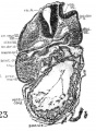 Wen1928-Fig23.jpg 806 × 1,103; 218 KB
 Wen1928-Fig24.jpg 730 × 1,000; 142 KB
 Wen1928-Fig25.jpg 660 × 1,000; 169 KB
 Wen1928-Fig28.jpg 1,200 × 899; 200 KB


