Category:Blood Vessel: Difference between revisions
From Embryology
(Created page with "The pages and media listed below relate to the topic of development of blood vessels, both artery and vein. Note development of lymphatic vessels have a separate category under...") |
No edit summary |
||
| Line 2: | Line 2: | ||
Note development of lymphatic vessels have a separate category under immune. | Note development of lymphatic vessels have a separate category under immune. | ||
[[Category:Cardiovascular]] | |||
Latest revision as of 18:03, 15 July 2012
The pages and media listed below relate to the topic of development of blood vessels, both artery and vein.
Note development of lymphatic vessels have a separate category under immune.
Pages in category 'Blood Vessel'
The following 43 pages are in this category, out of 43 total.
C
- Template:Capillary
- Template:CapillaryEM links
- Cardiovascular - Arterial Development
- Cardiovascular - Venous Development
- Cardiovascular System - Blood Vessel Development
- Cardiovascular System - Ductus Arteriosus
- Cardiovascular System - Ductus Venosus
- Cardiovascular System - Foramen Ovale
- Computed Tomography
P
- Paper - Development of the inferior vena cava (1929)
- Paper - On the development of the aortae cardinal and umbilical veins and the other blood vessels of vertebrate embryos from capillaries (1909)
- Paper - On the development of the blood-vessels of the brain in the human embryo (1905)
- Paper - On the origin of the pulmonary arteries in mammals
- Paper - The development of the principal arterial stems in the forelimb of the pig (1922)
- Paper - The development of the subcutaneous vascular plexus in the head of the human embryo (1923)
- Paper - The development of the vena cava inferior in man (1925)
- Paper - The origin of the heart and blood vessels in felis domestica (1924)
- Paper The development of the subcutaneous vascular plexus in the head of the human embryo (1923)
R
- Template:Ref-BacsichSmout1938
- Template:Ref-Bremer1902
- Template:Ref-Bremer1909
- Template:Ref-Clark1899b
- Template:Ref-Espinasse1933
- Template:Ref-Gladstone1929
- Template:Ref-Huntington1910a
- Template:Ref-Mall1905
- Template:Ref-McClure1925
- Template:Ref-Miller1903
- Template:Ref-Sabin1908
- Template:Ref-Stockard1915b
- Template:Ref-Wahl1915
- Template:Ref-Watson1924
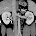
- Renal System - Abnormalities
- Template:Renal Vascular Anomalies
Media in category 'Blood Vessel'
The following 116 files are in this category, out of 116 total.
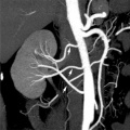
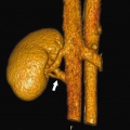
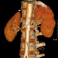 Accessory renal artery.jpg 800 × 798; 103 KB
 Aortic arch branching pattern abnormalities.jpg 656 × 1,404; 133 KB
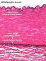
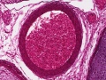
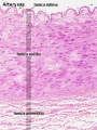 Artery histology 01.jpg 400 × 533; 80 KB
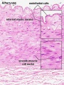 Artery histology 02.jpg 400 × 533; 78 KB
 Artery histology 03.jpg 400 × 533; 89 KB
 Artery histology 04.jpg 800 × 1,000; 93 KB
 Artery histology 05.jpg 400 × 533; 72 KB
 Artery histology 06.jpg 400 × 533; 91 KB
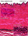
 Artery histology 11.jpg 1,280 × 1,024; 344 KB
 Artery histology 12.jpg 1,280 × 1,024; 206 KB
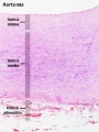
 Artery histology 13.jpg 1,280 × 1,024; 474 KB
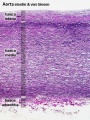
 Artery histology 14.jpg 1,280 × 1,024; 466 KB
 Artery histology 15.jpg 1,280 × 1,024; 343 KB
 Artery histology 16.jpg 1,280 × 1,024; 409 KB
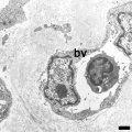
 Blood capillary EM 01.jpg 1,107 × 714; 260 KB
 Blood capillary EM 02.jpg 600 × 600; 99 KB
 Blood capillary EM 03.jpg 1,560 × 1,230; 441 KB
 Blood capillary EM 04.jpg 1,560 × 1,230; 462 KB
 Blood capillary EM 05.jpg 1,015 × 800; 205 KB
 Blood capillary EM 06.jpg 1,015 × 800; 216 KB
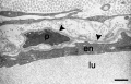
 Blood-thymus barrier EM01.jpg 1,280 × 1,747; 375 KB
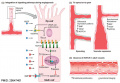
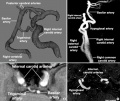
 Cerebral blood supply development 01.jpg 1,200 × 460; 67 KB
 Congdon1922-1-16.jpg 980 × 1,000; 157 KB
 Congdon1922-17.jpg 1,000 × 411; 55 KB
 Congdon1922-18-25.jpg 1,200 × 795; 179 KB
 Congdon1922-18.jpg 494 × 506; 29 KB
 Congdon1922-19.jpg 653 × 471; 31 KB
 Congdon1922-20.jpg 794 × 446; 43 KB
 Congdon1922-21.jpg 578 × 407; 27 KB
 Congdon1922-22.jpg 511 × 489; 27 KB
 Congdon1922-23.jpg 519 × 412; 26 KB
 Congdon1922-24.jpg 790 × 482; 40 KB
 Congdon1922-25.jpg 509 × 358; 25 KB
 Congdon1922-26.jpg 746 × 726; 54 KB
 Congdon1922-27-28.jpg 997 × 612; 68 KB
 Congdon1922-29.jpg 976 × 1,000; 81 KB
 Congdon1922-30.jpg 1,133 × 1,000; 176 KB
 Congdon1922-31.jpg 1,063 × 1,000; 93 KB
 Congdon1922-32.jpg 1,133 × 1,000; 132 KB
 Congdon1922-33.jpg 920 × 1,000; 107 KB
 Congdon1922-34.jpg 920 × 1,000; 122 KB
 Congdon1922-35.jpg 920 × 1,000; 97 KB
 Congdon1922-36.jpg 920 × 1,000; 113 KB
 Congdon1922-37.jpg 1,200 × 838; 163 KB
 Congdon1922-38.jpg 1,187 × 1,000; 165 KB
 Congdon1922-39.jpg 1,200 × 756; 138 KB
 Congdon1922-40.jpg 1,013 × 1,000; 110 KB
 Congdon1922-plate01.jpg 877 × 1,200; 145 KB
 Congdon1922-plate02.jpg 877 × 1,200; 191 KB
 Congdon1922-plate03.jpg 1,200 × 885; 182 KB
 Finley1923 fig01.jpg 494 × 968; 53 KB
 Finley1923 fig02.jpg 700 × 800; 77 KB
 Finley1923 fig03.jpg 600 × 512; 56 KB
 Finley1923 fig04.jpg 700 × 627; 61 KB
 Finley1923 fig05.jpg 674 × 800; 146 KB
 Finley1923 fig06.jpg 729 × 800; 95 KB
 Finley1923 fig07.jpg 314 × 800; 40 KB
 Finley1923 fig08.jpg 296 × 800; 32 KB
 Finley1923 fig09.jpg 456 × 800; 49 KB
 Finley1923 fig10.jpg 221 × 802; 17 KB
 Finley1923 fig11.jpg 432 × 800; 38 KB
 Finley1923 fig12.jpg 593 × 800; 48 KB
 Finley1923 fig13.jpg 594 × 800; 51 KB
 Finley1923 Plate 1.jpg 776 × 1,000; 151 KB
 Finley1923 Plate 2.jpg 864 × 1,200; 153 KB
 Gray0448.jpg 356 × 600; 70 KB
 Gray0460.jpg 818 × 551; 106 KB
 Gray0461.jpg 859 × 841; 82 KB
 Gray0462.jpg 822 × 800; 157 KB
 Gray0463.jpg 1,099 × 755; 134 KB
 Gray0464.jpg 993 × 961; 258 KB
 Gray0465.jpg 954 × 938; 207 KB
 Gray0473.jpg 499 × 418; 0 bytes
 Gray0477.jpg 597 × 560; 22 KB
 Gray0478.jpg 597 × 560; 21 KB
 Gray0479.jpg 597 × 560; 26 KB
 Gray0480.jpg 597 × 560; 31 KB
 McClure1925 fig01.jpg 1,000 × 1,585; 116 KB
 McClure1925 fig02.jpg 1,000 × 1,364; 159 KB
 McClure1925 fig03.jpg 1,000 × 1,563; 150 KB
 McClure1925 fig04.jpg 1,000 × 1,480; 258 KB
 McClure1925 fig05.jpg 1,000 × 1,548; 151 KB
 McClure1925 fig06.jpg 922 × 1,227; 196 KB
 McClure1925 fig07.jpg 1,000 × 1,509; 185 KB
 McClure1925 fig08.jpg 900 × 1,245; 193 KB
 McClure1925 fig09.jpg 1,000 × 1,474; 193 KB
 McClure1925 fig10.jpg 1,000 × 1,437; 311 KB
 McClure1925 fig11.jpg 900 × 1,047; 185 KB
 McClure1925 fig12.jpg 1,000 × 1,564; 184 KB
 McClure1925 fig13.jpg 1,000 × 1,368; 331 KB
 McClure1925 fig14.jpg 1,000 × 1,586; 211 KB
 McClure1925 fig15.jpg 1,000 × 1,301; 309 KB
 McClure1925 fig16.jpg 1,000 × 919; 212 KB
 McClure1925 fig17.jpg 1,000 × 1,470; 197 KB
 McClure1925 fig18.jpg 1,000 × 1,401; 158 KB
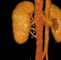
 Multiple renal arteries 01.jpg 496 × 496; 40 KB
 Notch and yolk sac blood vessels model.jpg 600 × 775; 97 KB
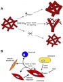
 NOTCH-endothelial-cartoon.jpg 1,280 × 888; 92 KB
 Stage 22 image 223.jpg 1,128 × 846; 366 KB
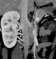
 Supernumerary renal vein 01.jpg 800 × 798; 72 KB
 Supernumerary renal vein 02.jpg 800 × 795; 89 KB
 Supernumerary renal vein 03.jpg 800 × 794; 80 KB
 Supernumerary renal vein 04.jpg 800 × 850; 76 KB
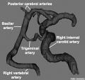
 Trigeminal artery 01.jpg 947 × 800; 102 KB
 Trigeminal artery 02.jpg 520 × 490; 35 KB
 Vasculature development 01 cartoon.jpg 599 × 900; 106 KB
 Vasculature development 02 cartoon.jpg 601 × 1,002; 93 KB
 Vein valve animation.gif 300 × 200; 54 KB
 Venule microvessel EM.jpg 600 × 626; 91 KB
 Woollard-plate01.jpg 744 × 1,000; 153 KB
 Woollard-plate02.jpg 788 × 1,000; 196 KB
 Woollard001.jpg 739 × 858; 142 KB
 Woollard002.jpg 699 × 873; 125 KB
 Woollard003.jpg 1,107 × 848; 159 KB
 Woollard004.jpg 1,037 × 717; 148 KB
 Woollard005.jpg 1,034 × 655; 144 KB


