Book - A Laboratory Text-Book of Embryology (1903): Difference between revisions
m (→Dedication) |
m (→Dedication) |
||
| Line 17: | Line 17: | ||
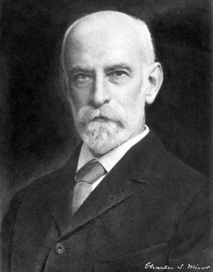
==Dedication== | ==Dedication== | ||
[[File:Henry Pickering Bowditch.jpg|left|thumb|Henry Pickering Bowditch (1840-1911)]] | |||
<center>To Henry Pickering Bowditch</center> | <center>To Henry Pickering Bowditch</center> | ||
| Line 25: | Line 24: | ||
<center>This Volume Is Dedicated By The Author</center> | <center>This Volume Is Dedicated By The Author</center> | ||
==Preface== | ==Preface== | ||
Revision as of 10:01, 5 April 2014
| Embryology - 3 May 2024 |
|---|
| Google Translate - select your language from the list shown below (this will open a new external page) |
|
العربية | català | 中文 | 中國傳統的 | français | Deutsche | עִברִית | हिंदी | bahasa Indonesia | italiano | 日本語 | 한국어 | မြန်မာ | Pilipino | Polskie | português | ਪੰਜਾਬੀ ਦੇ | Română | русский | Español | Swahili | Svensk | ไทย | Türkçe | اردو | ייִדיש | Tiếng Việt These external translations are automated and may not be accurate. (More? About Translations) |
Minot CS. A Laboratory Text-Book Of Embryology. (1903) Philadelphia:P. Blakiston's Son & Co.
| Online Editor |
|---|

|
| Historic Disclaimer - information about historic embryology pages |
|---|
|

A Laboratory Text-Book Of Embryology
Dedication

Preface
The accompanying volume is intended primarily for the use of students, taking a practical laboratory course in Embryology. The author's experience has led him to believe that the study of carefully selected sections of embryos, accompanied by directions and explanations of the significant structures in each section, offers many advantages. This conviction has determined the arrangement of the book. Attention is given chiefly to such points as serve to explain adult anatomical relations, to illustrate general biological principles, and to afford insight into pathological processes.
Portions of the text and many of the figures have been borrowed from the author's "Human Embryology." The woodcuts in Chapter IV were made by C. L. Albert Probst, of Braunschweig, after drawings by Dr. E. A. Locke. To both of these artists the author is indebted for their beautiful work. Much assistance has been rendered by Dr. F. T. Lewis, of the Harvard Medical School, to whom special acknowledgments are due for the reconstructions of the anatomy of the pig embryo of twelve millimeters and for invaluable help in the correction of the proofs.
Many of the illustrations are from the Harvard Embryologieal Collection, without which this work could not have been undertaken. The number of the embryo and of the section is given for all such illustrations.
The title was suggested by Dr. W. T. Porter's "A Laboratory Text-book of Physiology," and is adopted with his approval.
The author requests those who use this book to communicate to him any suggestions, which their experience may lead to, for improving it, in case it meets with sufficient favor to call for a new edition.
Charles Sedgwick Minot.
Cortina d'Ampezzo, August, 1902.
CONTENTS
Chapter I. — General Conceptions
The Vertebrate Type of Structure, IS The Principal Modifications of the Vertebrate Type, 22 Definition of Anlage, 25 A Summary of Embryological Development, 25 Cytomorphosis, . . 27 Comparison of Larval and Embryonic Types of Development, 32 Germ-layers 34 The Relations of Surface to Mass, 37 The Law of Unequal Growth, 38 Germ-cells 39 The Theory of Heredity, 40 The Law of Recapitulation,
Chapter II. — The Early Development of Mammals
The Spermatozoon The Fully Grown Ovum before Maturation, 45 Ovulation, 46 The Maturation of the Ovum, 47 Impregnation of the Ovum, 49 Segmentation of the Ovum 54 The Blastodermic Vesicle, 60 The Embryonic Shield, 62 Origin of the Mesoderm, 63 The Primitive Axis 65 The Notochordal Canal ". 66 The Notochord, 67 The Ultimate Fate of the Notochord 68 The Origin of the Nervous System, 69 The Structure of the Medullary Canal, 71 The Early History of the Mesoderm 74 Somatopleure and Splanchnopleure 76 The Embryonic coelom, 79 The Mesenchyma, 83 The Germ-cells, 84 The Yolk-sac, 85 The Origin of the Blood-vessels and Blood, 90 The Blood-corpuscles, . * 93 The Origin of the Heart 96 The Germinal Area, 97 The Main Vessels of the Area Vasculosa 97 The Ltver, 100 The Oral and Anal Plates, 100 The Excretory Organs, 101 The Allantois, 103 The Trophoblast, 106 The Growth of the Embryo, 107 The Umbilical Cord,
Chapter III. — The Human Embryo
Calculation of the Age of the Human Embryo, 112
The Classification of the Early Stages, 113
Hypothetical Development of the Blastodermic Vesicle in Primates, 116
Relations of the Embryo to the Uterus, Il8
Ovum of a Monkey in the Second Stage, 121
Human Embryo in the Second Stage 123
Embryo of a Gibbon in the Third Stage 127
Human Embryo in the Fourth Stage with Medullary Plate, 129
Human Embryo in the Fifth Stage with Open Medullary Groove 131
Human Embryo in the Sixth Stage with Medullary Canal, 132
Human Embryo in the Seventli Stage with One Gill-cleft, 135
Human Embryo in the Eighth Stage with Two Gill-clefts, 135
Human Embryo in the Ninth Stage with Three Gill-clefts 13S
Human Embryo in the Tenth Stage with Four Gill-clefts, 141
Human Embryo in the Eleventh Stage with Limb-buds, 142
Human Embryo of Twenty-six Days 143
Human Embryo of Twenty-eight Days, 143
Embryos of the Second, Third, and Fourth Months
Chapter IV. — Study of Pig Embryos
Methods of Obtaining Embryos, 157 The Making of Serial Sections, 158 Selection of the Planes of Section and the Stages for Practical Study, 158 The Study of the External Form, 159 Pig Embryo of 10 mm , 160 Pig Embryo of 12 mm. General Anatomy, 162 Pig Embryo of 15 mm. (External Form) 17° Pig Embryo of 20 mm. (External Form), 17 1 Pig Embryo of 12 mm. (Studied in Sections), 173 The Study of Transverse Sections, 1 75 Section through the Upper Part of the Otocyst, 175 Section through the Lower Part of the Otocyst, 181 Section through the First Gill-cleft, 184 Section through the Second Gill-cleft, 1S7 Section through the Third Gill-cleft 190 Section through the Fourth Gill-cleft 193 Section through the Anterior Limbs and Heart, I95 Section to show the Brachial Plexus, I99 Section through the Stomach and Liver, 201 The Study of Sagittal Sections, 205 Median Section of the Head, 205 Section of the Head through the Principal Ganglia, 208 The Study of Frontal Sections, 211 Section through the Trigeminal Roots 212 Section through the Acusttco-facial Ganglion, • 213 Section through the Otocyst 214 Section through the Dorsal Vertebne, 215 Pig Embryo of 9 mm. (Studied in Sections), 217 Transverse Section through the Branchial Arches, 217 Sagittal Section to the Right of the Median Plane, .... 219 Frontal Section through the Mid-brain and Fore-brain, 222 Frontal Section through the Umbilical Opening, 223 Frontal Section through the Second and Third Gill-clefts, 227 Pig Embryo of 6 mm. (Studied in Sections), 228 Pig Embryo of 17 mm. (Studied in Sections) 231 Transverse Section through the Lungs, 23 1 Section through the Wolffian Body and Genital Gland, 235 Section through the Kidney 237 Frontal Section of the Umbilical Cord, 239 Pig Embryo of 20 mm. (Studied in Sections), 240 Transverse Section through the Snout, 240 Transverse Section through the Lower Part of the Neck, 241 Transverse Section through the Lungs 244 Transverse Section through the Posterior Limbs, 247 Transverse Section through the Mammary Anlage, 249
Sagittal Section through the Right Lung and Kidney, 250 Frontal Sections of the Head 253 Pig Embryo of 24 mm. (Studied in Sections) 259 Frontal Section through the Eye, 259 Median Sagittal Section
Chapter V. — Study of Young Chick Embryos
Method of Obtaining Embryos, 269
Embryo Chick with Twenty-four Segments, 272
Embryo Chick with Twenty-eight Segments, 274
The Study of Transverse Sections, 274
Horizontal Section, • • 290
Histological Differentiation of Chick Embryo with Three Gill-clefts, 293
Embryo Chick with Seven Segments, 295
Examination in the Fresh State 295
Examination after Hardening, 297
Comparison with a Rabbit Embryo, 297
Longitudinal Section, 297
Study of Transverse Sections, 299
Chapter VI. — Study of the Blastodermic Vesicle and the Segmentation of the Ovum
Method of Obtaining Blastodermic Vesicles from the Rabbit, 305 Study of Rabbit Blastodermic Vesicles in Alcohol 306 The Maturation, Fertilization, and Segmentation of the Ovum in White Mice, 312 The First Polar Globule, 3 J 3 The Second Polar Globule, 3*3 The Single Polar Globule 3 '4 Fertilization, 3*5
Chapter VII. — Study of the Uterus and the Fcetal Appendages in Man
Histology of the Uterus 3'6 Menstruation, 316 Decidua Menstrualis, 3 X 7 The Pregnant Uterus : the Two Stages, 319 The Human Uterus Three Months Pregnant 319 Human Uterus Seven Months Pregnant 3 21 Decidua Vera of the First Stage in Section, 322 Decidua Reflexa of the First Stage, 325 Decidua Vera and Chorion Leeve of the Second Stage, 327 The Placenta in Situ, - 3 2 9 Decidua Serotina at Seven Months, . . . *. 334 The Human Placenta, 336 Histology of the Human Chorion, 34 1 The Chorion with Trophoblast, 342 The Chorionic Villi 345 The Structure of the Amnion, 349 The Umbilical Cord, 35 1 The Structure of the Human Yolk-sac, 354
Measuring the Length of Embryos, 35°
Methods of Reconstruction, 356
Reconstruction of Drawings, 356
Reconstruction with Wax-plates, 35^
Directions for Orienting Serial Sections, 360
Microtomes, 3°°
Methods of Hardening and Preserving, 363
Preservation in Alcohol, 3^5
Directions for Imbedding Specimens to be Microtomed, 3^5
Method of Mounting Paraffin Sections, 366
Methods of Staining, 367
| Historic Disclaimer - information about historic embryology pages |
|---|
|
Cite this page: Hill, M.A. (2024, May 3) Embryology Book - A Laboratory Text-Book of Embryology (1903). Retrieved from https://embryology.med.unsw.edu.au/embryology/index.php/Book_-_A_Laboratory_Text-Book_of_Embryology_(1903)
- © Dr Mark Hill 2024, UNSW Embryology ISBN: 978 0 7334 2609 4 - UNSW CRICOS Provider Code No. 00098G