Category:Male
This Embryology category shows pages and media related to Male embryos and male genital development.
Male Links: Y chromosome | testis | spermatozoa | meiosis | AMH | penis | prostate | seminal vesicle | genital abnormalities | hypospadias | puberty | Category:Y Chromosome
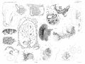
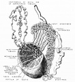
Male Embryos Carnegie Collection: 1163 | 1183 | 1199 | 1315 | 1474b | 1686 | 1705a | 1852 | 1945 | 2026 | 2114 | 217 | 256 | 2561 | 392 | 4160 | 417 | 431 | 4361 | 437 | 4570 | 5154 | 5609 | 5621A | 590 | 607 | 6150 | 657 | 6701 | 693 | 7254 | 75 | 7864 | 834 | 8394 | 840 | 847 | 895 | 948 | 950 | 966
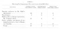
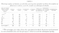
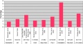
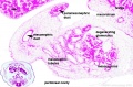
| Male preponderance | Female preponderance |
|---|---|
|
|
| Cardiac defects | |
|
|
| Table data[1] Links: abnormal development | cardiovascular abnormalities | USA | Male | Female | cleft lip and palate | |
Subcategories
This category has the following 46 subcategories, out of 46 total.
C
- Carnegie Embryo 1163
- Carnegie Embryo 1183
- Carnegie Embryo 1199
- Carnegie Embryo 1315
- Carnegie Embryo 1474b
- Carnegie Embryo 1686
- Carnegie Embryo 1705a
- Carnegie Embryo 1852
- Carnegie Embryo 1945
- Carnegie Embryo 2026
- Carnegie Embryo 2114
- Carnegie Embryo 217
- Carnegie Embryo 256
- Carnegie Embryo 2561
- Carnegie Embryo 392
- Carnegie Embryo 4160
- Carnegie Embryo 417
- Carnegie Embryo 431
- Carnegie Embryo 4361
- Carnegie Embryo 437
- Carnegie Embryo 4570
- Carnegie Embryo 5154
- Carnegie Embryo 5609
- Carnegie Embryo 5621A
- Carnegie Embryo 590
- Carnegie Embryo 607
- Carnegie Embryo 6150
- Carnegie Embryo 657
- Carnegie Embryo 6701
- Carnegie Embryo 693
- Carnegie Embryo 7254
- Carnegie Embryo 75
- Carnegie Embryo 7864
- Carnegie Embryo 834
- Carnegie Embryo 8394
- Carnegie Embryo 840
- Carnegie Embryo 847
- Carnegie Embryo 895
- Carnegie Embryo 948
- Carnegie Embryo 950
- Carnegie Embryo 966
D
T
Pages in category 'Male'
The following 161 pages are in this category, out of 161 total.
A
B
- Template:BGDB Sexual Differentiation - Abnormalities Interactive
- Template:BGDB Sexual Differentiation - Early Embryo Interactive
- Template:BGDB Sexual Differentiation - Fetal Interactive
- Template:BGDB Sexual Differentiation - Late Embryo Interactive
- Template:BGDB Sexual Differentiation - Postnatal Interactive
- Template:BGDB Sexual Differentiation - Sex Determination Interactive
- Template:Blood-testis barrier
- Template:Blood–testis barrier
- Book - A Laboratory Manual and Text-book of Embryology 8
- Book - Contributions to Embryology Carnegie Institution No.61
- Book - Manual of Human Embryology 19-4
- Template:Bulbourethral gland
C
- Template:CE1163
- Template:CE1183
- Template:CE1206
- Template:CE1474b
- Template:CE1686
- Template:CE1705a
- Template:CE1852
- Template:CE1936
- Template:CE2026
- Template:CE217
- Template:CE389a
- Template:CE5621A
- Template:CE590
- Template:CE607
- Template:CE693
- Template:CE834
- Template:CE847
- Template:CE948
- Template:CE950
- Template:CE955
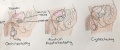
- Template:Cryptorchidism
- Template:Cryptorchidism table
D
H
I
L
M
P
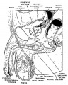
- Paper - A contribution to the development of the prostate in man (1909)
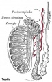
- Paper - A morphological study of testicular descent
- Paper - A note on a case of bifid penis (1924)
- Paper - Development and transition of the testis, normal and abnormal 1
- Paper - Development and transition of the testis, normal and abnormal 2
- Paper - Development and transition of the testis, normal and abnormal 3
- Paper - Development and transition of the testis, normal and abnormal 4
- Paper - Has a persistence of the Müllerian ducts any relation to the conditions of cryptorchidism?
- Paper - Hermaphroditism in a mole with male external genitals (1924)
- Paper - Notes on the development of the prepuce (1935)
- Paper - On the anlage of the bulbo-urethral and major vestibular glands in the human embryo (1915)
- Paper - On the phenomena of sex-differentiation (1892)
- Paper - Origin of the sex-cords and definitive spermatogonia in the male chick (1916)
- Paper - Testes descent 1909 - 1
- Paper - Testes descent 1909 - 2
- Paper - Testes descent 1909 - 3
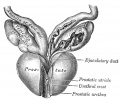
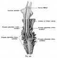
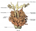
- Paper - The development of the human prostate gland with reference to the development of other structures at the neck of the urinary bladder (1912)
- Paper - The development of the seminal vesicles in man
- Template:Penis
- Penis Development
- Template:Peritubular myoid cell
- Template:Persistent Müllerian duct syndrome
- Template:Precocious puberty
- Template:Prepuce
- Template:Primary spermatocyte
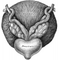
- Template:Prostate
- Template:Puberty
R
- Template:Ref-Andersen1932b
- Template:Ref-BascomOsterud1925
- Template:Ref-Bremer1911
- Template:Ref-Brown1924
- Template:Ref-CurlTromly1944
- Template:Ref-Eggerth1915
- Template:Ref-Evatt1909
- Template:Ref-Hart1909a
- Template:Ref-Hart1909b
- Template:Ref-Hart1909c
- Template:Ref-Hunter1935
- Template:Ref-Jirasek1971
- Template:Ref-Klinefelter1942
- Template:Ref-Lipschutz1924
- Template:Ref-Lockwood1887a
- Template:Ref-Lockwood1887b
- Template:Ref-Lockwood1888a
- Template:Ref-Lockwood1888b
- Template:Ref-Lowsley1912
- Template:Ref-Painter1923
- Template:Ref-Pohlman1904
- Template:Ref-Pryor1928
- Template:Ref-Siddiqi1937
- Template:Ref-Spaulding1921
- Template:Ref-Swift1916
- Template:Ref-Swyer1944
- Template:Ref-Watase1892
- Template:Ref-Watson1918
S
- Template:Seminal vesicle
- Seminal Vesicle Development
- Template:Seminal Vesicle Timeline table
- Template:Sertoli cell
- Sertoli cell
- Template:Sex bias
- Template:Sperm
- Template:Spermatogenesis
- Template:Spermatogonia
- Template:Spermatogonial stem cell
- Template:Spermatogonium
- Template:Spermatozoa
- Spermatozoa Development
- Template:Spermatozoon
- Template:Spermiogenesis
- Template:Sry
- Template:SRY
- Template:SSC
T
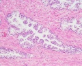
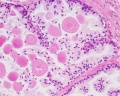
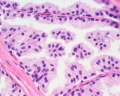
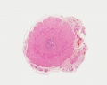
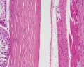
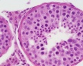
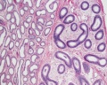
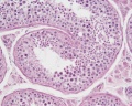
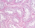
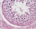
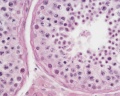
Media in category 'Male'
The following 200 files are in this category, out of 233 total.
(previous page) (next page) Bailey327.jpg 872 × 567; 89 KB
 Bailey332.jpg 637 × 356; 53 KB
 Bailey337.jpg 790 × 573; 74 KB
 Bailey338.jpg 940 × 473; 87 KB
 Bailey341.jpg 832 × 675; 69 KB
 Bailey343-344.jpg 1,185 × 630; 91 KB
 Bailey345-346.jpg 1,193 × 491; 79 KB
 Bailey347-348.jpg 1,198 × 526; 83 KB
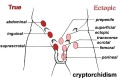 Cryptorchidism.jpg 600 × 390; 35 KB
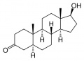 Dihydrotestosterone.jpg 500 × 368; 16 KB
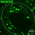
 Dog- spermatozoa NANOG expression.jpg 800 × 691; 109 KB
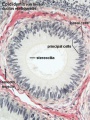
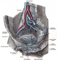
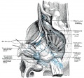
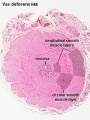 Ductus deferens 01.jpg 400 × 533; 76 KB
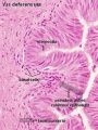 Ductus deferens 02.jpg 400 × 533; 80 KB
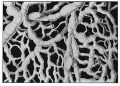
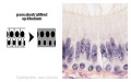 Epididymis histology 01.jpg 600 × 375; 20 KB
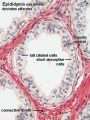 Epididymis histology 02.jpg 400 × 534; 71 KB
 Epididymis histology 03.jpg 400 × 533; 68 KB
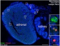 Fetal adrenal ectopic germ cells 03.jpg 899 × 700; 159 KB
 Fetal corpus cavernosum and corpus spongiosum 01.jpg 1,795 × 2,082; 919 KB
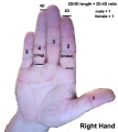 Finger length ratio - 2D4D.jpg 428 × 480; 26 KB
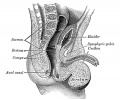
 Gray0585.jpg 752 × 800; 188 KB
 Gray0594.jpg 600 × 432; 83 KB
 Gray0619.jpg 800 × 741; 180 KB
 Gray1114.jpg 450 × 471; 47 KB
 Gray1119.jpg 700 × 807; 115 KB
 Gray1137.jpg 600 × 502; 64 KB
 Gray1152.jpg 597 × 600; 100 KB
 Gray1153.jpg 725 × 600; 100 KB
 Hamilton1959 fig01.jpg 802 × 1,000; 167 KB
 Historic-testis.jpg 509 × 800; 67 KB
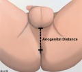 Human anogenital distance.jpg 570 × 499; 23 KB
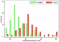 Human male anogenital distance graph.jpg 600 × 429; 28 KB
 Human Y chromosome 01.jpg 937 × 549; 167 KB
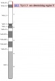 Human Y chromosome SRY region.jpg 351 × 517; 17 KB
 Human- spermatozoa NANOG expression 01.jpg 798 × 797; 79 KB
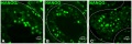 Human- spermatozoa NANOG expression.jpg 1,000 × 333; 77 KB
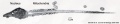 Human-spermatozoa EM01.jpg 1,000 × 204; 26 KB
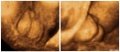 Hypospadia 3D ultrasound 01.jpg 1,150 × 497; 87 KB
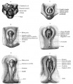
 Hypospadia classifications.jpg 500 × 410; 47 KB
 Keibel Mall 2 633.jpg 1,000 × 681; 74 KB
 Keibel Mall 2 635.jpg 1,000 × 1,073; 223 KB
 Keibel Mall 2 636.jpg 1,200 × 738; 119 KB
 Keibel Mall 2 642.jpg 1,200 × 644; 95 KB
 Keibel Mall 2 643.jpg 975 × 1,000; 95 KB
 Keibel Mall 2 656.jpg 1,200 × 746; 152 KB
 Keibel Mall 2 658a.jpg 1,127 × 1,200; 103 KB
 Keibel Mall 2 658b.jpg 895 × 1,200; 98 KB
 Keith1902 fig099.jpg 784 × 800; 105 KB
 Keith1902 fig104.jpg 800 × 601; 77 KB
 Klinefelter syndrome group response to startle.jpg 661 × 492; 73 KB
 Kollmann445.jpg 776 × 738; 112 KB
 Kollmann446.jpg 723 × 414; 38 KB
 Kollmann447.jpg 564 × 556; 42 KB
 Kollmann448.jpg 643 × 576; 43 KB
 Kollmann449.jpg 613 × 617; 44 KB
 Kollmann450.jpg 757 × 634; 98 KB
 Kollmann458.jpg 1,000 × 520; 120 KB
 Kollmann459.jpg 1,000 × 385; 50 KB
 Kollmann460.jpg 551 × 569; 40 KB
 Lockwood1887b fig23.jpg 500 × 423; 41 KB
 Lockwood1887b fig24.jpg 600 × 463; 82 KB
 Lockwood1887b fig25.jpg 715 × 1,000; 125 KB
 Lockwood1887b fig26.jpg 500 × 266; 38 KB
 Lockwood1887b fig27.jpg 500 × 378; 46 KB
 Lockwood1887b fig28.jpg 600 × 592; 43 KB
 Lockwood1887b fig29.jpg 800 × 656; 121 KB
 Lockwood1887b fig30.jpg 800 × 619; 69 KB
 Lockwood1887b fig31.jpg 800 × 576; 49 KB
 Lockwood1887b fig32.jpg 500 × 329; 55 KB
 Lockwood1887b fig33.jpg 600 × 617; 101 KB
 Lockwood1887b fig34.jpg 408 × 800; 43 KB
 Lockwood1887b fig35.jpg 800 × 600; 117 KB
 Lockwood1887b fig36.jpg 600 × 828; 53 KB
 Lockwood1887b fig37.jpg 800 × 739; 102 KB
 Lockwood1887b fig38.jpg 800 × 688; 65 KB
 Lockwood1887b fig39.jpg 800 × 1,102; 93 KB
 Lockwood1887b fig40.jpg 774 × 1,000; 92 KB
 Lockwood1887b fig41.jpg 800 × 547; 129 KB
 Lockwood1887b plate02.jpg 2,998 × 2,272; 1.21 MB
 Lockwood1888a fig47.jpg 1,000 × 712; 279 KB
 Lockwood1888a fig50.jpg 959 × 652; 85 KB
 Lockwood1888a plate07.jpg 1,280 × 985; 285 KB
 Lockwood1888b fig49.jpg 800 × 496; 69 KB
 Lockwood1888b fig51.jpg 800 × 725; 88 KB
 Lockwood1888b fig52.jpg 800 × 824; 60 KB
 Lowsley1912 fig01.jpg 990 × 620; 29 KB
 Lowsley1912 fig02.jpg 759 × 740; 102 KB
 Lowsley1912 fig03.jpg 1,897 × 1,601; 281 KB
 Lowsley1912 fig04.jpg 1,483 × 1,108; 343 KB
 Lowsley1912 fig05.jpg 1,641 × 1,083; 425 KB
 Lowsley1912 fig06.jpg 1,327 × 916; 237 KB
 Lowsley1912 fig07.jpg 1,437 × 1,435; 486 KB
 Lowsley1912 fig08.jpg 1,324 × 1,196; 312 KB
 Lowsley1912 plate01.jpg 1,904 × 2,412; 518 KB
 Lowsley1912 plate02.jpg 2,184 × 1,899; 402 KB
 Lowsley1912 plate03.jpg 1,682 × 2,166; 280 KB
 Lowsley1912 table01.jpg 1,836 × 904; 141 KB
 Lowsley1912 table02.jpg 1,817 × 1,029; 209 KB
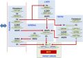
 Male genital molecular signaling 01.jpg 1,232 × 1,032; 94 KB
 Male histology 001.jpg 1,012 × 900; 378 KB
 Male histology 002.jpg 1,280 × 1,024; 490 KB
 Male histology 003.jpg 1,280 × 1,024; 703 KB
 Male histology 004.jpg 1,280 × 1,024; 540 KB
 Male puberty testicular volume graph.jpg 1,140 × 826; 126 KB
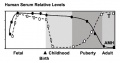 Male testosterone and AMH level graph.jpg 683 × 360; 31 KB
 Male urogenital sinus week 7to8 01.jpg 800 × 988; 238 KB
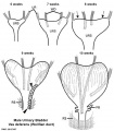 Male vas deferens and bladder week6to10.jpg 800 × 918; 86 KB
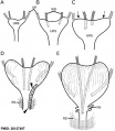 Mesonephric duct position week 6-11.jpg 697 × 800; 72 KB
 Model male androsterone synthesis.jpg 740 × 518; 92 KB
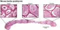
 Mouse epididymis development 01.jpg 1,200 × 909; 486 KB
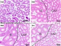
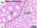
 Mouse epididymis development 02.jpg 600 × 451; 138 KB
 Mouse epididymis development 03.jpg 600 × 451; 119 KB
 Mouse epididymis development 04.jpg 600 × 451; 120 KB
 Mouse epididymis development 05.jpg 600 × 451; 112 KB
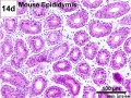
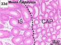
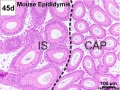
 Mouse- epididymis histology.jpg 751 × 383; 82 KB
 Mouse- spermatozoa NANOG expression.jpg 800 × 512; 111 KB
 Nelsen1953 fig002.jpg 1,200 × 1,037; 249 KB
 Nelsen1953 fig004.jpg 1,200 × 960; 200 KB
 Nelsen1953 fig005.jpg 1,200 × 709; 102 KB
 Nelsen1953 fig006.jpg 1,200 × 732; 224 KB
 Nelsen1953 fig007.jpg 1,200 × 1,311; 343 KB
 Newborn- cryptorchidism normal birthweight.jpg 800 × 428; 47 KB
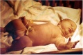 Newborn.jpg 570 × 381; 23 KB
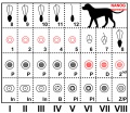
 Orchidometer.jpg 361 × 225; 14 KB
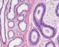
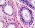
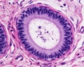
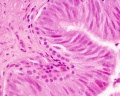
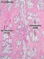 Prostate histology 01.jpg 300 × 400; 72 KB
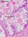 Prostate histology 02.jpg 300 × 400; 57 KB
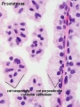 Prostate histology 03.jpg 300 × 400; 41 KB
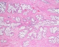 Prostate histology 04.jpg 1,280 × 1,024; 569 KB
 Prostate histology 05.jpg 1,280 × 1,024; 418 KB
 Prostate histology 06.jpg 1,280 × 1,024; 348 KB
 Prostate histology 07.jpg 1,280 × 1,024; 328 KB
 Prostate histology 08.jpg 1,280 × 1,024; 252 KB
 Prostate histology 09.jpg 1,019 × 764; 199 KB
 Prostate stem cell cartoon.png 907 × 2,789; 1.09 MB
 Rat blood–testis barrier 01.jpg 1,002 × 1,599; 221 KB
 Rat blood–testis barrier 02.jpg 1,002 × 853; 125 KB
 Reproductive surgeries in Males.jpeg 1,624 × 676; 354 KB
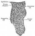
 Seminiferous-tubule-HEx40.jpg 400 × 500; 59 KB
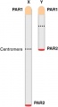 Sex chromosomes pseudoautosomal regions.jpg 217 × 400; 7 KB
 Small supernumerary marker chromosome 16.jpg 800 × 601; 58 KB
 Spaulding-fig05.jpg 778 × 744; 96 KB
 Spaulding-fig07.jpg 445 × 607; 35 KB
 Spaulding-fig08.jpg 445 × 607; 37 KB
 Spaulding-fig09.jpg 445 × 607; 37 KB
 Spaulding-fig10.jpg 445 × 607; 34 KB
 Spaulding-fig15.jpg 445 × 607; 38 KB
 Spaulding-fig16.jpg 445 × 607; 38 KB
 Spaulding-fig17.jpg 445 × 607; 36 KB
 Spaulding-fig18.jpg 445 × 607; 36 KB
 Spaulding-fig21.jpg 445 × 607; 33 KB
 Spaulding-fig24.jpg 443 × 604; 36 KB
 Spaulding-fig25.jpg 443 × 604; 33 KB
 Spaulding-fig26.jpg 443 × 604; 31 KB
 Spaulding-fig27.jpg 443 × 604; 32 KB
 Spaulding-fig28.jpg 443 × 604; 37 KB
 Spaulding-fig31.jpg 443 × 604; 31 KB
 Spaulding-fig32.jpg 443 × 604; 31 KB
 Spaulding-fig34.jpg 443 × 604; 30 KB
 Spaulding-fig42.jpg 430 × 599; 26 KB
 Spaulding-fig47.jpg 436 × 603; 32 KB
 Spaulding-fig48.jpg 445 × 604; 33 KB
 Spaulding-fig49.jpg 436 × 602; 26 KB
 Spaulding-fig50.jpg 430 × 599; 24 KB
 Spermatozoa histology 001.jpg 1,280 × 1,024; 366 KB
 Spermatozoa histology 002.jpg 1,280 × 1,024; 246 KB
 Spermatozoa histology 003.jpg 1,280 × 1,024; 166 KB
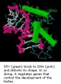 SRY and DNA.jpg 200 × 272; 19 KB
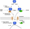 SRY nuclear import model.jpg 800 × 766; 79 KB
 Stage 22 image 194.jpg 1,000 × 671; 210 KB
 Stage 22 image 195.jpg 1,000 × 657; 265 KB
 Stage22 mesonephros.jpg 600 × 397; 83 KB
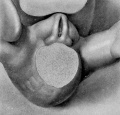
 Suprascrotal testis.jpg 1,000 × 751; 126 KB
 Testis histology 001.jpg 1,280 × 1,024; 574 KB
 Testis histology 002.jpg 1,280 × 1,024; 599 KB
 Testis histology 003.jpg 1,280 × 1,024; 183 KB
 Testis histology 004.jpg 1,280 × 1,024; 396 KB
 Testis histology 005.jpg 1,280 × 1,024; 266 KB
 Testis histology 006.jpg 1,280 × 1,024; 251 KB
 Testis histology 007.jpg 1,280 × 1,024; 256 KB
 Testis histology 008.jpg 1,280 × 1,024; 454 KB
 Testis histology 009.jpg 1,280 × 1,024; 339 KB
 Testis histology 010.jpg 1,280 × 1,024; 422 KB
 Testis histology 011.jpg 1,280 × 1,024; 245 KB
 Testis histology 012.jpg 1,280 × 1,024; 266 KB
 Testis histology 013.jpg 1,280 × 1,024; 418 KB
 Testis histology 014.jpg 1,280 × 1,024; 352 KB
 Testis histology 015.jpg 1,280 × 1,024; 281 KB
 Testis histology 016.jpg 1,280 × 1,024; 322 KB
 Testis histology 017.jpg 1,280 × 1,024; 283 KB
 Testis histology 018.jpg 1,280 × 1,024; 350 KB
 Testis histology 019.jpg 1,280 × 1,024; 239 KB
 Testis histology 02.jpg 246 × 481; 49 KB
 Testis histology 020.jpg 1,300 × 685; 334 KB
 Testis histology 021.jpg 1,200 × 962; 312 KB
 Testis histology 022.jpg 1,229 × 966; 311 KB
 Testis histology 023.jpg 600 × 375; 35 KB
 Testis histology 1.jpg 400 × 500; 113 KB
 Testis histology 2.jpg 400 × 500; 32 KB
 Testis histology.jpg 400 × 500; 54 KB
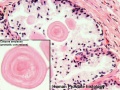
 Testis, young H&E reproductive system, male, convoluted seminiferous tubules x10.jpg 1,280 × 1,024; 396 KB
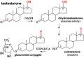 Testosterone metabolism.jpg 600 × 414; 36 KB


