Category:Fetal
From Embryology
Subcategories
This category has the following 71 subcategories, out of 71 total.
C
- Carnegie Embryo 1161
- Carnegie Embryo 1163
- Carnegie Embryo 1183
- Carnegie Embryo 131
- Carnegie Embryo 1318
- Carnegie Embryo 1388
- Carnegie Embryo 142
- Carnegie Embryo 145
- Carnegie Embryo 1455
- Carnegie Embryo 1474b
- Carnegie Embryo 1591
- Carnegie Embryo 1597b
- Carnegie Embryo 1656
- Carnegie Embryo 1686
- Carnegie Embryo 1705a
- Carnegie Embryo 172
- Carnegie Embryo 184
- Carnegie Embryo 1852
- Carnegie Embryo 2026
- Carnegie Embryo 211
- Carnegie Embryo 217
- Carnegie Embryo 2250a
- Carnegie Embryo 2250b
- Carnegie Embryo 23
- Carnegie Embryo 234a
- Carnegie Embryo 300
- Carnegie Embryo 34
- Carnegie Embryo 362
- Carnegie Embryo 3990
- Carnegie Embryo 4473
- Carnegie Embryo 4475
- Carnegie Embryo 448
- Carnegie Embryo 449
- Carnegie Embryo 458
- Carnegie Embryo 48
- Carnegie Embryo 538
- Carnegie Embryo 5652
- Carnegie Embryo 590
- Carnegie Embryo 607
- Carnegie Embryo 625
- Carnegie Embryo 6581
- Carnegie Embryo 662
- Carnegie Embryo 693
- Carnegie Embryo 7218
- Carnegie Embryo 834
- Carnegie Embryo 847
- Carnegie Embryo 858
- Carnegie Embryo 922
- Carnegie Embryo 928
- Carnegie Embryo 948
- Carnegie Embryo 95
- Carnegie Embryo 96
- Carnegie Embryo 972
Pages in category 'Fetal'
The following 200 pages are in this category, out of 270 total.
(previous page) (next page)2
A
B
- BGDA Practical - Fetal Development
- Talk:BGDA Practical - Fetal Development
- BGDA Practical - Fetal Development - Quiz
- BGDA Practical - Fetal Development Interactive
- BGDA Practical 12 - Abnormalities
- Template:BGDA Practical 12 - Abnormalities Interactive
- BGDA Practical 12 - Birth
- Template:BGDA Practical 12 - Birth Interactive
- BGDA Practical 12 - Embryo to Fetus
- Template:BGDA Practical 12 - Embryo to Fetus Interactive
- BGDA Practical 12 - Neonatal
- Template:BGDA Practical 12 - Neonatal Interactive
- BGDA Practical 12 - Second Trimester
- Template:BGDA Practical 12 - Second Trimester Interactive
- BGDA Practical 12 - Third Trimester
- Template:BGDA Practical 12 - Third Trimester Interactive
- Template:BGDALab12
- BGDB Gastrointestinal - Fetal
- Template:BGDB Practical 6 - Abnormalities Interactive
- Template talk:BGDB Practical 6 - Abnormalities Interactive
- Template:BGDB Practical 6 - Fetal Interactive
- Template:BGDB Practical 6 - Postnatal Interactive
- Template:BGDB Sexual Differentiation - Fetal Interactive
- Template:Birth weight
- Template:Birthweight
- Book - Human embryos of different ages examined in median sections - a contribution to the mechanics of development
- Book - Physiology of the Fetus
- Book - Physiology of the Fetus 1
- Book - Physiology of the Fetus 10
- Book - Physiology of the Fetus 11
- Book - Physiology of the Fetus 12
- Book - Physiology of the Fetus 13
- Book - Physiology of the Fetus 14
- Book - Physiology of the Fetus 15
- Book - Physiology of the Fetus 16
- Book - Physiology of the Fetus 2
- Book - Physiology of the Fetus 3
- Book - Physiology of the Fetus 4
- Book - Physiology of the Fetus 5
- Book - Physiology of the Fetus 6
- Book - Physiology of the Fetus 7
- Book - Physiology of the Fetus 8
- Book - Physiology of the Fetus 9
C
- Template:Canalicular stage
- Template:Carnegie Collection fetal table
- Template talk:Carnegie Collection fetal table
- Template:CE1161
- Template:CE1206
- Template:CE1282b
- Template:CE131
- Template:CE1318
- Template:CE1358
- Template:CE1388
- Template:CE142
- Template:CE1449
- Template:CE145
- Template:CE1455
- Template:CE1474b
- Template:CE1476
- Template:CE1561
- Template:CE1591
- Template:CE1597b
- Template:CE1656
- Template:CE1686
- Template:CE1702
- Template:CE1705a
- Template:CE1708
- Template:CE1716
- Template:CE1724
- Template:CE1742
- Template:CE1782
- Template:CE1811
- Template:CE1831
- Template:CE184
- Template:CE1840a
- Template:CE1845
- Template:CE1852
- Template:CE1858
- Template:CE190
- Template:CE1915
- Template:CE1980
- Template:CE2003
- Template:CE2066
- Template:CE2075
- Template:CE2079
- Template:CE2095
- Template:CE210
- Template:CE2118
- Template:CE2144
- Template:CE2163
- Template:CE217
- Template:CE2170
- Template:CE218
- Template:CE2185
- Template:CE219
- Template:CE2250a
- Template:CE2250b
- Template:CE2274
- Template:CE2328
- Template:CE234a
- Template:CE237
- Template:CE300
- Template:CE301
- Template:CE362
- Template:CE3669
- Template:CE389a
- Template:CE3990
- Template:CE4021
- Template:CE4229
- Template:CE4291
- Template:CE4473
- Template:CE4475
- Template:CE448
- Template:CE449
- Template:CE458
- Template:CE481
- Template:CE484
- Template:CE490
- Template:CE491
- Template:CE4985
- Template:CE508
- Template:CE509
- Template:CE538
- Template:CE5652
- Template:CE5712
- Template:CE5762
- Template:CE607
- Template:CE625
- Template:CE642
- Template:CE6581
- Template:CE662
- Template:CE6658
- Template:CE693
- Template:CE7218
- Template:CE834
- Template:CE84
- Template:CE847
- Template:CE858
- Template:CE886
- Template:CE922
- Template:CE928
- Template:CE948
- Template:CE95
- Template:CE9526
- Template:CE96
- Template:CE972
- Template:CE981
F
H
P
- Paper - A quantitative study of the fetal growth changes in the parts of the human stomach wall
- Paper - Blood supply of the otic capsule of a 150 mm (C.R.) human fetus
- Paper - Cell columns in the spinal cord of a human foetus of fourteen weeks (1941)
- Paper - Early fetal activity in mammals
- Paper - Fetal growth in man
- Paper - Ossification of the otic capsule in human fetuses
- Paper - Perichondrial ossification and the fate of the perichondrium with special reference to that of the otic capsule
- Paper - Significant superficial anastomoses in the arterial blood supply to the human brain (1959)
- Paper - Simple methods of correlating crown-rump and crown-heel lengths of the human fetus
Media in category 'Fetal'
The following 200 files are in this category, out of 313 total.
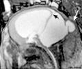
(previous page) (next page)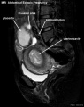 Abdominal ectopic pregnancy MRI.jpg 553 × 700; 45 KB
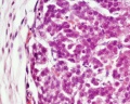 Adrenal histology 005.jpg 1,280 × 1,024; 298 KB
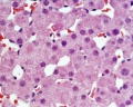 Adrenal histology 006.jpg 1,280 × 1,024; 313 KB
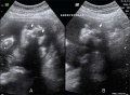 Anencephaly ultrasound.jpg 900 × 658; 108 KB
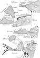 Anson1948 fig03.jpg 1,280 × 1,815; 358 KB
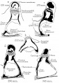 Anson1948 fig07.jpg 1,280 × 1,801; 219 KB
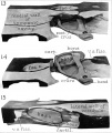 AnsonKarabinMartin1939 fig13-15.jpg 1,280 × 1,519; 171 KB
 Bailey094.jpg 376 × 689; 27 KB
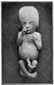 Bailey095.jpg 409 × 635; 52 KB
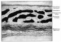 Bailey113.jpg 813 × 553; 128 KB
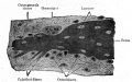 Bailey114.jpg 690 × 433; 63 KB
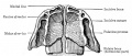 Bailey141.jpg 761 × 323; 66 KB
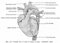 Bailey178.jpg 913 × 653; 125 KB
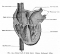 Bailey179.jpg 892 × 794; 114 KB
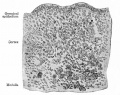 Bailey330.jpg 680 × 539; 109 KB
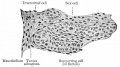 Bailey332.jpg 637 × 356; 53 KB
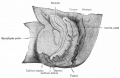 Bailey334.jpg 857 × 558; 77 KB
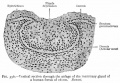 Bailey356.jpg 767 × 533; 103 KB
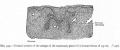 Bailey357.jpg 949 × 395; 88 KB
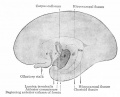 Bailey443.jpg 666 × 539; 49 KB
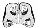 Bailey444.jpg 777 × 590; 127 KB
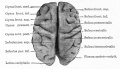 Bailey448.jpg 722 × 416; 55 KB
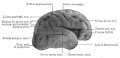 Bailey449.jpg 777 × 374; 45 KB
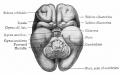 Bailey450.jpg 680 × 419; 45 KB
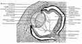 Bailey467.jpg 946 × 515; 159 KB
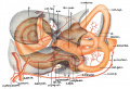 Bast1931 plate01.jpg 1,280 × 871; 113 KB
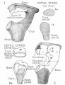 BeatonAnson1940 fig01-2.jpg 1,652 × 2,147; 652 KB
 BGDA2011-Embryo Lab 010611-01.mp3 ; 4.61 MB
- BGDA2011-Embryo Lab 010611-02.mp3 ; 3.23 MB
- BGDA2011-Embryo Lab 010611-03.mp3 ; 4.25 MB
- BGDA2011-Embryo Lab 010611-04.mp3 ; 1.79 MB
- BGDA2011-Embryo Lab 010611-05.mp3 ; 2.23 MB
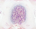 Bone histology 014.jpg 1,280 × 1,024; 541 KB
 Bone histology 015.jpg 1,280 × 1,024; 519 KB
 Bone histology 016.jpg 1,280 × 1,024; 379 KB
 Bone histology 017.jpg 1,280 × 1,024; 442 KB
 Bone histology 018.jpg 1,280 × 1,024; 336 KB
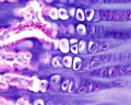 Bone histology 019.jpg 1,280 × 1,024; 275 KB
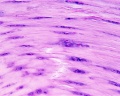 Bone histology 020.jpg 1,280 × 1,024; 272 KB
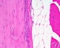 Bone histology 021.jpg 1,280 × 1,024; 254 KB
 Boyd1950 fig03a.jpg 600 × 872; 135 KB
 Boyd1950 fig03b.jpg 600 × 996; 152 KB
 Boyd1950 fig04.jpg 800 × 703; 102 KB
 Boyd1950 fig06.jpg 1,000 × 1,002; 272 KB
 Boyd1950 fig08.jpg 1,000 × 1,553; 341 KB
 Boyd1950 fig09.jpg 801 × 900; 163 KB
 Boyd1950 fig10.jpg 1,000 × 453; 114 KB
 Brain fissure development 01.jpg 1,157 × 502; 47 KB
 Brain fissure development 02.jpg 1,000 × 583; 52 KB
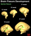 Brain fissure development 03.jpg 600 × 691; 33 KB
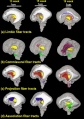 Brain tract development 01.jpg 720 × 1,023; 76 KB
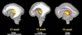 Brain tract development 02.jpg 1,000 × 424; 29 KB
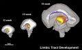 Brain tract development 06.jpg 1,000 × 605; 35 KB
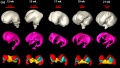 Brain ventricles and ganglia development 01.jpg 994 × 564; 45 KB
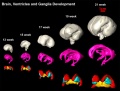 Brain ventricles and ganglia development 02.jpg 1,000 × 760; 49 KB
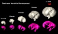 Brain ventricles and ganglia development 03.jpg 989 × 583; 36 KB
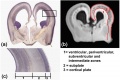 Brain week 17 histology.jpg 1,050 × 697; 61 KB
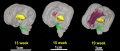 Brain-tract-development-03.jpg 1,000 × 424; 31 KB
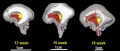 Brain-tract-development-04.jpg 1,000 × 424; 30 KB
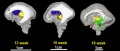 Brain-tract-development-05.jpg 1,000 × 424; 29 KB
 Crowder1957 fig07.jpg 848 × 642; 300 KB
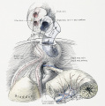 Cullen1916 fig14.jpg 1,280 × 1,293; 434 KB
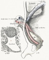 Cullen1916 fig15.jpg 1,000 × 1,227; 314 KB
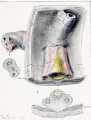 Cullen1916 fig16.jpg 1,280 × 1,686; 621 KB
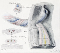 Cullen1916 fig17.jpg 1,280 × 1,134; 365 KB
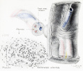 Cullen1916 fig19.jpg 1,280 × 1,098; 330 KB
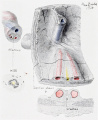 Cullen1916 fig22.jpg 1,224 × 1,501; 324 KB
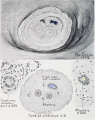 Cullen1916 fig23.jpg 1,000 × 1,266; 346 KB
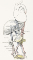 Cullen1916 fig33.jpg 1,154 × 2,052; 422 KB
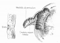 Cullen1916 fig92.jpg 885 × 637; 109 KB
 Dandy Walker malformation MRI 01.jpg 600 × 506; 37 KB
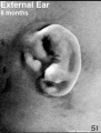 External Ear 5 months-icon.jpg 352 × 464; 63 KB
 Fawcett1911 fig06.jpg 1,000 × 680; 161 KB
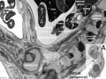 Fetal 10wk urogenital 1.jpg 800 × 600; 109 KB
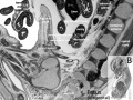 Fetal 10wk urogenital 2.jpg 800 × 600; 110 KB
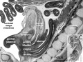 Fetal 10wk urogenital 3.jpg 800 × 600; 107 KB
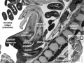 Fetal 10wk urogenital 4.jpg 800 × 600; 105 KB
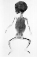 Fetal 5 month x-ray.jpg 1,500 × 2,298; 308 KB
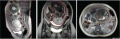 Fetal 9 month MRI 01.jpg 1,501 × 483; 145 KB
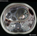 Fetal 9 month MRI 02.jpg 619 × 600; 67 KB
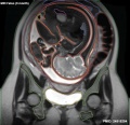 Fetal 9 month MRI 03.jpg 619 × 600; 64 KB
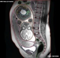 Fetal 9 month MRI 04.jpg 619 × 600; 54 KB
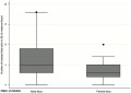 Fetal cells maternal blood graph.jpg 600 × 426; 18 KB
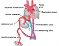 Fetal Circulation Pathway.jpg 1,371 × 1,069; 112 KB
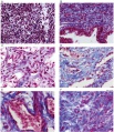 Fetal corpus cavernosum and corpus spongiosum 01.jpg 1,795 × 2,082; 919 KB
 Fetal ductus venosus pressure wave 01.jpg 706 × 755; 52 KB
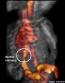 Fetal ductus venosus ultrasound 01.jpg 783 × 1,000; 68 KB
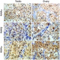 Fetal gonad retinoid receptor expression 01.jpg 1,004 × 1,000; 226 KB
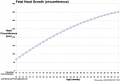 Fetal head growth circumference graph01.jpg 905 × 613; 58 KB
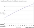 Fetal head growth circumference graph02.jpg 800 × 650; 44 KB
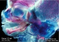 Fetal head lateral.jpg 632 × 447; 34 KB
 Fetal head medial.jpg 632 × 447; 34 KB
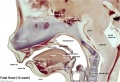 Fetal head section 01.jpg 1,200 × 821; 186 KB
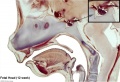 Fetal head section 02.jpg 1,200 × 821; 171 KB
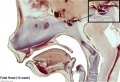 Fetal head section 03.jpg 1,200 × 821; 174 KB
 Fetal head section 04.jpg 1,200 × 821; 188 KB
 Fetal head section.jpg 1,200 × 821; 167 KB
 Fetal kidney MRI 01.jpg 797 × 880; 68 KB
 Fetal kidney MRI 02.jpg 797 × 880; 64 KB
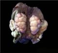 Fetal kidney.jpg 689 × 623; 43 KB
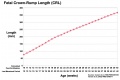 Fetal length change.jpg 972 × 648; 72 KB
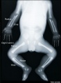 Fetal limb X-ray-01.jpg 685 × 931; 57 KB
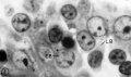 Fetal liver erythroblasts 01.jpg 905 × 534; 69 KB
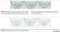 Fetal mandible and lower lip 01.jpg 1,028 × 557; 57 KB
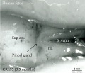 Fetal pineal gland 01.jpg 700 × 603; 67 KB
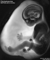 Fetal polyhydramnios MRI-01.jpg 768 × 932; 60 KB
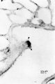 Fetal rabbit neuroepithelial body 01.jpg 793 × 1,200; 98 KB
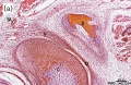 Fetal temporomandibular joint 01.jpg 600 × 391; 107 KB
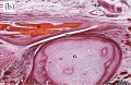 Fetal temporomandibular joint 02.jpg 600 × 390; 109 KB
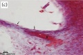 Fetal temporomandibular joint 03.jpg 600 × 393; 48 KB
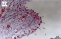 Fetal temporomandibular joint 04.jpg 600 × 390; 67 KB
 Fetal temporomandibular joint 05.jpg 600 × 392; 71 KB
 Fetal temporomandibular joint 06.jpg 600 × 389; 50 KB
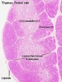 Fetal thymus.jpg 450 × 600; 122 KB
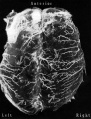 Gillilan1959-fig03.jpg 1,042 × 1,366; 317 KB
 Gilmour1941 fig02.jpg 1,000 × 526; 36 KB
 Gilmour1941 fig10.jpg 511 × 907; 105 KB
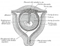 Gray0034.jpg 800 × 613; 98 KB
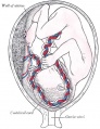 Gray0038.jpg 462 × 600; 45 KB
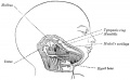 Gray0043.jpg 800 × 496; 50 KB
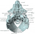 Gray0070.jpg 800 × 796; 182 KB
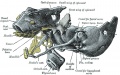 Gray0071.jpg 700 × 440; 104 KB
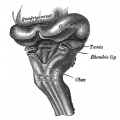 Gray0649.jpg 698 × 700; 72 KB
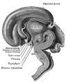 Gray0654.jpg 402 × 500; 39 KB
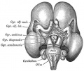 Gray0655.jpg 500 × 419; 39 KB
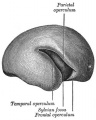 Gray0658.jpg 361 × 450; 29 KB
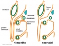 Greater-omentum.jpg 537 × 419; 48 KB
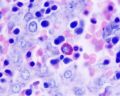 Histology-fetal liver HEx100.jpg 1,280 × 1,024; 214 KB
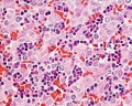 Histology-fetal liver HEx40.jpg 1,000 × 800; 281 KB
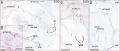 Human 15 weeks - terminal nerve and vomeronasal organ nerves.jpg 940 × 403; 306 KB
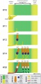 Human cochlea fetal development cartoon.jpg 592 × 1,200; 96 KB
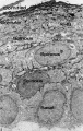 Human embryo skin 24 week EGA.jpg 596 × 939; 165 KB
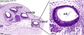 Human fetal cochlea 01.jpg 1,270 × 532; 266 KB
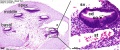 Human fetal cochlea 02.jpg 1,270 × 532; 271 KB
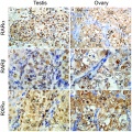 Human fetal gonad retinoid receptor expression.jpg 1,004 × 1,000; 447 KB
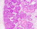 Human fetal kidney histology 01.jpg 1,280 × 1,024; 481 KB
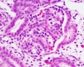 Human fetal kidney histology 02.jpg 1,280 × 1,024; 322 KB
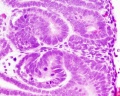 Human fetal kidney histology 03.jpg 1,280 × 1,024; 333 KB
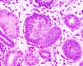 Human fetal kidney histology 04.jpg 1,280 × 1,024; 307 KB
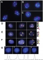 Human fetal neural aneuploidy.jpg 1,000 × 1,400; 134 KB
 Human fetal uterus myometrium.jpg 500 × 554; 86 KB
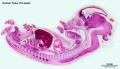 Human week 10 fetus 01.jpg 2,300 × 1,327; 448 KB
 Human week 10 fetus 02.jpg 800 × 462; 83 KB
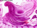 Human week 10 fetus 03.jpg 1,600 × 1,200; 370 KB
 Human week 10 fetus 04.jpg 1,600 × 1,200; 534 KB
 Human week 10 fetus 05.jpg 1,600 × 1,200; 612 KB
 Human week 10 fetus 06.jpg 1,200 × 900; 251 KB
 Human week 10 fetus 07.jpg 1,200 × 900; 283 KB
 Human week 10 fetus 08.jpg 1,200 × 900; 323 KB
 Human week 10 fetus 09.jpg 1,200 × 900; 345 KB
 Human week 10 fetus 10.jpg 1,200 × 900; 291 KB
 Human week 10 fetus 11.jpg 1,200 × 900; 304 KB
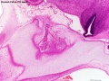 Human week 10 fetus 12.jpg 1,200 × 900; 349 KB
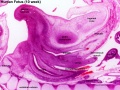 Human week 10 fetus 23.jpg 1,600 × 1,200; 393 KB
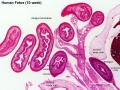 Human week 10 fetus 26.jpg 1,200 × 900; 262 KB
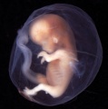 Human- fetal week 10 bf01.jpg 1,200 × 1,214; 119 KB
 Human- fetal week 10 bf02.jpg 1,200 × 900; 91 KB
 Human- fetal week 10 cerebellum A.jpg 347 × 284; 24 KB
 Human- fetal week 10 cerebellum B.jpg 347 × 284; 21 KB
 Human- fetal week 10 cerebellum C.jpg 347 × 284; 25 KB
 Human- fetal week 10 cerebellum D.jpg 347 × 284; 23 KB
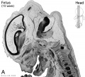 Human- fetal week 10 head A.jpg 600 × 544; 113 KB
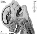 Human- fetal week 10 head A1.jpg 1,200 × 1,088; 159 KB
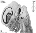 Human- fetal week 10 head B.jpg 600 × 544; 66 KB
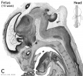 Human- fetal week 10 head C.jpg 600 × 544; 118 KB
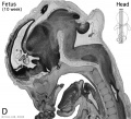 Human- fetal week 10 head D.jpg 600 × 544; 111 KB
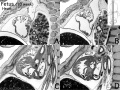 Human- fetal week 10 heart ABCD.jpg 600 × 450; 133 KB
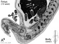 Human- fetal week 10 lower body A.jpg 600 × 450; 96 KB
 Human- fetal week 10 lower body B.jpg 600 × 450; 93 KB
 Human- fetal week 10 lower body C.jpg 600 × 450; 94 KB
 Human- fetal week 10 lower body D.jpg 600 × 450; 91 KB
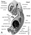 Human- fetal week 10 sagittal plane A.jpg 500 × 573; 96 KB
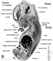 Human- fetal week 10 sagittal plane B.jpg 500 × 573; 99 KB
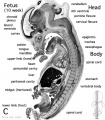 Human- fetal week 10 sagittal plane C.jpg 500 × 573; 98 KB
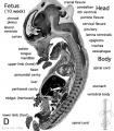 Human- fetal week 10 sagittal plane D.jpg 500 × 573; 105 KB
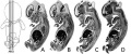 Human- fetal week 10 sagittal planes.jpg 600 × 250; 29 KB
 Human- fetal week 10 upper body A.jpg 600 × 450; 104 KB
 Human- fetal week 10 upper body B.jpg 600 × 450; 105 KB
 Human- fetal week 10 upper body C.jpg 600 × 450; 109 KB
 Human- fetal week 10 upper body D.jpg 600 × 450; 106 KB
 Human- fetal week 10 urogenital A.jpg 600 × 450; 109 KB
 Human- fetal week 10 urogenital B.jpg 600 × 450; 109 KB
 Human- fetal week 10 urogenital C.jpg 600 × 450; 105 KB
 Human- fetal week 10 urogenital D.jpg 600 × 450; 101 KB
 Hydronephrosis.jpg 600 × 345; 64 KB
 Keibel Mall 144.jpg 678 × 607; 40 KB
 Keibel Mall 148.jpg 604 × 909; 93 KB
 Keibel Mall 2 302.jpg 1,278 × 803; 149 KB
 Keibel Mall 2 429.jpg 1,280 × 652; 204 KB
 Keibel Mall 2 583.jpg 1,280 × 1,044; 259 KB
 Keibel Mall 2 584.jpg 464 × 800; 68 KB
 Keibel Mall 2 585.jpg 1,280 × 1,144; 277 KB
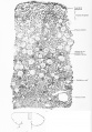 Keibel Mall 2 621.jpg 1,280 × 1,842; 589 KB
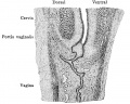 Keibel Mall 2 626.jpg 850 × 679; 143 KB
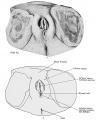 Keibel Mall 2 627.jpg 1,000 × 1,252; 169 KB
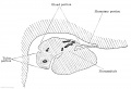 Keibel Mall 2 632.jpg 1,000 × 684; 100 KB
 Keibel Mall 2 643.jpg 975 × 1,000; 95 KB
 Keibel Mall 203-205.jpg 838 × 1,000; 129 KB
 Keibel Mall 209.jpg 458 × 990; 89 KB
 Keibel Mall 210.jpg 340 × 542; 41 KB


