File:Human embryo day 3.jpg
Human_embryo_day_3.jpg (400 × 409 pixels, file size: 7 KB, MIME type: image/jpeg)
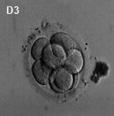
Human Embryo (day 3)
- early morula stage
- During day 3 there are still only a small number of blastomere cells (8) forming a solid ball of cells generated by mitotic cell division.
- Note the thick zona pellucid still surrounding the conceptus.
Image Links: Human oocyte to blastocyst | Germinal vesicle oocyte (GV) | Metaphase I oocyte | Metaphase II oocyte | Day 2 | Day 3 | Day 5 | Day 5 (label) | Day 5 (colour label)
Germinal vesicle oocyte
Metaphase I oocyte
Metaphase II oocyte
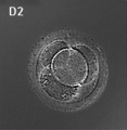
Day 2
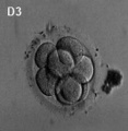
Day 3 - Morula
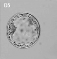
Day 5 - Blastocyst
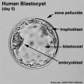
Day 5 (label)
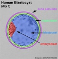
Day 5 (colour label)
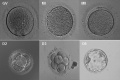
Human oocyte to blastocyst


- Links: Oocyte | Morula | Blastocyst | Carnegie stage 1 | Carnegie stage 2 | Carnegie stage 3 | Cell Division - Mitosis
Reference
<pubmed>19924284</pubmed>| PMC2773928 | PLoS One
PLoS One. 2009; 4(11): e7844.
Published online 2009 November 16. doi: 10.1371/journal.pone.0007844.
Copyright Zhang et al. This is an open-access article distributed under the terms of the Creative Commons Attribution License, which permits unrestricted use, distribution, and reproduction in any medium, provided the original author and source are credited.
Original file name: Pone.0007844.g004.jpg http://www.ncbi.nlm.nih.gov/pmc/articles/PMC2773928/figure/pone-0007844-g004/
File history
Yi efo/eka'e gwa ebo wo le nyangagi wuncin ye kamina wunga tinya nan
| Gwalagizhi | Nyangagi | Dimensions | User | Comment | |
|---|---|---|---|---|---|
| current | 17:04, 17 April 2012 | | 400 × 409 (7 KB) | Z8600021 (talk | contribs) | |
| 20:47, 2 Mayıs 2010 | 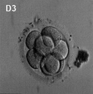 | 326 × 330 (15 KB) | S8600021 (talk | contribs) | Morphology of human embryo (day 3). MI - metaphase I oocyte Note - Original image has been modified to remove array data, colour and other embryo images. See also File:Human-oocyte_to_blastocyst.jpg Pone.0007844.g004.jpg http://www.ncbi.nlm.nih. |
You cannot overwrite this file.
File usage
The following 40 pages use this file:
- 2010 BGD Practical 3 - Early Cell Division
- 2010 BGD Tutorial - Applied Embryology and Teratology
- 2010 Lab 2
- 2010 Lecture 3
- Abnormal Development - Twinning
- BGDA Practical 3 - Early Cell Division
- BGDA Practical 3 - Week 1 Summary
- BGDA Practical 3 - Week 2 Summary
- BGD Tutorial - Applied Embryology and Teratology
- Carnegie Stages
- Carnegie stage 2
- Carnegie stage table
- Embryonic Development
- Lecture - Week 1 and 2 Development
- Morula Development
- Paper - A Human Embryo of Twenty-five Somites
- Paper - A Human Embryo of Twenty-seven Pairs of Somites, Embedded in Decidua
- Paper - Report upon the collection of human embryos at the Johns Hopkins University (1911)
- Paper - Two presomite human embryos
- Placenta - Membranes
- Preimplantation Genetic Diagnosis
- Preimplantation Genetic Screening
- Week 1
- Week 1 - Abnormalities
- Week 2
- Talk:Carnegie Stages
- File:Human-oocyte.jpg
- File:Human-oocyte to blastocyst.jpg
- File:Human embryo day 2.jpg
- File:Human embryo day 3.jpg
- File:Human embryo day 5.jpg
- File:Human embryo day 5 label.gif
- File:Human embryo day 5 label.jpg
- File:Human embryo day 5 label2.jpg
- File:Human oocyte-metaphase I.jpg
- File:Human oocyte-metaphase II.jpg
- Template:Carnegie stage table
- Template:Human oocyte to blastocyst
- Template:Monoygotic Twinning Table
- History:Paper - A Human Embryo of Twenty-seven Pairs of Somites, Embedded in Decidua


