File:Heart Tube Fusion.jpg
Original file (1,551 × 1,139 pixels, file size: 125 KB, MIME type: image/jpeg)
Early Human Embryo Heart Development
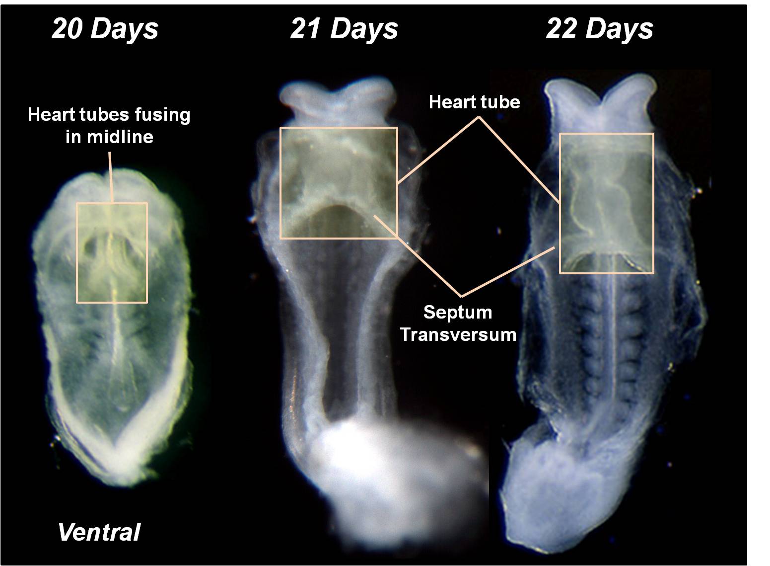
This is a ventral view of early human embryos between 20 to 22 days (GA week 5 to 6).
These bright-filed microscopy images show the early heart tubes fusing in the midline to form a single ventral primordial heart tube. This fusion event begins cranially and extends caudally.
- More information about human cardiac development can be found on the cardiovascular notes and heart tutorial pages.
- To see other views of the embryo during this period see the human embryo Carnegie stage 10 and Carnegie stage 11 pages.
- Links: Week 4 | Carnegie stage 10 | Carnegie stage 11 | Tutorial - Primordial Heart Tube | Cardiovascular
Image Source: Scanning electron micrographs of the Carnegie stages of the early human embryos are reproduced with the permission of Prof Kathy Sulik, from embryos collected by Dr. Vekemans and Tania Attié-Bitach. Images are for educational purposes only and cannot be reproduced electronically or in writing without permission.
Cite this page: Hill, M.A. (2026, October 4) Embryology Heart Tube Fusion.jpg. Retrieved from https://embryology.med.unsw.edu.au/embryology/index.php/File:Heart_Tube_Fusion.jpg
- © Dr Mark Hill 2026, UNSW Embryology ISBN: 978 0 7334 2609 4 - UNSW CRICOS Provider Code No. 00098G
File history
Yi efo/eka'e gwa ebo wo le nyangagi wuncin ye kamina wunga tinya nan
| Gwalagizhi | Nyangagi | Dimensions | User | Comment | |
|---|---|---|---|---|---|
| current | 11:41, 14 March 2010 | | 1,551 × 1,139 (125 KB) | Z3212774 (talk | contribs) | category:Heart ILP {{Template:SEM}} The primordial heart tubes fuse in the midline to form a single ventral heart tube. Fusion begins cranially and extends caudally. |
You cannot overwrite this file.
File usage
The following 15 pages use this file:
- 2009 Lecture 21
- 2010 BGD Lecture - Development of the Embryo/Fetus 1
- 2010 Lecture 21
- ANAT2341 Lab 4 - Early Cardiovascular Development
- BGDA Lecture - Development of the Embryo/Fetus 1
- BGDA Practical 7 - Week 4
- Cardiovascular - Arterial Development
- Cardiovascular - Venous Development
- Cardiovascular System Development
- Fetal ECHO Meeting 2012
- Human Embryo SEM
- Human System Development
- Intermediate - Primordial Heart Tube
- Lecture - Week 3 Development
- RPAH Cardiac Embryology 2014


