Category:Pituitary
From Embryology
This Embryology category shows content related to pituitary (hypophysis) development.
Pages in category 'Pituitary'
The following 50 pages are in this category, out of 50 total.
P
- Paper - A quantitative study of the hypophysis of the human anencephalic fetus (1927)
- Paper - Differentiation of pituicytes in the human foetus
- Paper - Experimental evidence regarding the role of the anterior pituitary in the development and regulation of the genital system
- Paper - Growth of the human prenatal hypophysis and the hypophyseal fossa (1927)
- Paper - Pharyngeal end of Rathke's pouch (1911)
- Paper - Sexual differences of the hypophyses and their determination by the gonads
- Paper - Some factors influencing the early development of the mammalian hypophysis (1935)
- Paper - The development of the hypophysis cerebri in man (1926)
- Paper - The development of the hypophysis cerebri of the rabbit
- Paper - The development of the mammalian pituitary and its morphological significance (1908)
- Paper - The histological appearances of the mammalian pituitary body (1908)
- Paper - The nerve supply to the pituitary body (1913)
- Template:Pituitary
- Template:Pituitary Vignette
- Template:Placode
- Template:Placodes
- Template:Posterior pituitary
R
- Template:Rathke's pouch
- Template:Ref-Andersen1971
- Template:Ref-Andriezen1894
- Template:Ref-Atwell1918
- Template:Ref-Atwell1926
- Template:Ref-AtwellSitler1918
- Template:Ref-Covell1927
- Template:Ref-Covell1927b
- Template:Ref-Espinasse1933
- Template:Ref-Gilbert1935
- Template:Ref-Herring1908a
- Template:Ref-Herring1908b
- Template:Ref-Hill1934
- Template:Ref-Pfeiffer1936
- Template:Ref-Shanklin1940
- Template:Ref-SmithEngle1927
Media in category 'Pituitary'
The following 113 files are in this category, out of 113 total.
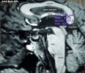 Adult human brain MRI01.jpg 700 × 607; 81 KB
 Atwell1918 fig01.jpg 600 × 432; 61 KB
 Atwell1918 fig02.jpg 800 × 582; 83 KB
 Atwell1918 fig03.jpg 800 × 604; 117 KB
 Atwell1918 fig04.jpg 409 × 550; 37 KB
 Atwell1918 fig05.jpg 704 × 550; 56 KB
 Atwell1918 fig06.jpg 600 × 641; 84 KB
 Atwell1918 fig07.jpg 425 × 378; 44 KB
 Atwell1918 fig08.jpg 638 × 553; 46 KB
 Atwell1918 fig09.jpg 559 × 635; 84 KB
 Atwell1918 fig10.jpg 627 × 703; 90 KB
 Atwell1918 fig11.jpg 665 × 904; 131 KB
 Atwell1918 fig12.jpg 453 × 603; 36 KB
 Atwell1918 fig13.jpg 625 × 800; 122 KB
 Atwell1918 fig14.jpg 800 × 788; 117 KB
 Atwell1918 fig15.jpg 701 × 900; 171 KB
 Atwell1918 fig18.jpg 1,000 × 1,193; 168 KB
 Atwell1918 fig19.jpg 316 × 515; 21 KB
 Atwell1918 fig20.jpg 702 × 515; 48 KB
 Atwell1918 fig21.jpg 800 × 477; 108 KB
 Atwell1918 fig22.jpg 1,000 × 887; 100 KB
 Atwell1918 fig23.jpg 1,000 × 953; 123 KB
 Atwell1918 fig24.jpg 506 × 399; 43 KB
 Atwell1918 fig25.jpg 532 × 496; 48 KB
 Atwell1918 fig26.jpg 566 × 498; 57 KB
 Atwell1918 fig27.jpg 900 × 802; 113 KB
 Atwell1918 fig28.jpg 1,000 × 886; 348 KB
 Atwell1918 fig29.jpg 791 × 780; 120 KB
 Atwell1918 fig30.jpg 601 × 451; 45 KB
 Atwell1918 fig31.jpg 1,000 × 700; 267 KB
 Atwell1918 fig32.jpg 510 × 303; 28 KB
 Atwell1918 fig33.jpg 387 × 336; 29 KB
 Atwell1918 fig34.jpg 353 × 406; 32 KB
 Atwell1918 fig35.jpg 1,200 × 849; 386 KB
 Atwell1918 fig38.jpg 1,000 × 377; 129 KB
 Atwell1918 fig39.jpg 600 × 709; 139 KB
 Drosophila and mouse placode similarity.jpg 499 × 1,086; 337 KB
 Embryonic and fetal pituitary.jpg 450 × 166; 14 KB
 Fetal head section 01.jpg 1,200 × 821; 186 KB
 Fetal head section 02.jpg 1,200 × 821; 171 KB
 Fetal head section 03.jpg 1,200 × 821; 174 KB
 Fetal head section.jpg 1,200 × 821; 167 KB
 Frazer1911 fig01.jpg 1,280 × 766; 187 KB
 Frazer1911 fig02.jpg 1,280 × 872; 112 KB
 Frazer1911 fig03.jpg 1,280 × 600; 153 KB
 Gray1180.jpg 1,200 × 712; 185 KB
 Gray1181.jpg 800 × 487; 77 KB
 Gray1182.jpg 800 × 982; 119 KB
 Herring1908b fig06.jpg 1,280 × 1,234; 283 KB
 Herring1908b fig07.jpg 1,280 × 974; 290 KB
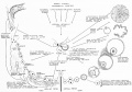
 Historic-pituitary.jpg 639 × 367; 54 KB
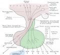
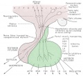
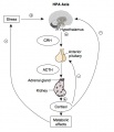 HPA axis.jpg 600 × 700; 46 KB
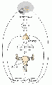
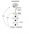 HPG female axis.jpg 600 × 700; 41 KB
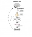 HPG male axis.jpg 600 × 700; 36 KB
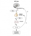 HPT axis.jpg 600 × 700; 35 KB
 Human week 10 fetus 10.jpg 1,200 × 900; 291 KB
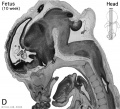 Human- fetal week 10 head D.jpg 600 × 544; 111 KB
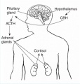
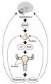
 Hypothalamus pituitary adrenal cartoon.jpg 653 × 600; 82 KB
 Hypothalamus pituitary adrenal pathway cartoon.jpg 490 × 516; 31 KB
 Hypothalamus pituitary cartoon.jpg 653 × 600; 81 KB
 Keith1902 fig015a.jpg 971 × 600; 74 KB
 Keith1902 fig117.jpg 561 × 800; 46 KB
 Keith1921 fig099.jpg 446 × 416; 39 KB
 Keith1921 fig101.jpg 1,144 × 652; 128 KB
 Keith1921 fig102.jpg 1,257 × 845; 172 KB
 Keith1921 fig103.jpg 758 × 495; 91 KB
 Kollmann355.jpg 861 × 460; 75 KB
 Kollmann356.jpg 867 × 584; 102 KB
 Martin Rathke.jpg 359 × 465; 20 KB
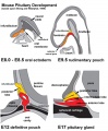 Mouse-pituitary development.jpg 660 × 800; 85 KB
 Mouse-pituitary Sox4 expression.jpg 596 × 448; 55 KB
 Nelsen1953 fig022.jpg 1,200 × 839; 207 KB
 Pituitary development animation.gif 600 × 400; 272 KB
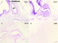
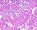
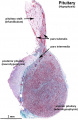
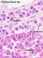 Pituitary histology 001.jpg 450 × 600; 72 KB
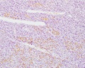
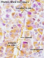 Pituitary histology 002.jpg 450 × 600; 81 KB
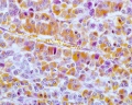
 Pituitary histology 003.jpg 450 × 600; 94 KB
 Pituitary histology 004.jpg 1,280 × 1,024; 342 KB
 Pituitary histology 005.jpg 1,280 × 1,024; 326 KB
 Pituitary histology 006.jpg 1,280 × 1,024; 450 KB
 Pituitary histology 007.jpg 1,280 × 1,024; 325 KB
 Pituitary histology 008.jpg 1,280 × 1,024; 340 KB
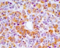
 Pituitary histology 009.jpg 466 × 610; 64 KB
 Pituitary histology 010.jpg 1,005 × 961; 249 KB
 Pituitary histology 011.jpg 900 × 1,388; 309 KB
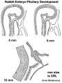 Pituitary rabbit development.jpg 374 × 500; 33 KB
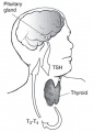 Pituitary thyroid pathway.jpg 454 × 632; 32 KB
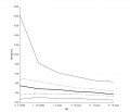 Postnatal thyrotropin levels graph.jpg 852 × 729; 35 KB
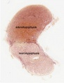
 Rathke cleft cyst 01.jpg 493 × 600; 46 KB
 Rugh 116.jpg 1,015 × 1,000; 270 KB
 Shanklin1940 fig01.jpg 1,000 × 657; 136 KB
 Shanklin1940 fig02.jpg 1,000 × 682; 131 KB
 Shanklin1940 fig04.jpg 884 × 1,018; 112 KB
 Shanklin1940 fig06.jpg 1,280 × 837; 92 KB
 Shanklin1940 fig07.jpg 1,280 × 1,080; 125 KB
 Shanklin1940 plate01.jpg 1,522 × 2,138; 362 KB
 Shanklin1940 plate02.jpg 1,411 × 2,131; 210 KB
 Stage 13 image 057.jpg 1,000 × 511; 99 KB
 Stage 13 image 058.jpg 1,000 × 481; 94 KB
 Stage 13 image 059.jpg 1,000 × 513; 92 KB
 Stage 22 image 158.jpg 1,000 × 646; 273 KB
 Stage 22 image 220.jpg 1,200 × 699; 334 KB
 Stage 22 image 221.jpg 1,200 × 699; 336 KB
 Stage17 bf11.jpg 1,375 × 2,048; 166 KB
 Streeter1957 fig08-19.jpg 800 × 1,102; 109 KB
 Streeter1957 fig08-20.jpg 800 × 1,102; 104 KB
 Streeter1957 fig08-21.jpg 800 × 1,102; 114 KB
 Streeter1957 fig08-22.jpg 800 × 1,102; 189 KB
 Streeter1957 fig08-23.jpg 800 × 1,102; 206 KB
 Streeter1957 fig08.jpg 1,280 × 1,712; 507 KB
 Streeter1957 plate02.jpg 1,500 × 2,001; 653 KB
 XXhpgaxis.gif 300 × 495; 22 KB
 XXhpgaxis.jpg 300 × 495; 24 KB


