File:Ureteral duplication 01.jpg
From Embryology
Size of this preview: 443 × 600 pixels. Other resolution: 600 × 812 pixels.
Original file (600 × 812 pixels, file size: 61 KB, MIME type: image/jpeg)
Ureteral Duplication
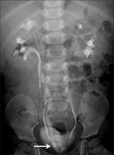
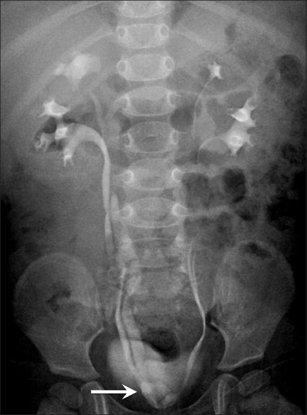
- Frontal radiograph from an intravenous urogram study shows bilateral complete ureteral duplication.
- The opening of the right upper moiety ureter is medial to the lower moiety ureter; there is ureterocele formation.
- Links: Renal System - Abnormalities
Reference
<pubmed>21042439</pubmed>| PMC2963757
Copyright
This is an open-access article distributed under the terms of the Creative Commons Attribution License, which permits unrestricted use, distribution, and reproduction in any medium, provided the original work is properly cited.
Original file name: Figure 10 IJRI-20-174-g010.jpg http://www.ncbi.nlm.nih.gov/pmc/articles/PMC2963757/figure/F0010/
File history
Yi efo/eka'e gwa ebo wo le nyangagi wuncin ye kamina wunga tinya nan
| Gwalagizhi | Nyangagi | Dimensions | User | Comment | |
|---|---|---|---|---|---|
| current | 23:29, 2 Mayıs 2011 | | 600 × 812 (61 KB) | S8600021 (talk | contribs) | ==Ureteral duplication== Ureteral duplication: Frontal radiograph from an intravenous urogram study shows bilateral complete ureteral duplication. The opening of the right upper moiety ureter is medial to the lower moiety ureter; there is ureterocele for |
You cannot overwrite this file.
File usage
The following 2 pages use this file: