File:Heart Tube Segments.jpg
Original file (1,082 × 771 pixels, file size: 63 KB, MIME type: image/jpeg)
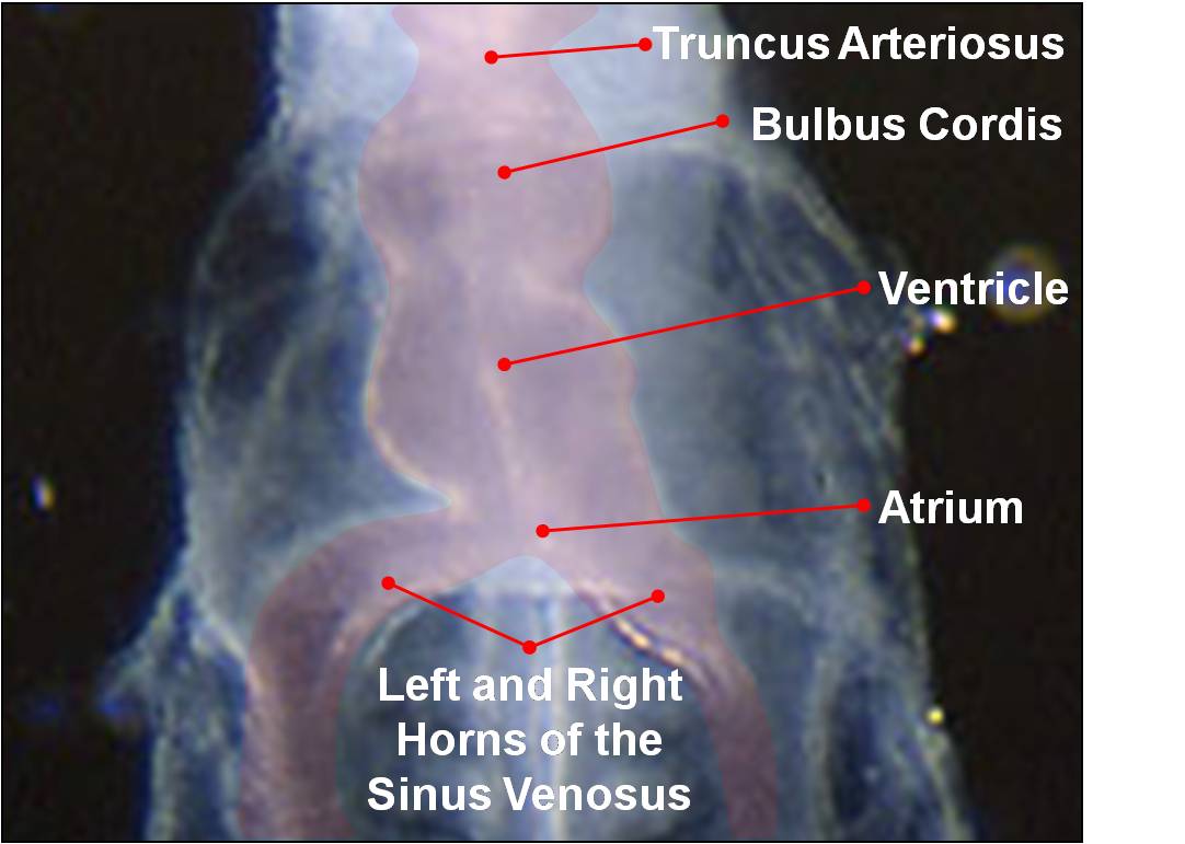
As the tubular heart grows it develops dilations and constrictions which form the truncus arteriosus, bulbus cordis, primitive ventricle, primitive atrium and sinus venosus.
Image Source: Scanning electron micrographs of the Carnegie stages of the early human embryos are reproduced with the permission of Prof Kathy Sulik, from embryos collected by Dr. Vekemans and Tania Attié-Bitach. Images are for educational purposes only and cannot be reproduced electronically or in writing without permission.
File history
Yi efo/eka'e gwa ebo wo le nyangagi wuncin ye kamina wunga tinya nan
| Gwalagizhi | Nyangagi | Dimensions | User | Comment | |
|---|---|---|---|---|---|
| current | 10:42, 14 March 2010 | | 1,082 × 771 (63 KB) | Z3212774 (talk | contribs) | category:Heart ILP {{Template:SEM}} As the tubular heart grows it develops dilations and constrictions which form the truncus arteriosus, bulbus cordis, primitive ventricle, primitive atrium and sinus venosus. |
You cannot overwrite this file.
File usage
The following file is a duplicate of this file (more details):
The following 19 pages use this file:
- 2010 Lab 4
- 2011 Lab 4
- ANAT2341 Lab 11 - Heart
- ANAT2341 Lab 4 - Early Cardiovascular Development
- B
- BGDA Practical 7 - Week 4
- Cardiovascular - Arterial Development
- Cardiovascular - Venous Development
- Cardiovascular System - Circulation Development
- Cardiovascular System - Coronary Circulation Development
- Cardiovascular System - Truncus Arteriosus
- Cardiovascular System Development
- Carnegie stage 10
- Fetal ECHO Meeting 2012
- Human Embryo SEM
- Intermediate - Primordial Heart Tube
- Lecture - Early Vascular Development
- Lecture - Heart Development
- RPAH Cardiac Embryology 2014