File:Respiratory histology 13.jpg
Respiratory_histology_13.jpg (450 × 600 pixels, file size: 102 KB, MIME type: image/jpeg)
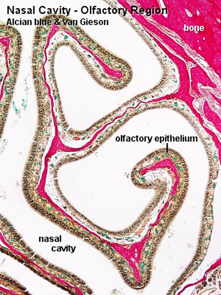
olfactory epithelium is formed by
- Olfactory cells
- Sustentacular cells - located mainly in the superficial cell layer of the epithelium (difficult to distinguish from olfactory cells).
- Basal cells - identified by their location.
Note - Cilia are not visible and goblet cells are absent from the olfactory epithelium.
Lightly stained rounded areas in the lamina propria represent bundles of olfactory axons in the lamina propria. Small mucous glands, olfactory glands or Bowman's glands, in the lamina propria moisturise the epithelium.
- Respiratory Histology: Bronchiole | Alveolar Duct | Alveoli | EM Alveoli septum | Alveoli Elastin | Trachea 1 | Trachea 2 | labeled lung | unlabeled lung | Respiratory Bronchiole | Lung Reticular Fibres | Nasal Inferior Concha | Nasal Respiratory Epithelium | Olfactory Region overview | Olfactory Region Epithelium | Histology Stains
Links: Histology | Histology Stains | Blue Histology images copyright Lutz Slomianka 1998-2009. The literary and artistic works on the original Blue Histology website may be reproduced, adapted, published and distributed for non-commercial purposes. See also the page Histology Stains.
Cite this page: Hill, M.A. (2026, October 10) Embryology Respiratory histology 13.jpg. Retrieved from https://embryology.med.unsw.edu.au/embryology/index.php/File:Respiratory_histology_13.jpg
- © Dr Mark Hill 2026, UNSW Embryology ISBN: 978 0 7334 2609 4 - UNSW CRICOS Provider Code No. 00098G
File history
Yi efo/eka'e gwa ebo wo le nyangagi wuncin ye kamina wunga tinya nan
| Gwalagizhi | Nyangagi | Dimensions | User | Comment | |
|---|---|---|---|---|---|
| current | 00:03, 29 February 2012 | | 450 × 600 (102 KB) | Z8600021 (talk | contribs) |
You cannot overwrite this file.
File usage
The following 10 pages use this file:


