Abnormal Development - Ectopic Implantation

Introduction

Human development during week 2 is about implantation and the endocrine signaling to block the normal menstrual cycle. The blastocyst implantation process should normally and does occur within the body of the uterus. There are a number of additional abnormal sites of implantation that are outside the uterine body, these are described as ectopic implantation or ectopic pregnancy. The most common form of human ectopic pregnancy is when implantation occurs within the uterine tube, described as a tubal pregnancy. Note that the endocrine signals blocking the menstrual cycle and indicating a pregnancy will still be released following this ectopic implantation. Ectopic pregnancies are therefore often identified by early ultrasound scans.
Ectopic pregnancy is also a high-risk maternal medical condition with an approximate incidence of 1.5 to 2 % in reported pregnancies. There is some indication that the incidence may be increasing (United States has increased from 4.5 per 1,000 pregnancies in 1970 to an estimated 19.7 per 1,000 pregnancies in 1992[1])
The risk factors for tubal ectopic pregnancy include: tubal damage by infection (particularly Chlamydia trachomatis) or surgery, smoking and in vitro fertilization therapy. Prolonged tubal damage is often described as pelvic inflammatory disease and "scarring" can affect the cilia-mediated transport of the blastocyst during the first week of development.
This is also the most common cause of pregnancy-related deaths in the first trimester. A recent United Kingdom enquiry into maternal deaths[2], identified ectopic pregnancy as the fourth most common cause of maternal death (73% of early pregnancy deaths).
Ectopic sites are named according to the anatomical location: Tubal (Ampullary, Isthmic, Cornual), Cervical and Ovarian.
- Links: Implantation | Week 2 | Quicktime - Ultrasound | Flash - Ultrasound | Ultrasound | Historic 1920 Paper
Some Recent Findings

|
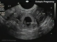
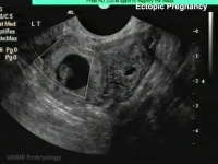
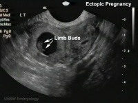
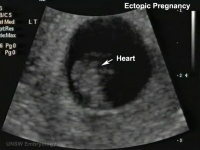
Ultrasound Ectopic Implantation
|
|
|
|
Ultrasound images of ectopic tubal pregnancy from movie.
- Links: Quicktime | Quicktime version | Flash version | Ectopic Implantation | Ultrasound
Computed Tomography Ectopic Implantation
Computed Tomography imaging findings of a 37-year-old woman with interstitial pregnancy.[6] Showing initial CT detection and a subsequent scan following rupture causing a hematoma around uterus and a massive hemoperitoneum.

Initial CT - gestational sac |

Follow-up CT - massive hemoperitoneum |

CT - hematoma around uterus |
- Links: Computed Tomography
Magnetic Resonance Imaging Abdominal Ectopic Implantation
2W SPAIR sagittal MRI of lower abdomen demonstrating the placental invasion.[7]
|

|
Statistics

Ectopic Pregnancies- United-States 1970-1992[8]

Ectopic Pregnancy Histology

CDC Image by Dr. Edwin P. Ewing, Jr., 1972

Ed Uthman Image (pathologist in Houston, Texas) section of ectopic (tubal) pregnancy about Carnegie stage 7 in Week 3.
Image version links: ExtraLarge 1712x1206px | Large 1024x721px | Medium 500x352px | Small 240x169px

Ed Uthman Image (pathologist in Houston, Texas) image of of ectopic (tubal) pregnancy about Carnegie stage 15 in Week 5.
Image version links: ExtraLarge 1874 x 2000px | Large 959 x 1024px | Medium 468 x 500px | Small 225 x 240px
Tubal Ectopic Pregnancy
| Chlamydia infections (Chlamydia trachomatis) are the most common bacterial sexually transmitted infection, often undiagnosed and asymptomatic. The infections can ascend the female genital tract, colonizing the endometrial mucosa, then the uterine tubes. This type of infection is described as pelvic inflammatory disease (PID). |
Interstitial Pregnancy
| (cornual pregnancy) A less common type 2 to 4% of ectopic pregnancies. The gestation develops in the uterine portion of the fallopian tube lateral to the round ligament. |  Interstitial ectopic pregnancy[9] |
Cervical Ectopic Pregnancy

This form of ectopic pregnancy is a rare high-risk condition and represents less than 1% of all ectopic pregnancies. The reported incidence varies between 1:1,000 to 1:18,000.
Rudimentary Horn Pregnancy
A rare types of ectopic pregnancy (about 1 in 76,000 pregnancies) in most cases the horn is non-communicating. Therefore fertilisation probably occurs by transperitoneal migration. This form untreated can also lead to uterine rupture.
Caesarean Scar Pregnancy
A rare types of ectopic pregnancy (about 1 in 2000 pregnancies), but probably increasing as caesarean rates rise. The gestation is completely surrounded by both myometrium and fibrous tissue of the caesarean section scar and separated from the endometrial cavity and endocervical canal.
References
- ↑ <pubmed>7823895</pubmed>
- ↑ Confidential Enquiry into Maternal Deaths (CEMD) Why Mothers Die 2000–2002 PDFPDF2
- ↑ <pubmed>22035883</pubmed>
- ↑ <pubmed>21748311</pubmed>
- ↑ <pubmed>20023297</pubmed>
- ↑ <pubmed>20046504</pubmed>| PMC2799642 | Korean J Radiol.
- ↑ <pubmed>19918376</pubmed>| Cases J.
- ↑ <pubmed>7823895</pubmed>
- ↑ <pubmed>20725587</pubmed>| German Medical Science
- ↑ <pubmed>22110520</pubmed>| PMC3205779
Reviews
<pubmed>20071358</pubmed> <pubmed>20023297</pubmed> <pubmed>16595714</pubmed>
Articles
<pubmed>7194809</pubmed> <pubmed>19978839</pubmed>
Search Pubmed
June 2010
- "ectopic pregnancy" All (14958) Review (1350) Free Full Text (1196)
- "tubal pregnancy" All (8010) Review (683) Free Full Text (630)
Search Pubmed: ectopic pregnancy | ectopic implantation | tubal pregnancy | tubal implantation
Glossary Links
- Glossary: A | B | C | D | E | F | G | H | I | J | K | L | M | N | O | P | Q | R | S | T | U | V | W | X | Y | Z | Numbers | Symbols | Term Link
Cite this page: Hill, M.A. (2024, June 27) Embryology Abnormal Development - Ectopic Implantation. Retrieved from https://embryology.med.unsw.edu.au/embryology/index.php/Abnormal_Development_-_Ectopic_Implantation
- © Dr Mark Hill 2024, UNSW Embryology ISBN: 978 0 7334 2609 4 - UNSW CRICOS Provider Code No. 00098G