Talk:ANAT2341 Lab 10 - Late Embryo: Difference between revisions
From Embryology
(Created page with "{| | Embryonic Development * Weeks 3 - 4 Eye Fields-Optic Vesicle * Weeks 5 - 6 Optic Cup, Lens Vesicle, Choroid Fissure, Hyaloid Artery * Weeks 7 - 8 Cornea, Anterior Chambe...") |
mNo edit summary |
||
| Line 10: | Line 10: | ||
|} | |} | ||
'''Week 4 Vision''' (data based upon<ref><pubmed>7364662</pubmed></ref> and other sources). | |||
* [[Carnegie_stage_14|Stage 14]] - (about 32 days) the lens placode is indented by the lens pit, cup-shaped and still communicates with the surface by a narrowing pore. | * [[Carnegie_stage_14|Stage 14]] - (about 32 days) the lens placode is indented by the lens pit, cup-shaped and still communicates with the surface by a narrowing pore. | ||
* [[Carnegie_stage_15|Stage 15]] - (about 33 days) the lens pit is closed. The lens vesicle and optic cup lie close to the surface ectoderm and appear to press against the surface. | * [[Carnegie_stage_15|Stage 15]] - (about 33 days) the lens pit is closed. The lens vesicle and optic cup lie close to the surface ectoderm and appear to press against the surface. | ||
| Line 25: | Line 20: | ||
* [[Carnegie_stage_20|Stage 20]] - The lens cavity is lost and a lens suture begins to form. The inner canthus is established. | * [[Carnegie_stage_20|Stage 20]] - The lens cavity is lost and a lens suture begins to form. The inner canthus is established. | ||
* [[Carnegie_stage_23|Stage 23]] - The retina comprises the pigmented layer, external limiting membrane, proliferative zone, external neuroblastic layer, transient fiber layer, internal neuroblastic layer, nerve fiber layer, and internal limiting membrane. Eyelids closure is complete (Note - shown as still open in the Kyoto embryo). | * [[Carnegie_stage_23|Stage 23]] - The retina comprises the pigmented layer, external limiting membrane, proliferative zone, external neuroblastic layer, transient fiber layer, internal neuroblastic layer, nerve fiber layer, and internal limiting membrane. Eyelids closure is complete (Note - shown as still open in the Kyoto embryo). | ||
[[Embryo Virtual Slides]]<br>{{SlideStage22-08}}<br>{{SlideStage22-08-eye}} | |||
Revision as of 08:46, 15 October 2015
|
Embryonic Development
|

|
Week 4 Vision (data based upon[1] and other sources).
- Stage 14 - (about 32 days) the lens placode is indented by the lens pit, cup-shaped and still communicates with the surface by a narrowing pore.
- Stage 15 - (about 33 days) the lens pit is closed. The lens vesicle and optic cup lie close to the surface ectoderm and appear to press against the surface.
- Stage 16 - (37 days) Growth of the lens body results in a D-shaped lens cavity. Perilental blood vessels (tunica vasculosa lentis) are visible. Prior to the development of the eyelids, one small sulcus or groove forms above the eye (eyelid groove) and another below it.
- Stages 17 - 19 - Retinal pigment is visible and the retinal fissure is largely closed. Eyelids grooves deepen, eyelid folds develop, first below, and then above, the eye.
- Stages 18 - Mesenchyme invades the region between the lens epithelium and the surface ectoderm.
- Stages 19 - 22 - the eyelid folds develop into the eyelids and cover more of the eye as the palpebral fissure takes shape. The upper and the lower eyelids meet at the outer canthus in Stage 19.
- Stage 20 - The lens cavity is lost and a lens suture begins to form. The inner canthus is established.
- Stage 23 - The retina comprises the pigmented layer, external limiting membrane, proliferative zone, external neuroblastic layer, transient fiber layer, internal neuroblastic layer, nerve fiber layer, and internal limiting membrane. Eyelids closure is complete (Note - shown as still open in the Kyoto embryo).
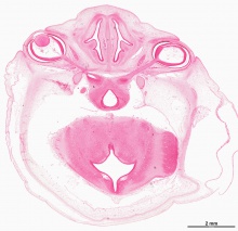
| Stage 22 - Eye and Nose
|
| Stage 22 | Embryo Slides |
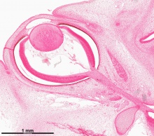
| Stage 22 - Eye
|
| Stage 22 | Embryo Slides |
- ↑ <pubmed>7364662</pubmed>


