Category:Carnegie Stage 9: Difference between revisions
mNo edit summary |
mNo edit summary |
||
| Line 26: | Line 26: | ||
<references/> | <references/> | ||
{| class="wikitable mw-collapsible mw-collapsed" | |||
! Piersol WH (1937) abstract | |||
|- | |||
| Piersol, W. H. 1937. A human embryo of two somites, in situ. Anat. Rec, 67 suppl, 39-40. (American Association of Anatomists, Fifty-Third Annual Session, University of Toronto, Toronto, Canada, March 25 to 27, 1937) | |||
(6) 88. A human embryo of two somites, in situ. W. H. Piersol, Department of Anatomy, University of Toronto. | |||
The embryo measures 2.03 x 0.72 mm. and has been sectioned sagittally. Contact between embryonic and maternal tissue is preserved over a considerable area. although elsewhere there is separation due to shrinkage. Numerous glands form 9. broad stratum spongiosum. Flattened in the stratum compaetum, their middle parts are much dilated; toward the muscularis they are again flattened. Frequently they contain degenerated detritus and occasionally blood. A few glandular remains occur in the peripheral parts of the decidua capsularis. Chorionic villi are longest and most numerous toward the decidua marginalis. Their tips may reach the maternal tissues or a spongy mass of trophoblaet may intervene. Cytotrophoblast predominates, frequently bordered and penetrated by plasmodiotrophoblast. On the chor-ionic wall the trophoblast may show only one layer of nuclei but two layers are the rule. On the villi there are two layers of nuclei, the inner ones frequently belonging to distinct cells. The pericardial cavity is present but contains no heart, nor are their blood vessels in any part of the embryo, the chorion, or its villi. The neurenterie canal is closed but its remains are evident. An endodermal allantois projects into the connecting stalk. Small blood islands are present in the wall of the yolk sac and larger ones in the connecting stalk; the latter are forming endothelium. | |||
|} | |||
[[Category:Carnegie Stage]] [[Category:Week 3]] [[Category:Human Embryo]] | [[Category:Carnegie Stage]] [[Category:Week 3]] [[Category:Human Embryo]] | ||
Revision as of 12:44, 5 September 2015
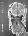
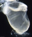
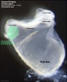
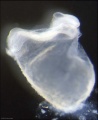
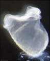
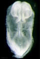
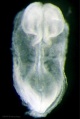
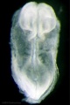
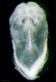
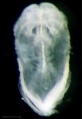
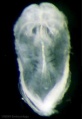
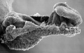
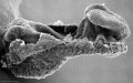
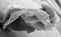
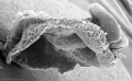
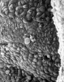
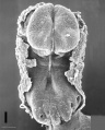
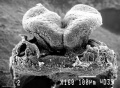
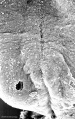
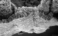
This Embryology category shows pages and media related to Carnegie stage 9 of embryonic development. In human development this stage occurs during week 3 (post-fertilisation) or gestational age GA week 5 (LMP).
- Links: Carnegie stage 9 | Week 3
| Carnegie Collection - Stage 9 | ||||||||||
|---|---|---|---|---|---|---|---|---|---|---|
| Serial No. | Pairs of somites | Size (mm) | Grade | Fixative | Embedding Medium | Plane | Thinness (µm) | Stain | Year | Notes |
| 1878 | 2-3 | Embryo, 1.38 Ch., 12x10.5x7.5 |
Good | Formalin | P | Coronal | 10 | (Stain - Haematoxylin Eosin) | 1917 | Described by Ingalls (1920).[1] |
| 5080 | 1 | Embryo, 1.5 Ch., 14.5 |
Poor | Formalin | P | Transverse | 10 | Al. coch. | 1926 | Studied by Davis (1927).[2] |
| 7650 | 2-3 | Embryo, 2-3 | Good | Alc & Bouin | C-P | Transverse | 6 | (Stain - Haematoxylin Eosin) | 1939 | Said to be female[3] |
Abbreviations
| ||||||||||
References
| ||||||||||
Embryo Examples
Based on O'Rahilly, R. and Müller (1987)[1] and listed in order of number of pairs of somites.
- 1 somite - Carnegie No. 5080. Studied and illustrated by Davis (1927, figs. 2–5 and 39–42) and Severn (1971, figs. 1–4). First pair of somites not separate rostrally and contain no myocoeles (Arey, 1938). Chorion, 14.5 x 1.5 mm. Embryonic disc, 1.5 mm. Reconstructed by Müller and O'Rahilly (1983, fig. 2).
- 1 somite - A specimen described briefly by Baginski and Borsuk (1967).
- 1-2 somites - (or more?) somites, Carnegie No. 7650. Reconstructed by Müller and O'Rahilly (1983, fig. 5).
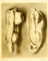
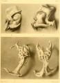
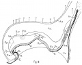
- 2 somites - Da 1 (Dann). An important specimen (fig. 9-2) possessing 2 pairs of somites (Studnicka, 1929; Florian and Völker, 1929; Arey, 1938), although featured originally as having only one. Described and illustrated in detail by Ludwig (1928). Removed from uterus. Chorion, 12 mm. Embryo, 1.8 mm in a straight line, 2.4 mm by flexible scale. Sectioned transversely at 8 μm. Stained with alum cochineal. Neurenteric canal present. Sections are housed in the Anatomisches Institut, Basel. Photographs of sections are in Carnegie Collection under No. 5982. Presumed age, about 21 days. Dorsal and median projections published (ibid., figs. 1 and 2; Florian and Völker, 1929, fig. 14). Reconstructed by Müller and O’Rahilly (1983, fig. 3).
- 2-3 somites - H3. Described by Wilson (1914), according to whom it “possessed probably two, possibly three, pairs of somites.” Chorion, 8.5 x 5.7 x 5 mm. Embryo, 1.43 mm. Sectioned obliquely (transversely) at 10 μm. Stained with hematoxylin. Fixation not adequate for reconstruction. The relatively longer primitive streak suggests that this embryo may be less advanced than No. 1878. Prechordal plate, or at least prechordal mesoderm, figured (Hill and Florian, 1931b). Presumed age, 18–21 days.
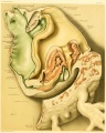
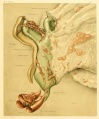
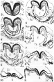
- 2/3 somites - Carnegie No. 1878 (figs. 9-3 to 9-7). An important specimen possessing 2 somites on the right side and 3 on the left. Florian (1934b) had certain difficulties and considered the embryo to be too small. Curettage. Chorion, 12 X 10.5 X 7.5 mm. Embryonic disc, 1.38 mm in a straight line. Described in detail and illustrated by Ingalls (1920) who believed that “the earliest recognizable stage of dextrocardia” is present, “to which might have been added later a more or less complete situs inversus viscerum”; at any rate, Davis (1927), who studied and illustrated the heart, considered that “the cardiac area is distorted.” Angiogenesis in chorion described by Hertig (1935). Primitive streak and node, 0.13 mm, according to Ingalls, but about 0.22 mm in fig. 15 of Florian and Völker (1929) and more than 0.3 mm in plate 5, fig. 9, of Bartelmez and Evans (1926). Neurenteric canal not patent but pit present (Bartelmez and Evans, 1926). Median projection published (ibid., plate 5, fig. 9; Florian and Völker, 1929, fig. 15; Müller and O’Rahilly, 1983, fig. 1).
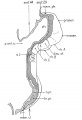
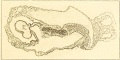
- 3 somites - T439 (Toronto). Possesses 3 pairs of somites (Arey, 1938), although considered originally as having only 2. Described by Piersol (1937).[2] Embryo (along surface), 2.03 x 0.72 mm. Sectioned sagitally. Neurenteric canal closed but its remains are identifiable. Primordial germ cells near allantois. Embryonic disc rostral to somites, including cardiac area, is retarded. Said to contain no blood vessels in any part of the embryo itself.
- 3 somites - Vant embryo. Described by Shaner (1945), who found “two to three pairs of somites.” Embryo (along curve), 1.5 mm. Thought to be 25 ± 2 days. Reconstructed again from original sections by Müller and O’Rahilly (1983, fig. 4).
- 3 somites - Gv (Madrid). Described by Jiménez Collado and Ruano Gil (1963). Heart described by Orts Llorca, Jiménez Collado, and Ruano Gil (1960). Tubal. Embryo, 1.81 mm. Sectioned at 7 μm. Stained with hematoxylin and eosin. Reconstructed. On the basis of its external characters, said to lie between stage 9 and stage 10. Presumed age, 21 ± 1 days. Madrid Collection
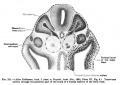
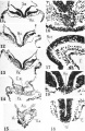
- 3 somites - No. 2008 (Prague). Excellent specimen (figs. 9-8 to 9-14) belonging to Dr. J. E. Jirásek. Embryo, 1.73 mm. Fixed in calcium formol. Sectioned transversely at 10 μm. Various stains used, including histochemical procedures. Should be published.
- 3 somites - (?), His embryo E. This 2.1 mm specimen is listed by Bartelmez and Evans (1926) between No. 1878 (2–3 somitic pairs) and No. 3709 (4 somitic pairs, stage 10).
References
- ↑ O'Rahilly, R. and Müller, F. Developmental Stages in Human Embryos. Carnegie Institution of Washington Publication 637 (1987).
- ↑ Piersol, W. H. 1937. A human embryo of two somites, in situ. Anat. Rec, 67 suppl, 39-40.
| Piersol WH (1937) abstract |
|---|
| Piersol, W. H. 1937. A human embryo of two somites, in situ. Anat. Rec, 67 suppl, 39-40. (American Association of Anatomists, Fifty-Third Annual Session, University of Toronto, Toronto, Canada, March 25 to 27, 1937)
(6) 88. A human embryo of two somites, in situ. W. H. Piersol, Department of Anatomy, University of Toronto. The embryo measures 2.03 x 0.72 mm. and has been sectioned sagittally. Contact between embryonic and maternal tissue is preserved over a considerable area. although elsewhere there is separation due to shrinkage. Numerous glands form 9. broad stratum spongiosum. Flattened in the stratum compaetum, their middle parts are much dilated; toward the muscularis they are again flattened. Frequently they contain degenerated detritus and occasionally blood. A few glandular remains occur in the peripheral parts of the decidua capsularis. Chorionic villi are longest and most numerous toward the decidua marginalis. Their tips may reach the maternal tissues or a spongy mass of trophoblaet may intervene. Cytotrophoblast predominates, frequently bordered and penetrated by plasmodiotrophoblast. On the chor-ionic wall the trophoblast may show only one layer of nuclei but two layers are the rule. On the villi there are two layers of nuclei, the inner ones frequently belonging to distinct cells. The pericardial cavity is present but contains no heart, nor are their blood vessels in any part of the embryo, the chorion, or its villi. The neurenterie canal is closed but its remains are evident. An endodermal allantois projects into the connecting stalk. Small blood islands are present in the wall of the yolk sac and larger ones in the connecting stalk; the latter are forming endothelium. |
Subcategories
This category has the following 4 subcategories, out of 4 total.
Pages in category 'Carnegie Stage 9'
The following 21 pages are in this category, out of 21 total.
C
P
- Paper - A human embryo of two somites in situ
- Paper - A human embryo of two to three pairs of somites (1945)
- Paper - Development of the human embryo during the period of somite formation with 2 to 16 pairs of somites
- Paper - Normal development of early human embryos: Observation of 90 specimens at Carnegie stages 7 to 13
Media in category 'Carnegie Stage 9'
The following 50 files are in this category, out of 50 total.
 Bartelmez1923 fig01.jpg 1,312 × 1,919; 245 KB
 Gray0031.jpg 1,371 × 1,309; 270 KB
 Ingalls1920FigureA.jpg 1,000 × 504; 66 KB
 Ingalls1920plate01.jpg 837 × 1,069; 53 KB
 Ingalls1920plate02.jpg 937 × 1,176; 99 KB
 Ingalls1920plate03.jpg 956 × 1,152; 70 KB
 Ingalls1920plate04.jpg 762 × 1,044; 52 KB
 Ingalls1920plate05.jpg 823 × 1,086; 160 KB
 Keibel Mall 221.jpg 650 × 460; 48 KB
 Shaner1945 fig02.jpg 1,280 × 1,026; 155 KB
 Shaner1945 plate01.jpg 600 × 1,076; 115 KB
 Shaner1945 plate02.jpg 1,000 × 1,531; 391 KB
 Shaner1945 plate03.jpg 1,280 × 1,977; 475 KB
 Stage 9 SEM1.jpg 347 × 450; 42 KB
 Stage9 bf1.jpg 886 × 1,000; 39 KB
 Stage9 bf1a.jpg 709 × 800; 28 KB
 Stage9 bf1b.jpg 532 × 600; 19 KB
 Stage9 bf1c.jpg 355 × 400; 11 KB
 Stage9 bf2-primordial germ cell region.jpg 814 × 1,000; 72 KB
 Stage9 bf2.jpg 814 × 1,000; 33 KB
 Stage9 bf2a.jpg 651 × 800; 24 KB
 Stage9 bf2b.jpg 488 × 600; 16 KB
 Stage9 bf2c.jpg 325 × 400; 9 KB
 Stage9 bf3.jpg 670 × 1,000; 27 KB
 Stage9 bf3a.jpg 536 × 800; 20 KB
 Stage9 bf3b.jpg 402 × 600; 14 KB
 Stage9 bf3c.jpg 268 × 400; 8 KB
 Stage9 bf4.jpg 686 × 1,000; 24 KB
 Stage9 bf4a.jpg 549 × 800; 18 KB
 Stage9 bf4b.jpg 412 × 600; 12 KB
 Stage9 bf4c.jpg 275 × 400; 7 KB
 Stage9 sem1.jpg 1,000 × 627; 80 KB
 Stage9 sem1a.jpg 800 × 502; 55 KB
 Stage9 sem1b.jpg 600 × 377; 33 KB
 Stage9 sem1c.jpg 400 × 251; 16 KB
 Stage9 sem2.jpg 1,000 × 612; 72 KB
 Stage9 sem2c.jpg 400 × 245; 16 KB
 Stage9 sem3c.jpg 313 × 400; 25 KB
 Stage9 sem4.jpg 804 × 1,000; 77 KB
 Stage9 sem4a.jpg 643 × 800; 56 KB
 Stage9 sem4b.jpg 482 × 600; 36 KB
 Stage9 sem5.jpg 1,359 × 1,000; 135 KB
 Stage9 sem5a.jpg 1,087 × 800; 100 KB
 Stage9 sem5b.jpg 815 × 600; 67 KB
 Stage9 sem6.jpg 627 × 1,000; 114 KB
 Stage9 sem6a.jpg 502 × 800; 80 KB
 Stage9 sem6b.jpg 377 × 600; 50 KB
 Stage9 sem7.jpg 1,000 × 626; 79 KB
 Stage9 sem7a.jpg 800 × 501; 57 KB
 Stage9 sem7b.jpg 600 × 376; 37 KB


