File:Thomson1919 fig02.jpg: Difference between revisions
(Z8600021 uploaded a new version of File:Thomson1919 fig02.jpg) |
mNo edit summary |
||
| Line 4: | Line 4: | ||
Fig. 2 shows an ovum in discus proligerus in Graafian follicle. Section 0.007 mm thick. Stained with Ehrlich’s acid haematoxylin and eosin. x 600. | Fig. 2 shows an ovum in discus proligerus in Graafian follicle. Section 0.007 mm thick. Stained with Ehrlich’s acid haematoxylin and eosin. x 600. | ||
'''Note''' - ''discus proligerus'' is an historic term for granulosa cells surrounding the oocyte and forming the cumulus oophorus. | |||
From a woman aged 22. Died of a femoral hernia. Same ovary as [[:File:Thomson1919 fig01.jpg|fig. 1.]] The figure shows that the egg is not spherical. The cells of the Corona radiata are here adherent to the outer side of the Zona pelluci(la by broader protoplasmic processes than in fig. 1, with arcaded spaces in between them, the summits of the arcades being directed to the Zona pellucida. The Zona pellucida consists clearly of two layers, the combined thickness of which amounts to from 0.008 to 0.O11 mm. The three deeply stained bodies seen overlying the Zona pellucida are nuclei which have got floated over it in the process of mounting. The inner zone is more homogeneous than the outer, it exhibits here and there evidence of a lamellar arrangement and at one or two points there seems to be distinct proof of the presence of cellular elements in its substance. At one pole it appears to be faintly striated radially. This layer of the Zona pellucida I have reason to believe is the ovular layer. The outer layer, continuous with the processes which pass to the cells of the Corona radiata is more open and fibrillar in structure. This I believe to be the ovarian layer of the Zona pellucida, the Zona thus‘ being a compound structure made up of ovular and ovarian elements. The cytoplasm, of granular appearance, is fairly uniform in density throughout. It is retracted from the inner surface of the Zona pellucida, the retraction cavity being slit-like for the most part, but increased in width towards one end, where it forms a considerable cavity, which is in part occupied by a ‘faintly granular translucent material capping the substance of the cytoplasm at this end of the egg. This is possibly of the nature of a coagulum. | |||
{{Thomson1919 figures}} | {{Thomson1919 figures}} | ||
Latest revision as of 13:43, 6 August 2015
Fig. 2. Human ovum in discus proligerus in propnase of first division
Fig. 2 shows an ovum in discus proligerus in Graafian follicle. Section 0.007 mm thick. Stained with Ehrlich’s acid haematoxylin and eosin. x 600.
Note - discus proligerus is an historic term for granulosa cells surrounding the oocyte and forming the cumulus oophorus.
From a woman aged 22. Died of a femoral hernia. Same ovary as fig. 1. The figure shows that the egg is not spherical. The cells of the Corona radiata are here adherent to the outer side of the Zona pelluci(la by broader protoplasmic processes than in fig. 1, with arcaded spaces in between them, the summits of the arcades being directed to the Zona pellucida. The Zona pellucida consists clearly of two layers, the combined thickness of which amounts to from 0.008 to 0.O11 mm. The three deeply stained bodies seen overlying the Zona pellucida are nuclei which have got floated over it in the process of mounting. The inner zone is more homogeneous than the outer, it exhibits here and there evidence of a lamellar arrangement and at one or two points there seems to be distinct proof of the presence of cellular elements in its substance. At one pole it appears to be faintly striated radially. This layer of the Zona pellucida I have reason to believe is the ovular layer. The outer layer, continuous with the processes which pass to the cells of the Corona radiata is more open and fibrillar in structure. This I believe to be the ovarian layer of the Zona pellucida, the Zona thus‘ being a compound structure made up of ovular and ovarian elements. The cytoplasm, of granular appearance, is fairly uniform in density throughout. It is retracted from the inner surface of the Zona pellucida, the retraction cavity being slit-like for the most part, but increased in width towards one end, where it forms a considerable cavity, which is in part occupied by a ‘faintly granular translucent material capping the substance of the cytoplasm at this end of the egg. This is possibly of the nature of a coagulum.
| Historic Disclaimer - information about historic embryology pages |
|---|
 |
- Human Ovum Links: Fig 1. Prophase I | Fig 2. | | Fig 3. | Plate 10 | Plate 11 | Plate 12
| Online Editor Notes |
|---|
|
Nature Obituary 1935 - Prof. Arthur Thomson (1858 - 1935)
Nature 135, 295-295 (23 February 1935) | doi:10.1038/135295a0 |
|
Reference
Cite this page: Hill, M.A. (2024, June 27) Embryology Thomson1919 fig02.jpg. Retrieved from https://embryology.med.unsw.edu.au/embryology/index.php/File:Thomson1919_fig02.jpg
- © Dr Mark Hill 2024, UNSW Embryology ISBN: 978 0 7334 2609 4 - UNSW CRICOS Provider Code No. 00098G
File history
Yi efo/eka'e gwa ebo wo le nyangagi wuncin ye kamina wunga tinya nan
| Gwalagizhi | Nyangagi | Dimensions | User | Comment | |
|---|---|---|---|---|---|
| current | 13:41, 6 August 2015 |  | 1,172 × 1,233 (354 KB) | Z8600021 (talk | contribs) | |
| 13:40, 6 August 2015 | 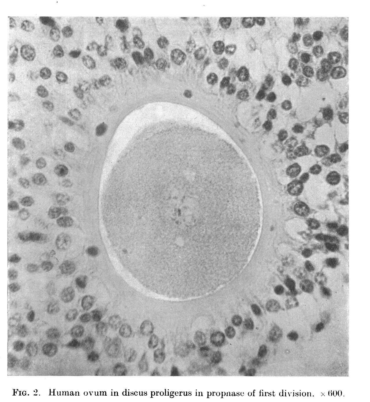 | 1,277 × 1,388 (524 KB) | Z8600021 (talk | contribs) | ==Fig. 2. == '''Note''' - ''discus proligerus'' is an historic term for granulosa cells surrounding the oocyte and forming the cumulus oophorus. {{Thomson1919 figures}} |
You cannot overwrite this file.
File usage
The following page uses this file: