Cartilage Histology: Difference between revisions
No edit summary |
No edit summary |
||
| Line 1: | Line 1: | ||
== Introduction == | |||
[[File:Endochondral_bone.jpg|thumb|300px|Developing Knee]] | [[File:Endochondral_bone.jpg|thumb|300px|Developing Knee]] | ||
Our adult skeleton forms from a larger number of developmental elements that are replaced and fuse. In development there are 2 separate signaling pathways for pattern formation and the formation of bone itself. Furthermore bone formation can be divided into 2 specific forms that occur in anatomically different regions. This practical class will describe the development and structure of bone and finish with a study of abnormalities associated with bone. | Our adult skeleton forms from a larger number of developmental elements that are replaced and fuse. In development there are 2 separate signaling pathways for pattern formation and the formation of bone itself. Furthermore bone formation can be divided into 2 specific forms that occur in anatomically different regions. This practical class will describe the development and structure of bone and finish with a study of abnormalities associated with bone. | ||
Revision as of 16:35, 5 September 2011
Introduction

Our adult skeleton forms from a larger number of developmental elements that are replaced and fuse. In development there are 2 separate signaling pathways for pattern formation and the formation of bone itself. Furthermore bone formation can be divided into 2 specific forms that occur in anatomically different regions. This practical class will describe the development and structure of bone and finish with a study of abnormalities associated with bone.
The image shown to the left shows a histological section through the developing lower limb at the level of a developing joint (knee), surrounding the developing bone is cartilage, skeletal muscles and connective tissue of the limb.
Lecture - Musculoskeletal Development and notes on Bone Development.
Chondroblasts and Chondrocytes
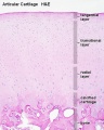
Articular cartilage
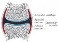
Synovial joint showing cartilage

- immature and mature cartilage forming cells located at articular cartilage regions.
- Interstitial growth - occurs mainly in immature cartilage. Chondroblasts in existing cartilage divide and form small groups of cells (isogenous groups) which produce matrix to become separated from each other by a thin partition of matrix.
- Appositional growth - occurs also in mature cartilage. Mesenchymal cells surrounding the cartilage in the deep part of the perichondrium (or the chondrogenic layer) differentiate into chondroblasts.
Endochondral ossification
Endochondral ossification slides: Developing bone | Bone, Developing (LS, Femur) Cat H&E
Blue Histology - endochondral | Dev Biology - endochondral ossification | endochondral ossification animation


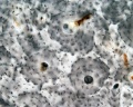
compact - low
compact - low
compact - med

compact - high
compact bone - low unstained
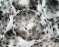
compact bone - high unstained
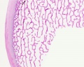
trabecular - overview HE
trabecular - low HE
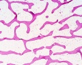
trabecular - med HE


- Bone Histology: Cartilage Histology | Histology Stains | Histology | cartilage | bone | bone timeline
- Trabecular bone trabecular | lamellar | trabecular - overview HE | trabecular - low HE | trabecular - med HE
- Endochondral ossification primary ossification | endochondral ossification
- Intramembranous ossification intramembranous - VG low | intramembranous - VG high | intramembranous - HE low | intramembranous - HE high
Human Fetal Head (12 week)


Histology Stains
Alizarin Red
- an anthraquinone derivative used to identify calcium in tissue sections
- calcium forms an Alizarin Red S-calcium complex in a chelation process and the end product is also birefringent.
- reaction can also identify magnesium, manganese, barium, strontium, and iron may interfere
- these elements usually in too low concentration to interfere with the staining
H&E
- acronym for hematoxylin and eosin stain
- hematoxylin - basic dye which colors basophilic structures with blue-purple hue (nucleus, DNA, RNA)
- eosin Y - acidic alcohol-based which colors eosinophilic structures bright pink (cytoplasm, extracellular matrix, protein)
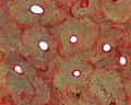
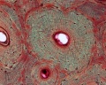
H&Van Gieson
- Van Gieson's Stain is a mixture of picric acid and acid fuchsin used for differential staining of collagen and other connective tissue.
- Nuclei - stains brownish black to black
- Collagen (fibrous connective tissue) - stains pink or deep red
- Muscle, Cytoplasm, RBC and Fibrin - stains yellow
- Links: Histology Stains
External Links
- Virtual Slidebox of Histology (USA) Skeletal system
- e-radiography Ossification
- UWA Blue Histology bone
Other Textbooks
- Anatomy of the Human Body (H. Gray, 1918.) historical anatomy text Osteology
- Molecular Biology of the Cell Bone Is Continually Remodeled by the Cells Within It | Image: Figure 22-52. Deposition of bone matrix by osteoblasts | Image: Figure 22-56. The development of a long bone
- Molecular Cell Biology Mutations in Collagen Reveal Aspects of Its Structure and Biosynthesis
- The Cell- A Molecular Approach Steroid Hormones and the Steroid Receptor Superfamily
- Clinical Methods: The History, Physical, and Laboratory Examinations 100. Alkaline Phosphatase and Gamma Glutamyltransferase
- Endocrinology: An Integrated Approach by Nussey, S.S. and Whitehead, S.A. Endocrinology: Definition and causes of osteoporosis
- Developmental Biology 6th ed. by Gilbert, Scott F. Figure 14.13. Schematic diagram of endochondral ossification | Aging: The Biology of Senescence
Search
- Pubmed ossification
Terms


- canaliculus - (plural, canaliculi) small channel in the bone matrix in which an osteocyte process lies and communicates with other osteocytes and the Haversian canal.
- haematopoiesis (Greek, haima = "blood"; poiesis = "to make") the process of blood cell formation.
- Haversian canal - the central canal of an osteon (Haversian system) in compact bone, within which blood vessels and nerves travel throughout the bone.
- Haversian system - (osteon) the historic name for the functional unit of compact bone. Consists of a central canal (Haversian canal) surrounded by lamellar bone matrix within which osteocytes reside.
- Howship's lacuna - (resorptive bay) the historic name for the shallow bay or cavity lying directly under an osteoclast. This is the site of bone matrix resorption.
- lacuna - (Latin, lacuna = “ditch, gap” diminutive form of lacus = “lake”) lacunae is the plural, cavity in bone or cartilage for cell.
- lamellar bone - the highly organized strong bone matrix deposited in concentric sheets with a low proportion of osteocytes. Many collagen fibers parallel to each other in the same layer.
- osteon - (Haversian system) the functional unit of compact bone. Consists of a central canal (Haversian canal) surrounded by lamellar bone matrix within which osteocytes reside.
- resorptive bay - (Howship's lacuna) the shallow bay or cavity lying directly under an osteoclast. This is the site of bone matrix resorption.
- suture - in the skull a form of articulation where the contiguous margins of the bones are united by a thin layer of fibrous tissue.
- woven bone - the first deposited weaker bone matrix with many osteocytes and a matrix disorganized structure. Replaced by lamellar bone. Seen in developing, healing and bone disease.
Glossary Links
- Glossary: A | B | C | D | E | F | G | H | I | J | K | L | M | N | O | P | Q | R | S | T | U | V | W | X | Y | Z | Numbers | Symbols | Term Link
Cite this page: Hill, M.A. (2024, June 14) Embryology Cartilage Histology. Retrieved from https://embryology.med.unsw.edu.au/embryology/index.php/Cartilage_Histology
- © Dr Mark Hill 2024, UNSW Embryology ISBN: 978 0 7334 2609 4 - UNSW CRICOS Provider Code No. 00098G