Category:Skull: Difference between revisions
From Embryology
(Created page with "The pages and media shown below relate to development of the skull.") |
No edit summary |
||
| Line 1: | Line 1: | ||
The pages and media shown below relate to development of the skull. | The pages and media shown below relate to development of the skull. | ||
[[Category:Head]] | |||
[[Category:Musculoskeletal]] | |||
Revision as of 23:16, 9 August 2012
The pages and media shown below relate to development of the skull.
Pages in category 'Skull'
The following 119 pages are in this category, out of 119 total.
A
B
F
J
M
P
- Palate Development
- Paper - A model of the left half of the human mandible at the 17 mm CRL stage
- Paper - Description of a reconstruction of the head of a thirty-millimetre embryo (1910)
- Paper - Development of the malleus of the human ear - Illustrated in atlas series
- Paper - Development of the otic capsule 2
- Paper - Further observations on the ossification of the human lower jaw
- Paper - Notes on the development of the human sphenoid (1910)
- Paper - On the development and morphology of the human sphenoid bone
- Paper - On the premature obliteration of sutures in the human skull (1915)
- Paper - Pharyngeal end of Rathke's pouch (1911)
- Paper - Preliminary note on the skull of a human fetus of 43 mm greatest length
- Paper - Some observations on the roof of the primordial human cranium (1923)
- Paper - Structure and development of the pig skull
- Paper - The cartilaginous skull of a human embryo twenty-one millimeters in length (1920)
- Paper - The chondrocranium of a 20 mm human embryo
- Paper - The development of the cochlear fenestra, fossula and secondary tympanic membrane
- Paper - The development of the human maxilla, vomer, and paraseptal cartilages (1911)
- Paper - The development of the otic capsule in the region of the vestibular aqueduct
- Paper - The developmental and adult anatomy of the air-cells in the petrous part of the temporal bone
- Paper - The fontanella metopica and its remnants in an adult skull (1918)
- Paper - The genesis and development of the nasolacrimal passages in man
- Paper - The lateral wall of the cavum nasi in man, with especial reference to the various developmental stages
- Paper - The Long Fox lecture - The development of the human skull (1910)
- Paper - The Monotreme Skull - A Contribution to Mammalian Morphogenesis
- Paper - The ossification of the human frontal bone with special reference to its presumed pre- and post-frontal elements
- Paper - The primordial cranium of miniopterus schreibersi at the 17 millimetre total length stage (1919)
- Paper - The skull of a human fetus of 40 mm 1
- Paper - The skull of a human fetus of 40 mm 2
- Template:Pharyngeal arch
R
- Template:Ref-AnsonBast1958e
- Template:Ref-AnsonBastRichany1956
- Template:Ref-Bolk1915
- Template:Ref-Covell1927
- Template:Ref-Fawcett1910lecture
- Template:Ref-Fawcett1910sphenoid
- Template:Ref-Fawcett1911
- Template:Ref-Fawcett1917
- Template:Ref-Fawcett1918
- Template:Ref-Fawcett1918a
- Template:Ref-Fawcett1919
- Template:Ref-Fawcett1923
- Template:Ref-Gilse1927
- Template:Ref-GladstoneWakeley1923
- Template:Ref-Hayes1922
- Template:Ref-Inman1937
- Template:Ref-InmanSaunders1937
- Template:Ref-Kernan1916
- Template:Ref-Lewis1920
- Template:Ref-Low1909
- Template:Ref-Macklin1914a
- Template:Ref-Macklin1914b
- Template:Ref-Macklin1921
- Template:Ref-Macklin1921a
- Template:Ref-Murray1943
- Template:Ref-Parker1874
- Template:Ref-RichanyBastAnson1956
- Template:Ref-Schaeffer1910a
- Template:Ref-Schaeffer1910b
- Template:Ref-Schaeffer1911
- Template:Ref-Schaeffer1912
- Template:Ref-Schultz1918
- Template:Ref-StelterBastAnson1960
- Template:Ref-Sutton1885
- Template:Ref-Terry1909
- Template:Ref-Tomes1853
- Template:Ref-Walusch1906
- Template:Ref-Watson1915
S
Media in category 'Skull'
The following 47 files are in this category, out of 247 total.
(previous page) (next page) Rugh 145.jpg 800 × 766; 152 KB
 Rugh 149.jpg 1,000 × 746; 196 KB
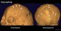
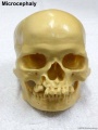 Skull - microcephaly 01.jpg 750 × 1,000; 75 KB
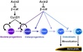 Skull - osteoblast lineage model.jpg 600 × 381; 20 KB
 Skull anterior.gif 200 × 205; 30 KB
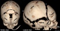
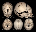
 Skull CT abnormal 01.jpg 1,000 × 549; 93 KB
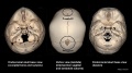
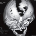
 Skull CT abnormal 02.jpg 1,000 × 900; 119 KB
 Skull CT abnormal 03.jpg 1,000 × 542; 82 KB
 Skull CT abnormal 04.jpg 1,000 × 646; 102 KB
 Skull CT abnormal 05.jpg 1,000 × 572; 88 KB
 Skull CT abnormal 06.jpg 1,000 × 541; 85 KB
 Skull CT abnormal 07.jpg 1,000 × 541; 64 KB
 Skull CT abnormal 08.jpg 1,000 × 516; 73 KB
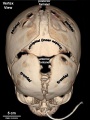
 Skull CT normal sutures 01.jpg 1,000 × 526; 89 KB
 Skull CT normal sutures 02.jpg 1,000 × 559; 92 KB
 Skull CT normal sutures 03.jpg 600 × 800; 63 KB
 Skull CT normal sutures.jpg 1,000 × 900; 138 KB
 Skull lateral view.gif 200 × 147; 22 KB
 Skull superior.gif 200 × 163; 22 KB
 Skull vault defect and midface hypoplasia.jpg 800 × 798; 81 KB
 Skull viscerocranium 01.mp4 ; 443 KB
 Stage 22 image 204.jpg 1,190 × 848; 363 KB
 Vesalius Skull.jpg 600 × 382; 44 KB
 Watson1915 Fig01.jpg 929 × 1,000; 171 KB
 Watson1915 Fig02.jpg 1,421 × 1,000; 245 KB
 Watson1915 Fig03.jpg 1,533 × 1,000; 276 KB
 Watson1915 Fig04.jpg 1,296 × 1,000; 197 KB
 Watson1915 Fig05.jpg 1,147 × 1,000; 127 KB
 Watson1915 Fig06.jpg 1,402 × 1,000; 226 KB
 Watson1915 Fig07.jpg 1,078 × 1,000; 235 KB
 Watson1915 Fig08.jpg 1,183 × 1,000; 211 KB
 Watson1915 Fig09.jpg 1,173 × 1,000; 253 KB
 Watson1915 Fig10.jpg 1,101 × 1,000; 0 bytes
 Watson1915 Fig11.jpg 1,060 × 1,000; 180 KB
 Watson1915 Fig12.jpg 1,119 × 1,000; 186 KB
 Watson1915 Fig13.jpg 1,168 × 1,000; 176 KB
 Watson1915 Fig14.jpg 1,256 × 1,000; 174 KB
 Watson1915 Fig15.jpg 1,132 × 1,000; 136 KB
 Watson1915 Fig16.jpg 1,187 × 1,000; 179 KB
 Watson1915 Fig17.jpg 1,193 × 1,000; 166 KB
 Watson1915 Fig18.jpg 1,186 × 1,000; 144 KB
 Watson1915 Fig19.jpg 1,354 × 1,000; 159 KB
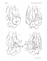 Watson1915 Plate01.jpg 1,552 × 2,000; 275 KB
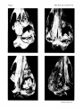 Watson1915 Plate02.jpg 1,552 × 2,000; 527 KB
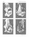 Watson1915 Plate03.jpg 1,552 × 2,000; 605 KB
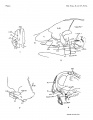 Watson1915 Plate04.jpg 1,552 × 2,000; 230 KB
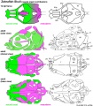 Zebrafish skull neural crest.jpg 815 × 933; 187 KB


