File:Spaulding01.jpg: Difference between revisions
From Embryology
(==Figure 1. Drawing of external genitalia of an embryo 16.8 mm. long== (Specimen No. 492), illustrating the genital-tubercle period. X 25. {{Template:Historic Disclaimer}} ==Reference== The development of the external genitalia in the human embryo By) |
mNo edit summary |
||
| (6 intermediate revisions by 2 users not shown) | |||
| Line 1: | Line 1: | ||
==Figure 1. Drawing of external genitalia of an embryo 16.8 mm. long== | ==Figure 1. Drawing of external genitalia of an embryo 16.8 mm. long== | ||
(Specimen No. | (Specimen No. {{CE492}}), illustrating the genital-tubercle period. X 25. | ||
{{Online Editor}} - CRL without knowing fixation shrinkage should be [[Carnegie stage 18]] or [[Carnegie stage 19]]. | |||
{{Spaulding1922}} | |||
[[Category:Carnegie Embryo 492]] | |||
Latest revision as of 12:57, 8 Haziran 2017
Figure 1. Drawing of external genitalia of an embryo 16.8 mm. long
(Specimen No. 492), illustrating the genital-tubercle period. X 25.
Online Editor - CRL without knowing fixation shrinkage should be Carnegie stage 18 or Carnegie stage 19.
- Figure Links: Text | Text Figure 1 | Text Figure 2 | Plate 1 | Fig. 1 | Fig. 2 | Fig. 3 | Fig. 4 | Fig. 5 | Fig. 6 | Plate 2 | Fig. 7 | Fig. 8 | Fig. 9 | Fig. 10 | Fig. 11 | Fig. 12 | Fig. 13 | Fig. 14 | Fig. 15 | Fig. 16 | Fig. 17 | Fig. 18 | Fig. 19 | Fig. 20 | Fig. 21 | Fig. 22 | Plate 3 | Fig. 23 | Fig. 24 | Fig. 25 | Fig. 26 | Fig. 27 | Fig. 28 | Fig. 29 | Plate 4 | Fig. 30 | Fig. 31 | Fig. 32 |Fig. 33 | Fig. 34 | Fig. 35 | Fig. 36 | Fig. 37 | Fig. 38 | Fig. 39 | Fig. 40 | Fig. 41 | Fig. 42 | Fig. 43 | Fig. 44 | Fig. 45 | Fig. 46 | Fig. 47 | Fig. 48 | Fig. 49 | Fig. 50 | Fig. 51 | Fig. 52 | Fig. 53 | Fig. 54
| Historic Disclaimer - information about historic embryology pages |
|---|
 |
Reference
Spaulding MH. The development of the external genitalia in the human embryo. (1921) Contrib. Embryol., Carnegie Inst. Wash. Publ. 81, 13: 69 – 88.
Cite this page: Hill, M.A. (2026, October 31) Embryology Spaulding01.jpg. Retrieved from https://embryology.med.unsw.edu.au/embryology/index.php/File:Spaulding01.jpg
- © Dr Mark Hill 2026, UNSW Embryology ISBN: 978 0 7334 2609 4 - UNSW CRICOS Provider Code No. 00098G
File history
Yi efo/eka'e gwa ebo wo le nyangagi wuncin ye kamina wunga tinya nan
| Gwalagizhi | Nyangagi | Dimensions | User | Comment | |
|---|---|---|---|---|---|
| current | 17:36, 26 March 2011 | 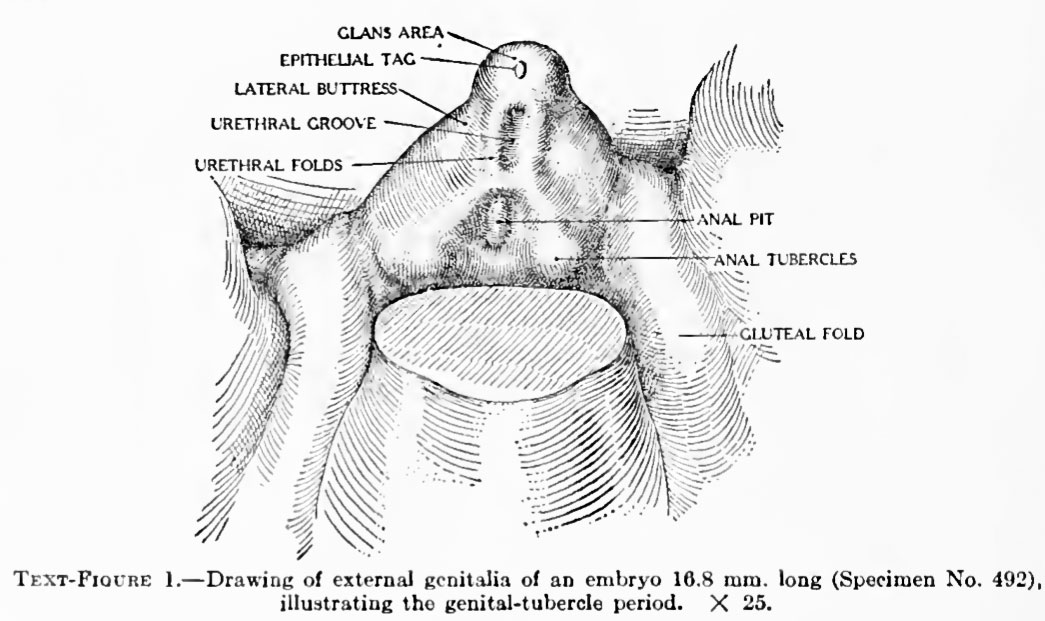 | 1,045 × 621 (111 KB) | S8600021 (talk | contribs) | ==Figure 1. Drawing of external genitalia of an embryo 16.8 mm. long== (Specimen No. 492), illustrating the genital-tubercle period. X 25. {{Template:Historic Disclaimer}} ==Reference== The development of the external genitalia in the human embryo By |
You cannot overwrite this file.
File usage
The following 3 pages use this file:


