File:Model human blastocyst development.jpg: Difference between revisions
No edit summary |
mNo edit summary |
||
| (2 intermediate revisions by 2 users not shown) | |||
| Line 1: | Line 1: | ||
==Proposed Model for Human Embryo Development | ==Proposed Model for Human Embryo Development== | ||
Human embryos begin life with a set of oocyte RNAs inherited from the mother. After fertilization, a subset of maternal RNAs specific to the egg (ESSP1) must be degraded as the transition from oocyte to embryo begins embryonic genome activation (EGA). | Human embryos begin life with a set of oocyte RNAs inherited from the mother. After fertilization, a subset of maternal RNAs specific to the egg (ESSP1) must be degraded as the transition from oocyte to embryo begins embryonic genome activation (EGA). | ||
| Line 16: | Line 16: | ||
:'''Links:''' [[Week_1#Genome_Expression|Week 1 Genome Expression]] | [[Blastocyst Day 3-6 Movie]] | |||
==Reference== | ===Reference=== | ||
<pubmed>20890283</pubmed>| [http://www.nature.com/nbt/journal/vaop/ncurrent/full/nbt.1686.html Nat Biotechnol.] | <pubmed>20890283</pubmed>| [http://www.nature.com/nbt/journal/vaop/ncurrent/full/nbt.1686.html Nat Biotechnol.] | ||
====Copyright==== | |||
Reprinted by permission from Macmillan Publishers Ltd: Nat Biotechnol. (Non-invasive imaging of human embryos before embryonic genome activation predicts development to the blastocyst stage. | Reprinted by permission from Macmillan Publishers Ltd: Nat Biotechnol. (Non-invasive imaging of human embryos before embryonic genome activation predicts development to the blastocyst stage. | ||
Wong CC, Loewke KE, Bossert NL, Behr B, De Jonge CJ, Baer TM, Pera RA. | Wong CC, Loewke KE, Bossert NL, Behr B, De Jonge CJ, Baer TM, Pera RA. Nat Biotechnol. 2010 Oct 3. [Epub ahead of print] PMID 20890283), copyright (2010) | ||
Nat Biotechnol. 2010 Oct 3. [Epub ahead of print] | |||
PMID | Original file name: Figure 6 Nbt.1686-F6.jpg http://www.nature.com/nbt/journal/vaop/ncurrent/fig_tab/nbt.1686_F6.html | ||
Licensee: Mark A Hill | Licensee: Mark A Hill | ||
| Line 34: | Line 36: | ||
Type Of Use: Web Site | Type Of Use: Web Site | ||
{{Footer}} | |||
[[Category:Human Embryo]] [[Category:Week 1]] [[Category:Zygote]] [[Category:Morula]] [[Category:Blastocyst]] [[Category:In Vitro Fertilization]] | [[Category:Human Embryo]] [[Category:Week 1]] [[Category:Zygote]] [[Category:Morula]] [[Category:Blastocyst]] [[Category:In Vitro Fertilization]] | ||
Latest revision as of 14:47, 6 October 2015
Proposed Model for Human Embryo Development
Human embryos begin life with a set of oocyte RNAs inherited from the mother. After fertilization, a subset of maternal RNAs specific to the egg (ESSP1) must be degraded as the transition from oocyte to embryo begins embryonic genome activation (EGA).
As development continues, other RNAs are partitioned equally to each blastomere (ESSP4). At EGA, ESSP2 genes are transcribed in a cell-autonomous manner.
During the cleavage divisions, embryonic blastomeres may arrest or progress independently.
'Feature extraction' indicates the three imaging parameters for predicting successful development to the blastocyst stage: cytokinesis, the time between 1st and 2nd mitoses, and the time between 2nd and 3rd mitoses.
Legend
- EGA - embryonic genome activation
- ESSP - embryonic stage–specific pattern, four unique embryonic stage–specific patterns (1-4)
- RNA - ribonucleic acid
Reference
<pubmed>20890283</pubmed>| Nat Biotechnol.
Copyright
Reprinted by permission from Macmillan Publishers Ltd: Nat Biotechnol. (Non-invasive imaging of human embryos before embryonic genome activation predicts development to the blastocyst stage. Wong CC, Loewke KE, Bossert NL, Behr B, De Jonge CJ, Baer TM, Pera RA. Nat Biotechnol. 2010 Oct 3. [Epub ahead of print] PMID 20890283), copyright (2010)
Original file name: Figure 6 Nbt.1686-F6.jpg http://www.nature.com/nbt/journal/vaop/ncurrent/fig_tab/nbt.1686_F6.html
Licensee: Mark A Hill
License Date: Oct 06, 2010
License Number: 2523401420894
Publication: Nature Biotechnology
Title: Non-invasive imaging of human embryos before embryonic genome activation predicts development to the blastocyst stage
Type Of Use: Web Site
Cite this page: Hill, M.A. (2024, June 27) Embryology Model human blastocyst development.jpg. Retrieved from https://embryology.med.unsw.edu.au/embryology/index.php/File:Model_human_blastocyst_development.jpg
- © Dr Mark Hill 2024, UNSW Embryology ISBN: 978 0 7334 2609 4 - UNSW CRICOS Provider Code No. 00098G
File history
Yi efo/eka'e gwa ebo wo le nyangagi wuncin ye kamina wunga tinya nan
| Gwalagizhi | Nyangagi | Dimensions | User | Comment | |
|---|---|---|---|---|---|
| current | 09:25, 12 October 2010 | 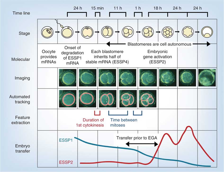 | 946 × 726 (84 KB) | S8600021 (talk | contribs) | ==Proposed Model for Human Embryo Development== Human embryos begin life with a set of oocyte RNAs inherited from the mother. After fertilization, a subset of maternal RNAs specific to the egg (ESSP1) must be degraded as the transition from oocyte to emb |
You cannot overwrite this file.
File usage
The following 5 pages use this file: