File:Ossification centre.jpg: Difference between revisions
No edit summary |
No edit summary |
||
| Line 7: | Line 7: | ||
Image and Text Source: UWA Blue Histology http://www.lab.anhb.uwa.edu.au/mb140/CorePages/Bone/Bone.htm | Image and Text Source: UWA Blue Histology http://www.lab.anhb.uwa.edu.au/mb140/CorePages/Bone/Bone.htm | ||
[[Category:Musculoskeletal]] [[Category:Histology]] | |||
Revision as of 17:47, 15 September 2009
Ossification centre
Histological areas of a gradual transition from connective tissue to bone. Light, pinkish bone matrix is found between the osteoblasts. Depending on the state of development of the bone, it is occasionally possible to find bone trabeculae which are lined by a layer of osteoblasts. These osteblasts are depositing the first lamellae on the already existing trabeculae. The trabeculae will therefore have a core of woven bone, which is surrounded by lamellar bone. Compare the shapes, sizes and frequencies of lacunae in lamellar and woven bone if both types of bone are present.
Original Image name: Imos10he.jpg
Image and Text Source: UWA Blue Histology http://www.lab.anhb.uwa.edu.au/mb140/CorePages/Bone/Bone.htm
File history
Click on a date/time to view the file as it appeared at that time.
| Date/Time | Thumbnail | Dimensions | User | Comment | |
|---|---|---|---|---|---|
| current | 15:57, 13 May 2012 | 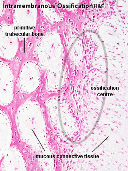 | 450 × 600 (101 KB) | Z8600021 (talk | contribs) | |
| 11:30, 11 September 2009 |  | 300 × 400 (66 KB) | S8600021 (talk | contribs) | Ossification centre Histological areas of a gradual transition from connective tissue to bone. Light, pinkish bone matrix is found between the osteoblasts. Depending on the state of development of the bone, it is occasionally possible to find bone trabec |
You cannot overwrite this file.
File usage
The following 14 pages use this file:
- 2009 Lecture 13
- 2010 BGD Lecture - Development of the Embryo/Fetus 2
- 2010 BGD Practical 6 - Week 7
- 2010 Lecture 13
- ANAT2241 Bone, Bone Formation and Joints
- ANAT2341 Lab 6 - Fetal
- BGDA Lecture - Development of the Embryo/Fetus 2
- BGDA Practical 7 - Week 7
- BGDB Face and Ear - Fetal
- Bone Development
- Bone Histology
- Lecture - Fetal Development
- Lecture - Musculoskeletal Development
- Musculoskeletal System - Bone Development