Bone Histology: Difference between revisions
| Line 73: | Line 73: | ||
* Early bone matrix deposited in development and during repair is '''woven''' rather than '''lamellar''' in appearance and structure. | * Early bone matrix deposited in development and during repair is '''woven''' rather than '''lamellar''' in appearance and structure. | ||
* In development, there are 2 distinct types of bone formation (intramembranous and endochondral) | * In development, there are 2 distinct types of bone formation (intramembranous and endochondral) | ||
== Bone Cells == | == Bone Cells == | ||
Revision as of 15:38, 5 September 2011

Introduction

Our adult skeleton forms from a larger number of developmental elements that are replaced and fuse. In development there are 2 separate signaling pathways for pattern formation and the formation of bone itself. Furthermore bone formation can be divided into 2 specific forms that occur in anatomically different regions. This practical class will describe the development and structure of bone and finish with a study of abnormalities associated with bone.
The image shown to the left shows a histological section through the developing lower limb at the level of a developing joint (knee), surrounding the developing bone are skeletal muscles and connective tissue of the limb.
Note: This current page contains both additional information and images to the practical class set. These are provided for educational information and study purposes only. For more development background see the Science Lecture - Musculoskeletal Development and notes on Bone Development.
Objectives
- Understand the general microanatomy of bone
- Understand bone cell types (location, structure, function)
- Understand the histology of compact and spongy bone
- Understand the 2 forms of developmental bone formation
Practical Audio

Files below are Quicktime audio files recorded 2009 Wednesday 12 - 2 PM class (Podcast MP3 versions to follow).
Part 1 - Adult Bone Structure | Part 2 - Bone Structure | Part 3 - Developing Endochondral Ossification | Part 4 - Developing Intramembranous Ossification
- MH - Please note "perichondrium" instead of "periosteum" error somewhere in the above audio. Open in a separate tab to play the audio in the background.
Textbook
Histology and Cell Biology: An Introduction to Pathology, A.L. Kierszenbaum, 2002 - Connective Tissue, Chapter 4 pp118-129; Osteogenesis, Chapter 5 pp131-145
Slides
UNSW Virtual Slidebox Virtual Slidebox Phase 1
Virtual Slidebox of Histology Decalcified rib, bone marrow | Developing bone | Paget's disease of bone
Bone Structure

Terminology
- Diaphysis - shaft
- Epiphysis - expanded ends
- Metaphysis - connecting region (between diaphysis and epiphysial line)
- Medullary Cavity - (marrow) cavity within the bone.
More? Terms
Compact bone
- (dense) no spaces or hollows in the bone matrix visible to the eye.
- forms the thick-walled tube of the shaft (or diaphysis) of long bones, which surrounds the marrow cavity (or medullary cavity). A thin layer of compact bone also covers the epiphyses of long bones.
Trabecular bone
- (cancellous or spongy bone) consists of delicate bars (spicules) and sheets of bone, trabeculae
- branch and intersect to form a sponge-like network
- ends of long bones (or epiphyses) consist mainly of trabecular bone.
Periosteum
Connective tissue covering the surface of bone (except articular surfaces).

Endosteum
Connective tissue lining inner surface of bone.
Bone Growth
- Appositional growth occurs at either the periosteum (outer surface), or the endosteum (inner surface).
- Osteoblasts secrete osteoid, a pre-bone material composed mainly of type I collagen that becomes mineralized.
- Early bone matrix deposited in development and during repair is woven rather than lamellar in appearance and structure.
- In development, there are 2 distinct types of bone formation (intramembranous and endochondral)
Bone Cells
Osteoblasts

- derive from osteogenic stem cells the osteoprogenitor cells that differentiate to form pre-osteoblast then osteoblasts maturing to an osteocyte
- osteoprogenitor cells - "resting cell" line the inner and outer surfaces of bone
Osteocytes
- mature bone-forming cells embedded in lacunae within the bone matrix
- osteoblasts and osteocytes - secrete organic matrix of bone (osteoid), converted into osteocytes when become embedded in matrix (which calcifies soon after deposition)
Osteoclasts


- bone-resorbing multinucleated macrophage-like cells
- origin- fusion of monocytes or macrophages, Blood macrophage precursor, Attach to bone matrix
- seal a small segment of extracellular space (between plasma membrane and bone surface), HCl and lysosomes secreted into this space by osteoclasts dissolves calcium phosphate crystals (give bone rigidity and strength)
- Resorptive bay - (Howship's lacuna) shallow bay lying directly under an osteoclast.
- do not mistake for megakaryocytes, found in bone marrow not associated with bone matrix.
- megakaryocytes are also multi-niucleated and form platelets
Bone Marrow

- red marrow - mainly haematopoietic (myeloid) tissue, newborn has all red marrow
- yellow marrow - mainly fat cells, found in diaphysis region of long bones
- stromal cells - all other support cells not involved in haematopoiesis
Chondroblasts and Chondrocytes
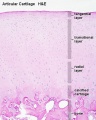
Articular cartilage
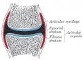
Synovial joint showing cartilage

- immature and mature cartilage forming cells located at articular cartilage regions.
- Interstitial growth - occurs mainly in immature cartilage. Chondroblasts in existing cartilage divide and form small groups of cells (isogenous groups) which produce matrix to become separated from each other by a thin partition of matrix.
- Appositional growth - occurs also in mature cartilage. Mesenchymal cells surrounding the cartilage in the deep part of the perichondrium (or the chondrogenic layer) differentiate into chondroblasts.
Bone Matrix
The bone matrix has 2 major components.
- Organic portion composed of mainly collagen Type 1 (about 95%) and amorphous ground substance.
- Inorganic portion (50% dry weight of the matrix) composed of hydroxyapatite crystals, calcium, phosphorus, bicarbonate, nitrate, Mg, K, Na.
- storage calcium and phosphate
- regulate blood calcium levels
Haversian Systems

- also called osteons
- Volkmann's canals - interconnect Haversian systems
Lamellae
- concentric - surrounding each Haversian System
- interstitial - bony plates that fill in between the haversian systems.
- circumferential - layers of bone that underlie the periosteum and endosteum
Cells
- osteocytes extending cytoplasmic processes into canaliculi
- Additional Histology images: low | medium | high
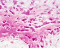
Endochondral ossification
Endochondral ossification slides: Developing bone | Bone, Developing (LS, Femur) Cat H&E
Blue Histology - endochondral | Dev Biology - endochondral ossification | endochondral ossification animation


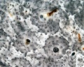
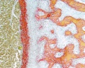
compact bone - low unstained
compact bone - high unstained

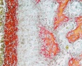
compact - low
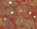
compact - low
compact - med

compact - high


- Bone Histology: Cartilage Histology | Histology Stains | Histology | cartilage | bone | bone timeline
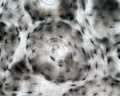
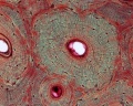
- Trabecular bone trabecular | lamellar | trabecular - overview HE | trabecular - low HE | trabecular - med HE
- Endochondral ossification primary ossification | endochondral ossification
- Intramembranous ossification intramembranous - VG low | intramembranous - VG high | intramembranous - HE low | intramembranous - HE high
Intramembranous Ossification
Intramembranous ossification slides: Head (Neonatal) Rat H& Van Gieson
Blue histology - intramembranous | intramembranous ossification animation


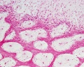
intramembranous - HE low
intramembranous - HE high
intramembranous - VG low
intramembranous - VG high


- Bone Histology: Cartilage Histology | Histology Stains | Histology | cartilage | bone | bone timeline
- Trabecular bone trabecular | lamellar | trabecular - overview HE | trabecular - low HE | trabecular - med HE
- Endochondral ossification primary ossification | endochondral ossification
- Intramembranous ossification intramembranous - VG low | intramembranous - VG high | intramembranous - HE low | intramembranous - HE high
Human Fetal Head (12 week)


Histology Stains
Alizarin Red
- an anthraquinone derivative used to identify calcium in tissue sections
- calcium forms an Alizarin Red S-calcium complex in a chelation process and the end product is also birefringent.
- reaction can also identify magnesium, manganese, barium, strontium, and iron may interfere
- these elements usually in too low concentration to interfere with the staining
H&E
- acronym for hematoxylin and eosin stain
- hematoxylin - basic dye which colors basophilic structures with blue-purple hue (nucleus, DNA, RNA)
- eosin Y - acidic alcohol-based which colors eosinophilic structures bright pink (cytoplasm, extracellular matrix, protein)
H&Van Gieson
- Van Gieson's Stain is a mixture of picric acid and acid fuchsin used for differential staining of collagen and other connective tissue.
- Nuclei - stains brownish black to black
- Collagen (fibrous connective tissue) - stains pink or deep red
- Muscle, Cytoplasm, RBC and Fibrin - stains yellow
- Links: Histology Stains
External Links
- Virtual Slidebox of Histology (USA) Skeletal system
- e-radiography Ossification
- UWA Blue Histology bone
Other Textbooks
- Anatomy of the Human Body (H. Gray, 1918.) historical anatomy text Osteology
- Molecular Biology of the Cell Bone Is Continually Remodeled by the Cells Within It | Image: Figure 22-52. Deposition of bone matrix by osteoblasts | Image: Figure 22-56. The development of a long bone
- Molecular Cell Biology Mutations in Collagen Reveal Aspects of Its Structure and Biosynthesis
- The Cell- A Molecular Approach Steroid Hormones and the Steroid Receptor Superfamily
- Clinical Methods: The History, Physical, and Laboratory Examinations 100. Alkaline Phosphatase and Gamma Glutamyltransferase
- Endocrinology: An Integrated Approach by Nussey, S.S. and Whitehead, S.A. Endocrinology: Definition and causes of osteoporosis
- Developmental Biology 6th ed. by Gilbert, Scott F. Figure 14.13. Schematic diagram of endochondral ossification | Aging: The Biology of Senescence
Search
- Pubmed ossification
Terms


- canaliculus - (plural, canaliculi) small channel in the bone matrix in which an osteocyte process lies and communicates with other osteocytes and the Haversian canal.
- haematopoiesis (Greek, haima = "blood"; poiesis = "to make") the process of blood cell formation.
- Haversian canal - the central canal of an osteon (Haversian system) in compact bone, within which blood vessels and nerves travel throughout the bone.
- Haversian system - (osteon) the historic name for the functional unit of compact bone. Consists of a central canal (Haversian canal) surrounded by lamellar bone matrix within which osteocytes reside.
- Howship's lacuna - (resorptive bay) the historic name for the shallow bay or cavity lying directly under an osteoclast. This is the site of bone matrix resorption.
- lacuna - (Latin, lacuna = “ditch, gap” diminutive form of lacus = “lake”) lacunae is the plural, cavity in bone or cartilage for cell.
- lamellar bone - the highly organized strong bone matrix deposited in concentric sheets with a low proportion of osteocytes. Many collagen fibers parallel to each other in the same layer.
- osteon - (Haversian system) the functional unit of compact bone. Consists of a central canal (Haversian canal) surrounded by lamellar bone matrix within which osteocytes reside.
- resorptive bay - (Howship's lacuna) the shallow bay or cavity lying directly under an osteoclast. This is the site of bone matrix resorption.
- suture - in the skull a form of articulation where the contiguous margins of the bones are united by a thin layer of fibrous tissue.
- woven bone - the first deposited weaker bone matrix with many osteocytes and a matrix disorganized structure. Replaced by lamellar bone. Seen in developing, healing and bone disease.
Glossary Links
- Glossary: A | B | C | D | E | F | G | H | I | J | K | L | M | N | O | P | Q | R | S | T | U | V | W | X | Y | Z | Numbers | Symbols | Term Link
Cite this page: Hill, M.A. (2024, June 27) Embryology Bone Histology. Retrieved from https://embryology.med.unsw.edu.au/embryology/index.php/Bone_Histology
- © Dr Mark Hill 2024, UNSW Embryology ISBN: 978 0 7334 2609 4 - UNSW CRICOS Provider Code No. 00098G