Category:Female: Difference between revisions
From Embryology
mNo edit summary |
mNo edit summary |
||
| Line 1: | Line 1: | ||
This {{Embryology}} category shows pages and media related to {{Female}} development. | This {{Embryology}} category shows pages and media related to {{Female}} development. | ||
<br><br> | <br><br> | ||
{{female links}} | {{female links}} | ||
| Line 9: | Line 7: | ||
{{Genital Links}} | {{Genital Links}} | ||
<br> | |||
'''Female Embryos [[Carnegie Collection]]''': {{CE108}} | {{CE128}} | {{CE1388}} | {{CE1455}} | {{CE1535}} | {{CE1597b}} | {{CE2023}} | {{CE2250a}} | {{CE2250b}} | {{CE227}} | {{CE2937}} | {{CE4090}} | {{CE4205}} | {{CE4289}} | {{CE45}} | {{CE464}} | {{CE4960}} | {{CE5422}} | {{CE630}} | {{CE632}} | {{CE6512}} | {{CE6531}} | {{CE6573}} | {{CE7425}} | {{CE86}} | {{CE875}} | {{CE9226}} | '''Female Embryos [[Carnegie Collection]]''': {{CE108}} | {{CE128}} | {{CE1388}} | {{CE1455}} | {{CE1535}} | {{CE1597b}} | {{CE2023}} | {{CE2250a}} | {{CE2250b}} | {{CE227}} | {{CE2937}} | {{CE4090}} | {{CE4205}} | {{CE4289}} | {{CE45}} | {{CE464}} | {{CE4960}} | {{CE5422}} | {{CE630}} | {{CE632}} | {{CE6512}} | {{CE6531}} | {{CE6573}} | {{CE7425}} | {{CE86}} | {{CE875}} | {{CE9226}} | ||
<br> | |||
==Female Bias Anomalies== | ==Female Bias Anomalies== | ||
Revision as of 08:57, 23 April 2020
This Embryology category shows pages and media related to Female development.
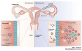
| Female | X | X inactivation | ovary | corpus luteum | oocyte | uterus | vagina | reproductive cycles | menstrual cycle | Category:Female |
Additional Female Links: X Chromosome | X Inactivation | Trisomy X | ovary | oocyte | meiosis | uterus | vagina | mammary gland | menstrual cycle | corpus luteum | puberty | genital abnormalities | Category:X Chromosome
Female Embryos Carnegie Collection: 108 | 128 | 1388 | 1455 | 1535 | 1597b | 2023 | 2250a | 2250b | 227 | 2937 | 4090 | 4205 | 4289 | 45 | 464 | 4960 | 5422 | 630 | 632 | 6512 | 6531 | 6573 | 7425 | 86 | 875 | 9226
Female Bias Anomalies
| Male preponderance | Female preponderance |
|---|---|
|
|
| Cardiac defects | |
|
|
| Table data[1] Links: abnormal development | cardiovascular abnormalities | USA | Male | Female | cleft lip and palate | |
- atrial septal defects (ASD) are a group of common (1% of cardiac) congenital anomolies defects occurring in a number of different forms and more often in.
Subcategories
This category has the following 39 subcategories, out of 39 total.
C
- Carnegie Embryo 108
- Carnegie Embryo 128
- Carnegie Embryo 1388
- Carnegie Embryo 1455
- Carnegie Embryo 1535
- Carnegie Embryo 1597b
- Carnegie Embryo 2023
- Carnegie Embryo 2250a
- Carnegie Embryo 2250b
- Carnegie Embryo 227
- Carnegie Embryo 2937
- Carnegie Embryo 4090
- Carnegie Embryo 4205
- Carnegie Embryo 4289
- Carnegie Embryo 45
- Carnegie Embryo 464
- Carnegie Embryo 4960
- Carnegie Embryo 5422
- Carnegie Embryo 630
- Carnegie Embryo 632
- Carnegie Embryo 6512
- Carnegie Embryo 6531
- Carnegie Embryo 6573
- Carnegie Embryo 7425
- Carnegie Embryo 86
- Carnegie Embryo 875
- Carnegie Embryo 9226
- Corpus Luteum
D
U
V
Pages in category 'Female'
The following 129 pages are in this category, out of 129 total.
B
- Template:BGDB Sexual Differentiation - Abnormalities Interactive
- Template:BGDB Sexual Differentiation - Early Embryo Interactive
- Template:BGDB Sexual Differentiation - Fetal Interactive
- Template:BGDB Sexual Differentiation - Late Embryo Interactive
- Template:BGDB Sexual Differentiation - Postnatal Interactive
- Template:BGDB Sexual Differentiation - Sex Determination Interactive
- Book - A Laboratory Manual and Text-book of Embryology 8
- Book - Contributions to Embryology Carnegie Institution No.61
- Book - Manual of Human Embryology 19-4
- Template:Braune 1877 Plate 2
C
E
F
G
M
O
P
- Paper - Anatomy, pathology and development of the hymen
- Paper - Another case of hermaphroditism in man (1924)
- Paper - Development of the human ovary from birth to sexual maturity
- Paper - Development of the mammary gland
- Paper - Development of the vagina in the human fetus
- Paper - On the anlage of the bulbo-urethral and major vestibular glands in the human embryo (1915)
- Paper - On the origin and phylogenetic significance of the female genital passages
- Paper - On the phenomena of sex-differentiation (1892)
- Paper - Some points in the nomenclature of the external genitalia of the female
- Paper - The development of the human vagina
- Paper - The development of the hymen
- Paper - The development of the prostate gland in the human female and homologies of the urethra and vagina of the sexes
- Paper - The Internal Genital Organs of a Female Foetus of 15 cm Length
- Paper - The physiological descent of the ovaries in the human foetus
- Template:Paramesonephric duct
- Template:PCOS
- Template:Polycystic ovarian syndrome
- Template:Polycystic ovary syndrome
- Template:Prader Stages table
- Template:Precocious puberty
- Template:Preovulatory follicle
- Template:Primary follicle
- Template:Primordial follicle
- Template:Progesterone
- Template:Puberty
R
- Rabbit Ovulation Movie
- Template:Ref-Barrington1915
- Template:Ref-Baxter1933
- Template:Ref-Bulmer1957
- Template:Ref-Evatt1911
- Template:Ref-Fleming1927
- Template:Ref-Gellhorn1904
- Template:Ref-Girgis1924
- Template:Ref-Hart1893
- Template:Ref-Hart1896
- Template:Ref-Hart1896b
- Template:Ref-Hart1897
- Template:Ref-Hart1909d
- Template:Ref-Jones1914
- Template:Ref-Koff1933
- Template:Ref-McKeown1935
- Template:Ref-Pryor1928
- Template:Ref-Simkins1932
- Template:Ref-Skene1898
- Template:Ref-Spaulding1921
- Template:Ref-Watase1892
- Template:Ref-Wichmann1914
- Template:Ref-Wood-Jones1913
- Template:RU 486
- Template:RU486
U
Media in category 'Female'
The following 143 files are in this category, out of 143 total.
 Adult female fibroblast and lymphocyte nuclear DNA 01.jpg 1,095 × 1,200; 197 KB
 Adult female fibroblast nuclear DNA 01.jpg 2,186 × 1,000; 219 KB
 Adult mesentery 1.jpg 1,000 × 1,311; 184 KB
 Bailey310.jpg 688 × 365; 41 KB
 Bailey328.jpg 704 × 795; 85 KB
 Bailey329.jpg 838 × 381; 70 KB
 Bailey330.jpg 680 × 539; 109 KB
 Bailey331.jpg 890 × 782; 118 KB
 Bailey334.jpg 857 × 558; 77 KB
 Bailey335.jpg 590 × 468; 55 KB
 Bailey339.jpg 802 × 602; 111 KB
 Bailey342.jpg 777 × 561; 55 KB
 Bailey343-344.jpg 1,185 × 630; 91 KB
 Bailey345-346.jpg 1,193 × 491; 79 KB
 Bailey347-348.jpg 1,198 × 526; 83 KB
 Barr-murray.jpg 100 × 113; 3 KB
 Braune 1877 plate 2 fig1.jpg 960 × 1,000; 181 KB
 Braune 1877 plate 2 fig2.jpg 806 × 1,000; 200 KB
 Braune 1877 plate 2 fig3.jpg 893 × 1,000; 208 KB
 Braune 1877 plate 2 fig4.jpg 809 × 1,000; 213 KB
 Braune 1877 plate 2 fig5.jpg 771 × 1,000; 169 KB
 Braune 1877 plate 2 fig6.jpg 690 × 1,000; 144 KB
 Braune 1877 plate 2 fig7.jpg 893 × 1,000; 241 KB
 Braune 1877 plate 29B.jpg 868 × 1,200; 322 KB
 Braune 1877 plate 2A.jpg 780 × 1,200; 263 KB
 Braune 1877 plate 2B.jpg 801 × 1,200; 264 KB
 Braune 1877 plate 30.jpg 857 × 1,200; 295 KB
 Braune 1877 plate 31.jpg 870 × 1,200; 299 KB
 BrauneB1.jpg 1,200 × 485; 139 KB
 BrauneB2.jpg 1,200 × 485; 143 KB
 BrauneC1.jpg 1,200 × 461; 143 KB
 Bulmer1957 plate01.jpg 1,280 × 1,808; 353 KB
 Caudal duplication syndrome.jpg 700 × 599; 47 KB
 Cullen1916 fig12.jpg 1,280 × 1,246; 560 KB
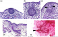
 Female genital and ureter abnormality 01.jpg 766 × 732; 86 KB
 Female genital and ureter abnormality 02.jpg 766 × 733; 78 KB
 Female genital and ureter abnormality 03.jpg 766 × 762; 79 KB
 Female genital tract chlamydia trachomatis infection 01.jpg 804 × 500; 78 KB
 Female reproductive tract Wnt4.jpg 1,000 × 563; 79 KB
 Female- OHVIRA syndrome 01.jpg 340 × 1,000; 76 KB
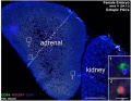 Fetal adrenal ectopic germ cells 04.jpg 899 × 700; 147 KB
 Fleming1927-fig01.jpg 953 × 1,000; 167 KB
 Fleming1927-fig02.jpg 1,000 × 685; 53 KB
 Fleming1927-fig03.jpg 1,200 × 1,908; 602 KB
 Fleming1927-fig03a.jpg 1,179 × 493; 150 KB
 Fleming1927-fig03b.jpg 1,179 × 577; 171 KB
 Fleming1927-fig03c.jpg 1,179 × 800; 266 KB
 Fleming1927-fig04.jpg 800 × 899; 103 KB
 Gray0540.jpg 1,355 × 1,000; 261 KB
 Gray0589.jpg 900 × 534; 134 KB
 Gray0607.jpg 800 × 623; 126 KB
 Gray0620.jpg 706 × 600; 127 KB
 Gray1108.jpg 590 × 400; 73 KB
 Gray1109.jpg 464 × 487; 56 KB
 Gray1112.jpg 550 × 548; 51 KB
 Gray1113.jpg 600 × 385; 68 KB
 Gray1119.jpg 700 × 807; 115 KB
 Gray1138.jpg 656 × 472; 57 KB
 Gray1139.jpg 600 × 524; 73 KB
 Gray1163.jpg 600 × 442; 91 KB
 Gray1166.jpg 750 × 750; 194 KB
 Gray1170.jpg 1,000 × 718; 170 KB
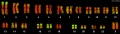 Human female karyotype 01.jpg 2,000 × 562; 118 KB
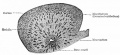
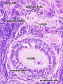
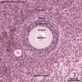 Human infant ovary follicle 01.jpg 800 × 800; 107 KB
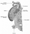
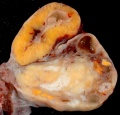
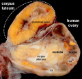
 Human ovary - corpus luteum 01.jpg 1,024 × 979; 162 KB
 Human ovary - corpus luteum 02.jpg 837 × 800; 119 KB
 Human ovary - corpus luteum 11.jpg 1,024 × 979; 89 KB
 Human ovary - corpus luteum 21.jpg 1,024 × 979; 89 KB
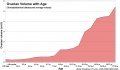 Human ovary postnatal growth.jpg 800 × 467; 40 KB
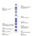 Human X chromosome.jpg 400 × 450; 25 KB
 Infant ovary.jpg 943 × 571; 108 KB
 Keibel Mall 2 621.jpg 1,280 × 1,842; 589 KB
 Keibel Mall 2 623.jpg 1,549 × 1,709; 437 KB
 Keibel Mall 2 626.jpg 850 × 679; 143 KB
 Keibel Mall 2 627.jpg 1,000 × 1,252; 169 KB
 Keibel Mall 2 628.jpg 1,000 × 512; 65 KB
 Keibel Mall 2 641.jpg 1,041 × 518; 62 KB
 Keibel Mall 2 645.jpg 957 × 1,000; 98 KB
 Keibel Mall 2 646.jpg 1,280 × 1,307; 181 KB
 Keibel Mall 2 658a.jpg 1,127 × 1,200; 103 KB
 Keibel Mall 2 658c.jpg 1,000 × 1,019; 90 KB
 Keith1902 fig081.jpg 818 × 800; 113 KB
 Keith1921 fig490.jpg 1,200 × 656; 214 KB
 Kollmann451.jpg 796 × 826; 102 KB
 Kollmann452.jpg 687 × 606; 74 KB
 Kollmann453.jpg 664 × 566; 65 KB
 Kollmann454.jpg 729 × 641; 84 KB
 Kollmann455.jpg 746 × 509; 102 KB
 Kollmann457.jpg 777 × 598; 60 KB
 Kollmann461.jpg 721 × 528; 100 KB
 Kollmann462.jpg 758 × 429; 63 KB
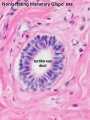 Lactiferous duct 01.jpg 450 × 600; 58 KB
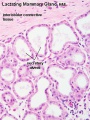 Mammary gland 01.jpg 450 × 600; 75 KB
 Mammary involution.jpg 600 × 427; 54 KB
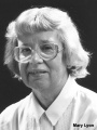 Mary Lyon.jpg 324 × 432; 23 KB
 Mouse mammary development 01.jpg 1,200 × 773; 147 KB
 Nelsen1953 fig030.jpg 1,200 × 995; 414 KB
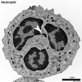 Neutrophil EM01.jpg 999 × 1,000; 402 KB
 OHVIRA syndrome 02.jpg 700 × 669; 72 KB
 OHVIRA syndrome 03.jpg 700 × 669; 65 KB
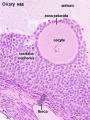 Ova20he.jpg 450 × 600; 96 KB
 Ovary follicle 01.jpg 450 × 600; 112 KB
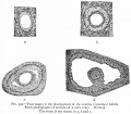
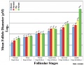 Ovary follicle size graph.jpg 1,057 × 820; 102 KB
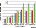 Ovary oocyte size graph.jpg 1,057 × 820; 114 KB
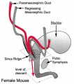 Paramesonephric duct.jpg 423 × 478; 40 KB
 Persistent cloaca perineum.jpg 600 × 800; 57 KB
 Septate uterus ultrasound 01.jpg 1,280 × 525; 95 KB
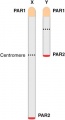 Sex chromosomes pseudoautosomal regions.jpg 217 × 400; 7 KB
 Spaulding-fig06.jpg 778 × 744; 85 KB
 Spaulding-fig11.jpg 445 × 607; 36 KB
 Spaulding-fig12.jpg 445 × 607; 25 KB
 Spaulding-fig13.jpg 445 × 607; 36 KB
 Spaulding-fig14.jpg 445 × 607; 35 KB
 Spaulding-fig19.jpg 445 × 607; 36 KB
 Spaulding-fig20.jpg 445 × 607; 37 KB
 Spaulding-fig22.jpg 445 × 607; 32 KB
 Spaulding-fig23.jpg 443 × 604; 37 KB
 Spaulding-fig29.jpg 443 × 604; 29 KB
 Spaulding-fig30.jpg 443 × 604; 27 KB
 Spaulding-fig33.jpg 443 × 604; 29 KB
 Spaulding-fig35.jpg 443 × 604; 39 KB
 Spaulding-fig36.jpg 443 × 604; 28 KB
 Spaulding-fig37.jpg 443 × 604; 27 KB
 Spaulding-fig38.jpg 443 × 604; 34 KB
 Spaulding-fig39.jpg 436 × 603; 31 KB
 Spaulding-fig40.jpg 445 × 604; 31 KB
 Spaulding-fig41.jpg 446 × 605; 27 KB
 Spaulding-fig43.jpg 436 × 603; 29 KB
 Spaulding-fig44.jpg 445 × 604; 27 KB
 Spaulding-fig45.jpg 440 × 599; 31 KB
 Spaulding-fig46.jpg 430 × 599; 26 KB
 Spaulding-fig51.jpg 429 × 599; 32 KB
 Spaulding-fig52.jpg 439 × 601; 33 KB
 Spaulding-fig53.jpg 436 × 596; 28 KB
 Spaulding-fig54.jpg 430 × 599; 27 KB
 UK Growth Chart-A4 Girls NICM.pdf ; 138 KB
 Unicornate uterus.jpg 250 × 241; 10 KB
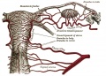
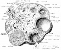
 Urogenital female.jpg 578 × 600; 40 KB
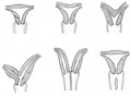 Uterine abnormalities.jpg 400 × 294; 15 KB
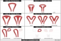 Uterine anomalies ESHRE-ESGE classification.jpg 1,000 × 684; 93 KB
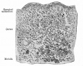
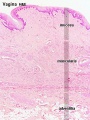 Vagina histology 01.jpg 450 × 600; 92 KB
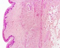 Vagina histology 02.jpg 1,280 × 1,024; 555 KB


