Manual of Human Embryology II - Figures: Difference between revisions
mNo edit summary |
mNo edit summary |
||
| Line 4: | Line 4: | ||
==XIV. The Development of the Nervous System== | ==XIV. The Development of the Nervous System== | ||
===I. Histogenesis of Nervous Tissue=== | |||
[[Book_-_Manual_of_Human_Embryology_14-1|I. Histogenesis of Nervous Tissue]] | |||
<gallery> | |||
Keibel_Mall_2_001.jpg|Fig. 1. Three early stages in the development of the wall of the neural tube, | |||
Keibel_Mall_2_009.jpg|Fig. 2. Wall of the neurfl tube in a human embryo about two weeks old, showing its syncytial character. | |||
Keibel_Mall_2_009.jpg|Fig. 3. Diagram showing the differentiation of the cells of the wall of the neural tube | |||
Keibel_Mall_2_009.jpg|Fig. 4. Development of neuroglia framework. | |||
Keibel_Mall_2_009.jpg|Fig. 5. Combined drawings, after Golgi and Benda methods, of the spinal cord of fetal pig, 20 cm. long | |||
Keibel_Mall_2_009.jpg|Fig. 6. Section of spinal cord of suckling pig of two weeks | |||
Keibel_Mall_2_009.jpg|Fig. 7. Neuroglia fibres in adult human spinal cord, showing their relation to 'the protoplasm of the neuroglia cell and its processes. | |||
Keibel_Mall_2_009.jpg|Fig. 8. Diagram showing distribution of neuroblasts in human embryo of four weeks. | |||
Keibel_Mall_2_009.jpg|Fig. 9. Cluster of neuroblasts from nucleus of origin of n. oculomotorius, showing characteristic shape and grouping of cells. | |||
Keibel_Mall_2_010.jpg|Fig. 10. Section through floor of mid-brain of human embryo one month old. | |||
Keibel_Mall_2_019.jpg|Fig. 11. Isolated ganglion-cells, from embryonic spinal cord of frog, and growing in clotted lymph. | |||
Keibel_Mall_2_019.jpg|Fig. 12. Three views, taken at intervals of Ik and 8i hours, of the same living nerve-fibres growing from a mass of spinal-cord tissue (frog embryo) out into clotted lymph. | |||
Keibel_Mall_2_019.jpg|Fig. 13. Transverse .sections through dorsal region of human embryos showing three stages in the development of the ganglion crest and the anlage of the spinal ganglia. | |||
Keibel_Mall_2_019.jpg|Fig. 14. Section through spinal ganglion of human embryo 18 mm. long, about 6 weeks old . | |||
Keibel_Mall_2_019.jpg|Fig. 15. Section through cervical spinal ganglion of human fetus 8.5 cm. long, about 3 months old, showing large ganglion-cells with eccentric nuclei. | |||
Keibel_Mall_2_019.jpg|Fig. 16. Section through sixth cervical ganglion of human fetus 10.5 cm. long, about 4 months old. | |||
Keibel_Mall_2_019.jpg|Fig. 17. Isolated cells teased from spinal ganglia of embryo pigs 20-40 mm. long, showing the variation in the form of the early ganglion-cells | |||
Keibel_Mall_2_019.jpg|Fig. 18. Teased preparations from spinal ganglia of pig, showing development of sheath and capsule cells. | |||
Keibel_Mall_2_019.jpg|Fig. 19. Isolated fibres showing development of medullary sheath. | |||
Keibel_Mall_2_021.jpg|Fig. 20. Isolated fibres of the sciatic nerve of sheep fetus 15 cm. long, treated with osmic acid and showing development of the nerve-sheath. | |||
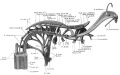
Keibel_Mall_2_021.jpg|Fig. 21. Section through hind-brain of new-born child, showing myelinization of fifth, sixth, seventh, and eighth cranial nerves and associated fibre tracts | |||
</gallery> | |||
<gallery> | <gallery> | ||
Keibel_Mall_2_030.jpg|Fig. 30. Profile views of the brains of human embryos as seen during the third (A), fourth (B), and eighth (C) weeks | Keibel_Mall_2_030.jpg|Fig. 30. Profile views of the brains of human embryos as seen during the third (A), fourth (B), and eighth (C) weeks | ||
Keibel_Mall_2_031.jpg| | |||
Keibel_Mall_2_032.jpg| | |||
Keibel_Mall_2_033.jpg| | |||
Keibel_Mall_2_034.jpg| | |||
Keibel_Mall_2_035.jpg| | |||
Keibel_Mall_2_036.jpg| | |||
Keibel_Mall_2_037.jpg| | |||
Keibel_Mall_2_038.jpg| | |||
Keibel_Mall_2_039.jpg| | |||
Keibel_Mall_2_040.jpg| | |||
</gallery> | </gallery> | ||
Revision as of 11:44, 24 January 2014
Figures
XIV. The Development of the Nervous System
I. Histogenesis of Nervous Tissue
I. Histogenesis of Nervous Tissue
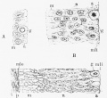
Fig. 1. Three early stages in the development of the wall of the neural tube,
- Keibel Mall 2 009.jpg
Fig. 2. Wall of the neurfl tube in a human embryo about two weeks old, showing its syncytial character.
- Keibel Mall 2 009.jpg
Fig. 3. Diagram showing the differentiation of the cells of the wall of the neural tube
- Keibel Mall 2 009.jpg
Fig. 4. Development of neuroglia framework.
- Keibel Mall 2 009.jpg
Fig. 5. Combined drawings, after Golgi and Benda methods, of the spinal cord of fetal pig, 20 cm. long
- Keibel Mall 2 009.jpg
Fig. 6. Section of spinal cord of suckling pig of two weeks
- Keibel Mall 2 009.jpg
Fig. 7. Neuroglia fibres in adult human spinal cord, showing their relation to 'the protoplasm of the neuroglia cell and its processes.
- Keibel Mall 2 009.jpg
Fig. 8. Diagram showing distribution of neuroblasts in human embryo of four weeks.
- Keibel Mall 2 009.jpg
Fig. 9. Cluster of neuroblasts from nucleus of origin of n. oculomotorius, showing characteristic shape and grouping of cells.
- Keibel Mall 2 010.jpg
Fig. 10. Section through floor of mid-brain of human embryo one month old.
- Keibel Mall 2 019.jpg
Fig. 11. Isolated ganglion-cells, from embryonic spinal cord of frog, and growing in clotted lymph.
- Keibel Mall 2 019.jpg
Fig. 12. Three views, taken at intervals of Ik and 8i hours, of the same living nerve-fibres growing from a mass of spinal-cord tissue (frog embryo) out into clotted lymph.
- Keibel Mall 2 019.jpg
Fig. 13. Transverse .sections through dorsal region of human embryos showing three stages in the development of the ganglion crest and the anlage of the spinal ganglia.
- Keibel Mall 2 019.jpg
Fig. 14. Section through spinal ganglion of human embryo 18 mm. long, about 6 weeks old .
- Keibel Mall 2 019.jpg
Fig. 15. Section through cervical spinal ganglion of human fetus 8.5 cm. long, about 3 months old, showing large ganglion-cells with eccentric nuclei.
- Keibel Mall 2 019.jpg
Fig. 16. Section through sixth cervical ganglion of human fetus 10.5 cm. long, about 4 months old.
- Keibel Mall 2 019.jpg
Fig. 17. Isolated cells teased from spinal ganglia of embryo pigs 20-40 mm. long, showing the variation in the form of the early ganglion-cells
- Keibel Mall 2 019.jpg
Fig. 18. Teased preparations from spinal ganglia of pig, showing development of sheath and capsule cells.
- Keibel Mall 2 019.jpg
Fig. 19. Isolated fibres showing development of medullary sheath.
- Keibel Mall 2 021.jpg
Fig. 20. Isolated fibres of the sciatic nerve of sheep fetus 15 cm. long, treated with osmic acid and showing development of the nerve-sheath.
- Keibel Mall 2 021.jpg
Fig. 21. Section through hind-brain of new-born child, showing myelinization of fifth, sixth, seventh, and eighth cranial nerves and associated fibre tracts

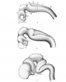
Fig. 30. Profile views of the brains of human embryos as seen during the third (A), fourth (B), and eighth (C) weeks
- Keibel Mall 2 031.jpg
- Keibel Mall 2 033.jpg
- Keibel Mall 2 034.jpg
- Keibel Mall 2 035.jpg
- Keibel Mall 2 036.jpg
- Keibel Mall 2 037.jpg
- Keibel Mall 2 038.jpg
- Keibel Mall 2 039.jpg
- Keibel Mall 2 040.jpg


| Historic Disclaimer - information about historic embryology pages |
|---|
 |
Glossary Links
- Glossary: A | B | C | D | E | F | G | H | I | J | K | L | M | N | O | P | Q | R | S | T | U | V | W | X | Y | Z | Numbers | Symbols | Term Link
Cite this page: Hill, M.A. (2024, June 1) Embryology Manual of Human Embryology II - Figures. Retrieved from https://embryology.med.unsw.edu.au/embryology/index.php/Manual_of_Human_Embryology_II_-_Figures
- © Dr Mark Hill 2024, UNSW Embryology ISBN: 978 0 7334 2609 4 - UNSW CRICOS Provider Code No. 00098G