Book - Manual of Human Embryology - Figures: Difference between revisions
mNo edit summary |
|||
| (47 intermediate revisions by the same user not shown) | |||
| Line 7: | Line 7: | ||
==I. The Germ-Cells== | ==I. The Germ-Cells== | ||
[[Book - Manual of Human Embryology|'''Manual of Human Embryology''']]''':''' [[Book - Manual of Human Embryology_1|The Germ Cells]] | [[Book - Manual of Human Embryology|'''Manual of Human Embryology''']]''':''' [[Book - Manual of Human Embryology_1|The Germ Cells]] | ||
<gallery> | <gallery> | ||
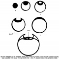
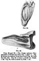
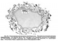
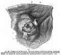
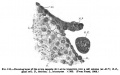
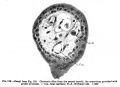
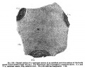
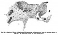
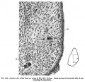
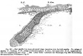
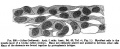
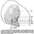
File:Keibel_Mall_001.jpg|1 | File:Keibel_Mall_001.jpg|Fig. 1. A Fresh Ovum from an Ovarian Follicle | ||
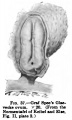
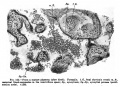
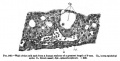
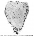
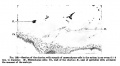
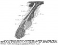
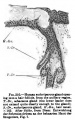
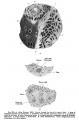
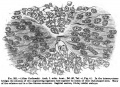
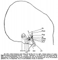
File:Keibel_Mall_002.jpg|2 | File:Keibel_Mall_002.jpg|Fig. 2. Almost Mature Human Ovum | ||
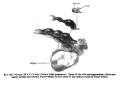
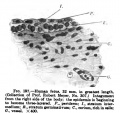
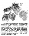
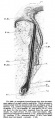
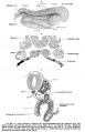
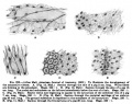
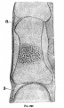
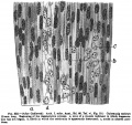
File:Keibel_Mall_003.jpg|3 | File:Keibel_Mall_003.jpg|Fig. 3. Four stages in the spermatogenesis of man | ||
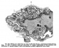
File:Keibel_Mall_004.jpg|4 | File:Keibel_Mall_004.jpg|Fig. 4. A Typical Human Spermium | ||
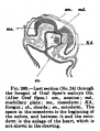
File:Keibel_Mall_005.jpg|5 | File:Keibel_Mall_005.jpg|Fig. 5. Diagram of a Human Spermium according to Meves | ||
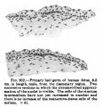
File:Keibel_Mall_006.jpg|6 | File:Keibel_Mall_006.jpg|Fig. 6. Abnormal Human Spermia | ||
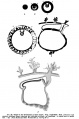
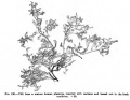
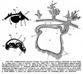
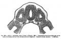
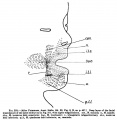
File:Keibel_Mall_007.jpg|7 | File:Keibel_Mall_007.jpg|Fig. 7. Diagram of the development of the primitive germ cells | ||
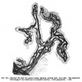
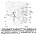
File:Keibel_Mall_008.jpg|8 | File:Keibel_Mall_008.jpg|Fig. 8. Diagram showing a comparison of the testis and the ovary | ||
</gallery> | </gallery> | ||
==III. Segmentation== | ==III. Segmentation== | ||
[[Book - Manual of Human Embryology|'''Manual of Human Embryology''']]''':''' [[Book - Manual of Human Embryology_3|Segmentation]] | [[Book - Manual of Human Embryology|'''Manual of Human Embryology''']]''':''' [[Book - Manual of Human Embryology_3|Segmentation]] | ||
<gallery> | <gallery> | ||
File:Keibel_Mall_009.jpg|9 | File:Keibel_Mall_009.jpg|Fig. 9. Ovum from a Monkey | ||
</gallery> | </gallery> | ||
==IV. Young Human Ova and Embryos up to the Formation of the First Primitive Segment== | ==IV. Young Human Ova and Embryos up to the Formation of the First Primitive Segment== | ||
[[Book - Manual of Human Embryology|'''Manual of Human Embryology''']]''':''' [[Book - Manual of Human Embryology_4|First Primitive Segment]] | [[Book - Manual of Human Embryology|'''Manual of Human Embryology''']]''':''' [[Book - Manual of Human Embryology_4|First Primitive Segment]] | ||
<gallery> | <gallery> | ||
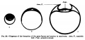
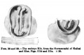
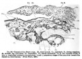
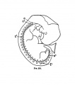
File:Keibel_Mall_010.jpg|10 | File:Keibel_Mall_010.jpg|Fig. 10. Diagram of the Ovum of Teacher and Bryce | ||
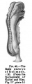
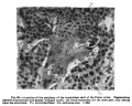
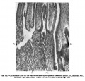
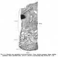
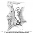
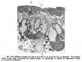
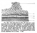
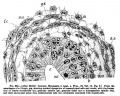
File:Keibel_Mall_011.jpg|11 | File:Keibel_Mall_011.jpg|Fig. 11. Section through the Basal Portion of the Ovum | ||
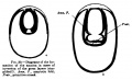
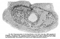
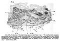
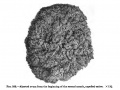
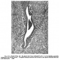
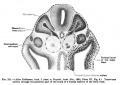
File:Keibel_Mall_012.jpg|12 | File:Keibel_Mall_012.jpg|Fig. 12. Figure of the Embryonic Shield of the Frassi Ovum | ||
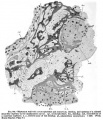
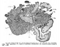
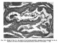
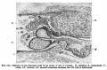
File:Keibel_Mall_013.jpg|13 | File:Keibel_Mall_013.jpg|Fig. 13. Section of the Frassi Ovum | ||
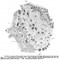
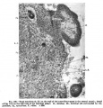
File:Keibel_Mall_014.jpg|14 | File:Keibel_Mall_014.jpg|Fig. 14. Section of the Frassi Ovum | ||
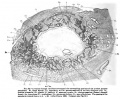
File:Keibel_Mall_014a.jpg|14a | File:Keibel_Mall_014a.jpg|Fig. 14a. Section of the Frassi Ovum | ||
File:Keibel_Mall_015.jpg|15 | File:Keibel_Mall_015.jpg|Fig. 15. Section of the Frassi Ovum | ||
File:Keibel_Mall_016.jpg|16 | File:Keibel_Mall_016.jpg|Fig. 16. Section of the Frassi Ovum | ||
File:Keibel_Mall_017.jpg|17 | File:Keibel_Mall_017.jpg|Fig. 17. Section of the Frassi Ovum | ||
File:Keibel_Mall_018.jpg|18 | File:Keibel_Mall_018.jpg|Fig. 18. Section of the Yolk Sack of the Frassi Ovum | ||
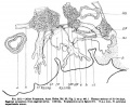
File:Keibel_Mall_019.jpg|19 | File:Keibel_Mall_019.jpg|Fig. 19. The Glaevecke Embryo of Graf Spee | ||
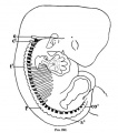
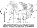
File:Keibel_Mall_020.jpg|20 | File:Keibel_Mall_020.jpg|Fig. 20. Median Sagittal Section of the Glaevecke Embryo of Graf Spee | ||
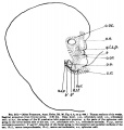
File:Keibel_Mall_021.jpg|21 | File:Keibel_Mall_021.jpg|Fig. 21. Three Views of the Glaevecke Embryo of Graf Spee | ||
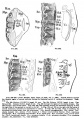
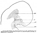
File:Keibel_Mall_022-026.jpg|22 | File:Keibel_Mall_022-026.jpg|Fig. 22. to Fig. 26. Section of the Embryo | ||
File:Keibel_Mall_022.jpg|22 | File:Keibel_Mall_022.jpg|Fig. 22. Section of the Embryo | ||
File:Keibel_Mall_022a.jpg| | File:Keibel_Mall_022a.jpg|Fig. 22. Section of the Embryo | ||
File:Keibel_Mall_023.jpg|23 | File:Keibel_Mall_023.jpg|Fig. 23. Section of the Embryo | ||
File:Keibel_Mall_024.jpg|24 | File:Keibel_Mall_024.jpg|Fig. 24. Section of the Embryo | ||
File:Keibel_Mall_025.jpg|25 | File:Keibel_Mall_025.jpg|Fig. 25. Section of the Embryo | ||
File:Keibel_Mall_026.jpg| | File:Keibel_Mall_026.jpg|Fig. 2. 6Section of the Embryo | ||
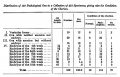
File:Keibel_Mall_table01.jpg|Table I | File:Keibel_Mall_table01.jpg|Table I | ||
File:Keibel_Mall_table02.jpg|Table II | File:Keibel_Mall_table02.jpg|Table II | ||
| Line 67: | Line 70: | ||
<gallery> | <gallery> | ||
File:Keibel_Mall_034.jpg|34 | File:Keibel_Mall_034.jpg|34 | ||
File:Keibel_Mall_table_034.jpg|His embryo table | |||
File:Keibel_Mall_035.jpg|35 | File:Keibel_Mall_035-036.jpg|35-36 | ||
File:Keibel_Mall_037.jpg|37 | File:Keibel_Mall_037.jpg|37 | ||
File:Keibel_Mall_038.jpg|38 | File:Keibel_Mall_038-039.jpg|38-39 | ||
File:Keibel_Mall_040.jpg|40 | File:Keibel_Mall_040.jpg|40 | ||
File:Keibel_Mall_041-042.jpg|41-42 | File:Keibel_Mall_041-042.jpg|41-42 | ||
| Line 88: | Line 89: | ||
File:Keibel_Mall_053.jpg|53 | File:Keibel_Mall_053.jpg|53 | ||
File:Keibel_Mall_054-056.jpg|54-56 | File:Keibel_Mall_054-056.jpg|54-56 | ||
File:Keibel_Mall_057.jpg | File:Keibel_Mall_057.jpg | ||
File:Keibel_Mall_058.jpg | File:Keibel_Mall_058.jpg | ||
File:Keibel_Mall_059-060.jpg|59-60 | File:Keibel_Mall_059-060.jpg|59-60 | ||
File:Keibel_Mall_061-062.jpg|61-62 Head | |||
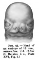
File:Keibel_Mall_063.jpg|63 Head | |||
File:Keibel_Mall_061-062.jpg|61-62 | File:Keibel_Mall_064.jpg|64 Head | ||
File:Keibel_Mall_065.jpg|65 Head | |||
File:Keibel_Mall_063.jpg|63 | |||
File:Keibel_Mall_064.jpg | |||
File:Keibel_Mall_065.jpg | |||
File:Keibel_Mall_066-071.jpg|66-71 | File:Keibel_Mall_066-071.jpg|66-71 | ||
File:Keibel_Mall_072.jpg|72 Head | |||
File:Keibel_Mall_073.jpg|73 Right Hand | |||
File:Keibel_Mall_074.jpg|74 | |||
File:Keibel_Mall_075-076.jpg|75-76 | |||
File:Keibel_Mall_077.jpg|77 Right Foot | |||
File:Keibel_Mall_078.jpg|78 Middle Finger | |||
File:Keibel_Mall_072.jpg | |||
File:Keibel_Mall_073.jpg | |||
File:Keibel_Mall_074.jpg | |||
File:Keibel_Mall_075.jpg | |||
File:Keibel_Mall_077.jpg | |||
File:Keibel_Mall_078.jpg | |||
</gallery> | </gallery> | ||
| Line 134: | Line 121: | ||
File:Keibel_Mall_099.jpg|99 | File:Keibel_Mall_099.jpg|99 | ||
File:Keibel_Mall_100.jpg|100 | File:Keibel_Mall_100.jpg|100 | ||
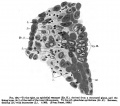
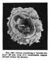
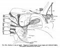
File:Keibel_Mall_101.jpg|101 | File:Keibel_Mall_101.jpg|101 Pregnancy of the first month | ||
File:Keibel_Mall_102.jpg|102 | File:Keibel_Mall_102.jpg|102 | ||
File:Keibel_Mall_103.jpg|103 | File:Keibel_Mall_103.jpg|103 | ||
| Line 140: | Line 127: | ||
File:Keibel_Mall_105.jpg|105 | File:Keibel_Mall_105.jpg|105 | ||
File:Keibel_Mall_106.jpg|106 | File:Keibel_Mall_106.jpg|106 | ||
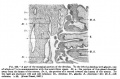
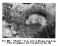
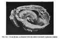
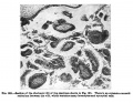
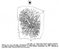
File:Keibel_Mall_107.jpg|107 | File:Keibel_Mall_107.jpg|107 Section through the chorion of an aborted ovum of one month | ||
File:Keibel_Mall_108.jpg|108 | File:Keibel_Mall_108.jpg|108 Section through the chorion of an aborted ovum of one month | ||
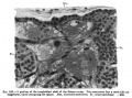
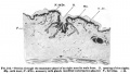
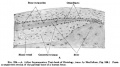
File:Keibel_Mall_109.jpg|109 | File:Keibel_Mall_109.jpg|109 Basal Ectoderm on the wall of the intervillous space in the second month | ||
File:Keibel_Mall_110.jpg|110 | File:Keibel_Mall_110.jpg|110 | ||
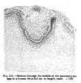
File:Keibel_Mall_111.jpg|111 | File:Keibel_Mall_111.jpg|111 | ||
| Line 179: | Line 166: | ||
[[Book - Manual of Human Embryology|'''Manual of Human Embryology''']]''':''' [[Book - Manual of Human Embryology_8|Human Embryo and Fetus Age]] | [[Book - Manual of Human Embryology|'''Manual of Human Embryology''']]''':''' [[Book - Manual of Human Embryology_8|Human Embryo and Fetus Age]] | ||
<gallery> | <gallery> | ||
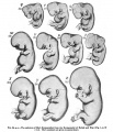
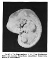
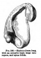
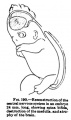
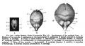
File:Keibel_Mall_141.jpg|141 | File:Keibel_Mall_141.jpg|Fig. 141 Human Embryo No. 163 | ||
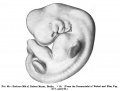
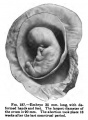
File:Keibel_Mall_142.jpg|142 | File:Keibel_Mall_142.jpg|Fig. 142 Human Embryo No. 144 | ||
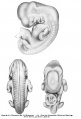
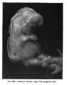
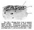
File:Keibel_Mall_143.jpg|143 | File:Keibel_Mall_143.jpg|Fig. 143 Human Embryo No. 22 | ||
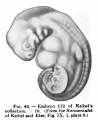
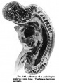
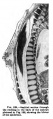
File:Keibel_Mall_144.jpg|144 | File:Keibel_Mall_144.jpg|Fig. 144 Human Embryo No. 131 | ||
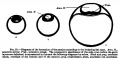
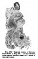
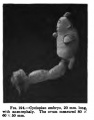
File:Keibel_Mall_145.jpg|145 | File:Keibel_Mall_145.jpg|Fig. 145 Human Embryo Length | ||
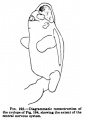
File:Keibel_Mall_146.jpg| | File:Keibel_Mall_146.jpg|Fig. 145 Human Embryo and Fetal Length | ||
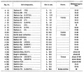
File:Keibel_Mall_Table-embryo_and_fetal_age.jpg|Table - embryo and fetal ages | File:Keibel_Mall_Table-embryo_and_fetal_age.jpg|Table - embryo and fetal ages | ||
File:Keibel_Mall_147.jpg|147 | File:Keibel_Mall_147.jpg|Fig. 147 Human Embryo Sizes | ||
File:Keibel_Mall_148.jpg|148 | File:Keibel_Mall_148.jpg|Fig. 148 Human Embryo and Fetal Size | ||
File:Keibel Mall Table-embryo length.jpg|Table - embryo length | File:Keibel Mall Table-embryo length.jpg|Table - embryo length | ||
</gallery> | </gallery> | ||
| Line 286: | Line 273: | ||
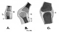
File:Keibel_Mall_224B.jpg|224B | File:Keibel_Mall_224B.jpg|224B | ||
File:Keibel_Mall_224C.jpg|224C | File:Keibel_Mall_224C.jpg|224C | ||
File: | File:Keibel_Mall_225A.jpg|225A | ||
File: | File:Keibel_Mall_225B.jpg|225B | ||
File: | File:Keibel Mall 226-228.jpg|226-228 | ||
Keibel Mall 227.jpg|227 | |||
Keibel Mall 228.jpg|228 | |||
</gallery> | |||
===Axial Skeleton=== | |||
<gallery> | |||
Keibel Mall 229-230.jpg|229-230 | |||
Keibel Mall 231-232.jpg|231-232 | |||
Keibel Mall 231.jpg|231 | |||
Keibel Mall 232.jpg|232 | |||
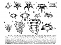
Keibel Mall 233-236.jpg|233-236 | |||
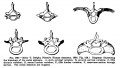
File:Keibel_Mall_237-239.jpg|237-239 | |||
File:Keibel_Mall_237-239-no-legend.jpg|237-239-no-legend | |||
Keibel Mall 237.jpg|237 | |||
Keibel Mall 238.jpg|238 | |||
Keibel Mall 239.jpg|239 | |||
Keibel Mall 240-247.jpg|240-247 | |||
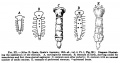
File:Keibel_Mall_248-259.jpg|248-259 | |||
File:Keibel_Mall_248-259-no-legend.jpg|248-259-no-legend | |||
Keibel Mall 248-259.jpg|248-259 | |||
Keibel Mall 248-250.jpg|248-250 | |||
Keibel Mall 251-253.jpg|251-253 | |||
Keibel Mall 254-256.jpg|254-256 | |||
Keibel Mall 257-259.jpg|257-259 | |||
File:Keibel_Mall_260-263.jpg|260-263 | |||
File:Keibel_Mall_260.jpg|260 | |||
File:Keibel_Mall_261.jpg|261 | |||
File:Keibel_Mall_262.jpg|262 | |||
File:Keibel_Mall_263.jpg|263 | |||
File:Keibel_Mall_264-265.jpg|264-265 | |||
File:Keibel_Mall_264.jpg|264 | |||
File:Keibel_Mall_265.jpg|265 | |||
File:Keibel_Mall_266.jpg|266 | |||
File:Keibel_Mall_267.jpg|267 | |||
Keibel Mall 268.jpg|268 | |||
Keibel Mall 269.jpg|269 | |||
Keibel Mall 270.jpg|270 | |||
Keibel Mall 271.jpg|271 | |||
Keibel Mall 272.jpg|272 | |||
File:Keibel Mall 272-no-legend.jpg|272-no-legend | |||
Keibel Mall 273.jpg|273 | |||
File:Keibel Mall 273-no-legend.jpg|273-no-legend | |||
</gallery> | </gallery> | ||
===Limb Skeleton=== | |||
{{KM Skeleton}} | |||
<gallery> | <gallery> | ||
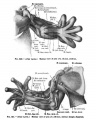
File:Keibel Mall 274-278.jpg|274-278 | File:Keibel Mall 274-278.jpg|274-278 | ||
File:Keibel Mall 279-284.jpg|279-284 | File:Keibel Mall 279-284.jpg|279-284 | ||
File:Keibel_Mall_285.jpg|285 | File:Keibel_Mall_285-288.jpg|285-288 | ||
File:Keibel_Mall_289.jpg|289 | File:Keibel_Mall_289.jpg|289 | ||
File:Keibel_Mall_290.jpg|290 | File:Keibel_Mall_290.jpg|290 | ||
| Line 305: | Line 331: | ||
File:Keibel_Mall_292.jpg|292 | File:Keibel_Mall_292.jpg|292 | ||
File:Keibel_Mall_293.jpg|293 | File:Keibel_Mall_293.jpg|293 | ||
File:Keibel_Mall_Table-Skeleton_1.jpg| | File:Keibel_Mall_Table-Skeleton_1.jpg|Bone Ossification Inferior Extremity | ||
File:Keibel_Mall_Table-Skeleton_2.jpg| | File:Keibel_Mall_Table-Skeleton_2.jpg|Bone Ossification Inferior Extremity | ||
File:Keibel_Mall_294-297.jpg|294-297 | File:Keibel_Mall_294-297.jpg|294-297 | ||
File:Keibel_Mall_294.jpg|294 | File:Keibel_Mall_294.jpg|294 | ||
| Line 321: | Line 347: | ||
File:Keibel Mall 305-306.jpg|305-306 | File:Keibel Mall 305-306.jpg|305-306 | ||
File:Keibel_Mall_307.jpg|307 | File:Keibel_Mall_307.jpg|307 | ||
File:Keibel_Mall_Table-Skeleton_4.jpg|Table- | File:Keibel_Mall_Table-Skeleton_4.jpg|Table- Bone Ossification Superior Extremity | ||
File:Keibel_Mall_Table-Skeleton_5.jpg|Table- | File:Keibel_Mall_Table-Skeleton_5.jpg|Table- Bone Ossification Superior Extremity | ||
</gallery> | </gallery> | ||
| Line 355: | Line 381: | ||
File:Keibel_Mall_329.jpg|329 | File:Keibel_Mall_329.jpg|329 | ||
File:Keibel_Mall_330.jpg|330 | File:Keibel_Mall_330.jpg|330 | ||
File:Keibel_Mall_331.jpg|331 | |||
File:Keibel_Mall_332.jpg|332 | |||
File:Keibel_Mall_333.jpg|333 | |||
File:Keibel_Mall_334.jpg|334 | |||
File:Keibel_Mall_335.jpg|335 | |||
File:Keibel_Mall_336.jpg|336 | |||
File:Keibel_Mall_337.jpg|337 | |||
File:Keibel_Mall_338.jpg|338 | |||
File:Keibel_Mall_339.jpg|339 | |||
File:Keibel_Mall_340-345.jpg|340-345 | |||
File:Keibel_Mall_346.jpg|346 | |||
File:Keibel_Mall_347.jpg|347 | |||
File:Keibel_Mall_348.jpg|348 | |||
File:Keibel_Mall_349.jpg|349 | |||
File:Keibel_Mall_350-351.jpg|350-351 | |||
File:Keibel_Mall_352.jpg|352 | |||
File:Keibel_Mall_353.jpg|353 | |||
File:Keibel_Mall_354.jpg|354 | |||
File:Keibel_Mall_355.jpg|355 | |||
File:Keibel_Mall_356.jpg|356 | |||
File:Keibel_Mall_357.jpg|357 | |||
File:Keibel_Mall_358.jpg|358 | |||
File:Keibel_Mall_359.jpg|359 | |||
File:Keibel_Mall_360.jpg|360 | |||
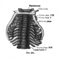
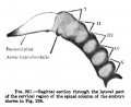
File:Keibel_Mall_361.jpg|361 | |||
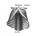
File:Keibel_Mall_362.jpg|362 | |||
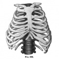
File:Keibel_Mall_363.jpg|363 | |||
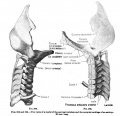
File:Keibel_Mall_364.jpg|364 | |||
File:Keibel_Mall_365.jpg|365 | |||
File:Keibel_Mall_366.jpg|366 | |||
File:Keibel_Mall_367.jpg|367 | |||
File:Keibel_Mall_368.jpg|368 | |||
File:Keibel_Mall_369.jpg|369 | |||
File:Keibel_Mall_370.jpg|370 | |||
File:Keibel_Mall_371.jpg|371 | |||
File:Keibel_Mall_372.jpg|372 | |||
File:Keibel_Mall_373.jpg|373 | |||
File:Keibel_Mall_374.jpg|374 | |||
File:Keibel_Mall_375.jpg|375 | |||
File:Keibel_Mall_376.jpg|376 | |||
File:Keibel_Mall_377.jpg|377 | |||
File:Keibel_Mall_378.jpg|378 | |||
File:Keibel_Mall_379.jpg|379 | |||
</gallery> | </gallery> | ||
| Line 360: | Line 429: | ||
[[Book - Manual of Human Embryology|'''Manual of Human Embryology''']]''':''' [[Book - Manual of Human Embryology_13|Coelom and Diaphragm]] | [[Book - Manual of Human Embryology|'''Manual of Human Embryology''']]''':''' [[Book - Manual of Human Embryology_13|Coelom and Diaphragm]] | ||
<gallery> | <gallery> | ||
File:Keibel_Mall_380.jpg | File:Keibel_Mall_380.jpg|380 | ||
File:Keibel_Mall_381.jpg|381 | |||
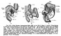
File:Keibel_Mall_382-386.jpg|382-386 | |||
File:Keibel_Mall_387.jpg|387 | |||
File:Keibel_Mall_388-392.jpg|388-392 | |||
File:Keibel_Mall_393-394.jpg|393-394 | |||
File:Keibel_Mall_395.jpg|395 | |||
File:Keibel_Mall_396-397.jpg|396-397 | |||
File:Keibel_Mall_396.jpg|396 | |||
File:Keibel_Mall_397.jpg|397 | |||
File:Keibel_Mall_398.jpg|398 | |||
File:Keibel_Mall_399.jpg|399 | |||
File:Keibel_Mall_400.jpg|400 | |||
File:Keibel_Mall_401.jpg|401 | |||
File:Keibel_Mall_402.jpg|402 | |||
File:Keibel_Mall_403-406.jpg|403-406 | |||
File:Keibel_Mall_403.jpg|403 | |||
File:Keibel_Mall_404.jpg|404 | |||
File:Keibel_Mall_405.jpg|405 | |||
File:Keibel_Mall_406.jpg|406 | |||
File:Keibel_Mall_407.jpg|407 | |||
File:Keibel_Mall_408-409.jpg|408-409 | |||
File:Keibel_Mall_408.jpg|408 | |||
File:Keibel_Mall_409.jpg|409 | |||
File:Keibel_Mall_410-411.jpg|410-411 | |||
File:Keibel_Mall_410.jpg|410 | |||
File:Keibel_Mall_411.jpg|411 | |||
File:Keibel_Mall_412-413.jpg|412-413 | |||
File:Keibel_Mall_412.jpg|412 | |||
File:Keibel_Mall_413.jpg|413 | |||
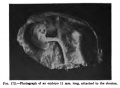
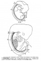
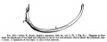
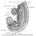
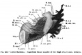
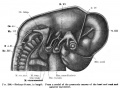
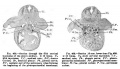
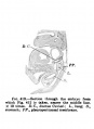
File:Keibel_Mall_414.jpg|Fig. 414. Embryo 11 mm long | |||
File:Keibel_Mall_415-416.jpg|415-416 | |||
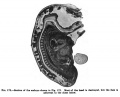
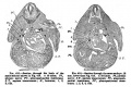
File:Keibel_Mall_415.jpg|Fig. 415. Section through the body of the same embryo shown in Fig 414 | |||
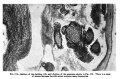
File:Keibel_Mall_416.jpg|Fig. 416. Section through the same embryo 0.18 mm lower than Fig 415 | |||
File:Keibel_Mall_417.jpg|Fig. 417. Section 0.46 mm lower than Fig. 415 | |||
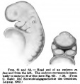
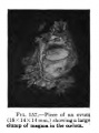
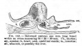
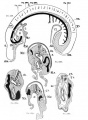
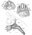
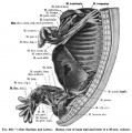
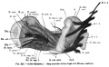
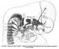
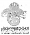
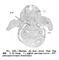
File:Keibel_Mall_418.jpg|Fig. 418. Sagittal section through an embryo 14 mm long | |||
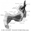
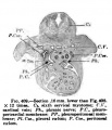
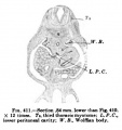
File:Keibel_Mall_419.jpg|Fig. 419. Sagittal section through an embryo 16 mm long | |||
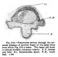
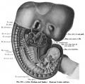
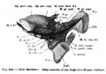
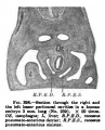
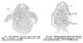
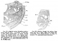
File:Keibel_Mall_420.jpg|Fig. 420. Transverse section through an embryo 19 mm long | |||
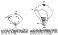
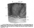
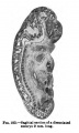
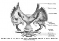
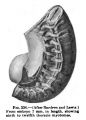
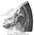
File:Keibel_Mall_421.jpg|Fig. 421. Cast of the lesser peritoneal cavity encircling the intestine, from a human embryo 5 mm long | |||
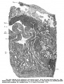
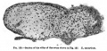
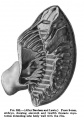
File:Keibel_Mall_422.jpg|Fig. 422. Cast of the lesser peritoneal cavity from an embryo 11.5 mm long | |||
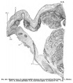
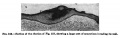
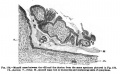
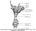
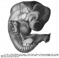
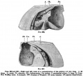
File:Keibel_Mall_423.jpg|Fig. 423. Right lung of an adult man showing the bursa infracardia | |||
</gallery> | </gallery> | ||
Latest revision as of 21:35, 14 November 2013
| Historic Disclaimer - information about historic embryology pages |
|---|
 |
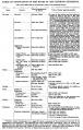
Figures
I. The Germ-Cells
Manual of Human Embryology: The Germ Cells
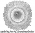
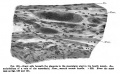
Fig. 1. A Fresh Ovum from an Ovarian Follicle
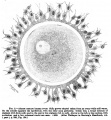
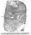
Fig. 2. Almost Mature Human Ovum
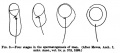
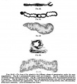
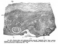
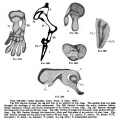
Fig. 3. Four stages in the spermatogenesis of man
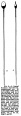
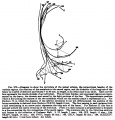
Fig. 4. A Typical Human Spermium
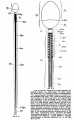
Fig. 5. Diagram of a Human Spermium according to Meves
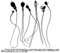
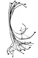
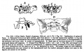
Fig. 6. Abnormal Human Spermia
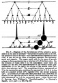
Fig. 7. Diagram of the development of the primitive germ cells
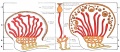
Fig. 8. Diagram showing a comparison of the testis and the ovary


III. Segmentation
Manual of Human Embryology: Segmentation
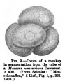
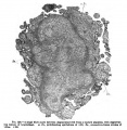
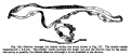
Fig. 9. Ovum from a Monkey

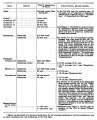
IV. Young Human Ova and Embryos up to the Formation of the First Primitive Segment
Manual of Human Embryology: First Primitive Segment
Fig. 10. Diagram of the Ovum of Teacher and Bryce
Fig. 11. Section through the Basal Portion of the Ovum
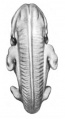
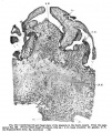
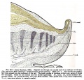
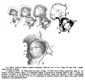
Fig. 12. Figure of the Embryonic Shield of the Frassi Ovum
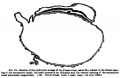
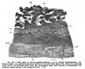
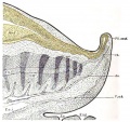
Fig. 13. Section of the Frassi Ovum
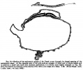
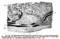
Fig. 14. Section of the Frassi Ovum
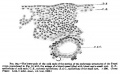
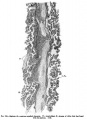
Fig. 14a. Section of the Frassi Ovum
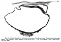
Fig. 15. Section of the Frassi Ovum
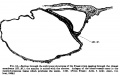
Fig. 16. Section of the Frassi Ovum
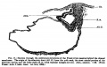
Fig. 17. Section of the Frassi Ovum
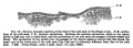
Fig. 18. Section of the Yolk Sack of the Frassi Ovum
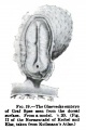
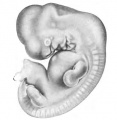
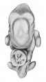
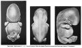
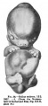
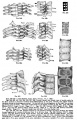
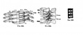
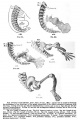
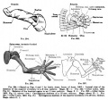
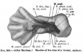
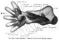
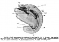
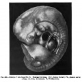
Fig. 19. The Glaevecke Embryo of Graf Spee
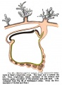
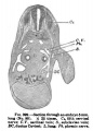
Fig. 20. Median Sagittal Section of the Glaevecke Embryo of Graf Spee
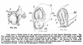
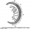
Fig. 21. Three Views of the Glaevecke Embryo of Graf Spee
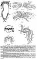
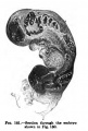
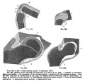
Fig. 22. to Fig. 26. Section of the Embryo
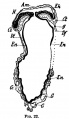
Fig. 22. Section of the Embryo
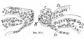
Fig. 22. Section of the Embryo
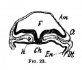
Fig. 23. Section of the Embryo
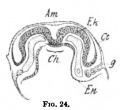
Fig. 24. Section of the Embryo
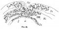
Fig. 25. Section of the Embryo
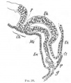
Fig. 2. 6Section of the Embryo
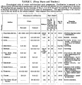
Table I
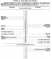
Table II


V. The Formation of the Germ Layers and the Gastrulation Problem
Manual of Human Embryology: Gastrulation
27
28
29
30
31
32
33


VI. Summary of the Development of the Human Embryo and the Differentiation of its External Form
Manual of Human Embryology: External Form
34
His embryo table
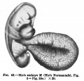
35-36
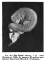
37
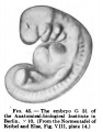
38-39
40
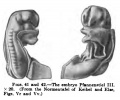
41-42
43
44
45
46
47
48
49-51
49
50
51
52
53
54-56
59-60
61-62 Head
63 Head
64 Head
65 Head
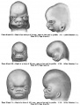
66-71
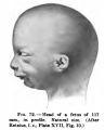
72 Head
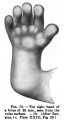
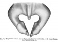
73 Right Hand
74
75-76

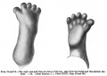
77 Right Foot
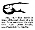
78 Middle Finger


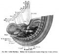
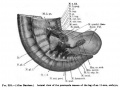
VII. The Development of the Egg Membranes and the Placenta - Menstruation
Manual of Human Embryology: Placenta
79-82
83-87
88-91
92
Embryo Table
93
94
95
96
97
98
99
100
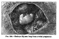
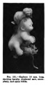
101 Pregnancy of the first month
102
103
104
105
106

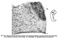
107 Section through the chorion of an aborted ovum of one month
108 Section through the chorion of an aborted ovum of one month
109 Basal Ectoderm on the wall of the intervillous space in the second month
110
111
112
113
114

115
116
117
118
119
120
121
122
123
124
125
126
127
128
129
130
131
132
133
134
135
136
137
138
139
140


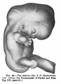
VIII. Determination of the Age of Human Embryos and Fetuses
Manual of Human Embryology: Human Embryo and Fetus Age
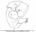
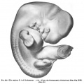
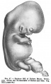
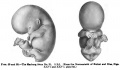
Fig. 141 Human Embryo No. 163
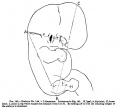
Fig. 142 Human Embryo No. 144

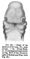
Fig. 143 Human Embryo No. 22
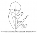
Fig. 144 Human Embryo No. 131
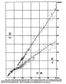
Fig. 145 Human Embryo Length

Fig. 145 Human Embryo and Fetal Length
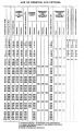
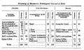
Table - embryo and fetal ages
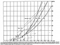
Fig. 147 Human Embryo Sizes
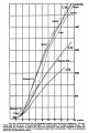
Fig. 148 Human Embryo and Fetal Size
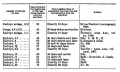
Table - embryo length


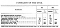
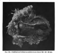
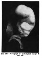
IX. The Pathology of the Human Ovum
Manual of Human Embryology: Ovum Pathology
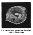
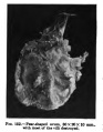
150
151
152
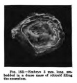
153
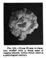
154
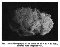
155
156
157
158
159
160
161
162
163
164
165
166
167
168
169
170
171
172
173
174
175
176
177
178
179
180
181
- Keibel Mall 182.jpg
182
183
184
185
186
187
188
189
190
191
192
193
194
195
194-195


X. The Development of the Integument
Manual of Human Embryology: Integument
196
197
198
199
200
201
202
203-205
206
207
208
209
210
211
212
213
214
215
216
217
218


XI. Development of the Skeleton and of the Connective Tissues
- Skeleton and Connective Tissues: Connective Tissue Histogenesis | Skeletal Morphogenesis | Chorda Dorsalis | Vertebral Column and Thorax | Limb Skeleton | Skull Hyoid Bone Larynx
219
220
221
222
223
224
224A
224B
224C
225A
225B
226-228
227
228


Axial Skeleton
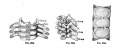
229-230
231-232
231
232
233-236

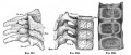
237-239
237-239-no-legend
237
238
239
240-247
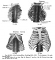
248-259
248-259-no-legend
248-259
248-250
251-253
254-256
257-259
260-263
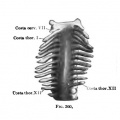
260
261
262
263
264-265
264
265
266
267
268
269
270
271
272
272-no-legend
273
273-no-legend


Limb Skeleton
- Limb Images: 274-278 Spinal Column and Lower Limb | 279-284 Lower Limb | 285-288 Knee | 289 Os Coxae | 290 Femur | 291 Tibia | 292 Fibula | 293 Foot | 294 | 295 | 296 | 297 | 298-299 | 300 Forearm and Hand | 301 Upper Limb Joints | 302 Clavicle | Upper Limb Ossification 1 | Upper Limb Ossification 2 | Bone Development Timeline
- Skeleton and Connective Tissues: Connective Tissue Histogenesis | Skeletal Morphogenesis | Chorda Dorsalis | Vertebral Column and Thorax | Limb Skeleton | Skull Hyoid Bone Larynx
274-278
279-284
285-288
289
290
291
292
293
Bone Ossification Inferior Extremity
Bone Ossification Inferior Extremity
294-297
294
295
296
297
298-299
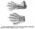
300
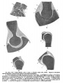
301
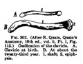
302
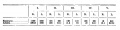
Table-Skeleton
303
304
305-306
307
Table- Bone Ossification Superior Extremity
Table- Bone Ossification Superior Extremity


Skull
308
309
310
311
312
313
314
315
316
317
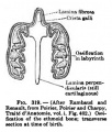
318
319
320
321
322
323
324


XII. The Development of the Muscular System
Manual of Human Embryology: Muscular System
325
326
327
328
329
330
331
332
333
334
335
336
337
338
339
340-345
346
347
348
349
350-351
352
353
354
355
356
357
358
359
360
361
362
363
364

365
366

367
368

369
370
371
372
373
374
375
376
377
378
379


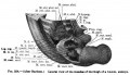
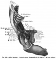
XIII. Coelom and Diaphragm
Manual of Human Embryology: Coelom and Diaphragm
380
381
382-386
387
388-392
393-394
395
396-397
396
397
398
399
400
- Keibel Mall 401.jpg
401
402
403-406
- Keibel Mall 403.jpg
403
- Keibel Mall 404.jpg
404
- Keibel Mall 405.jpg
405
- Keibel Mall 406.jpg
406
- Keibel Mall 407.jpg
407
408-409
408
409
410-411
410
411
412-413
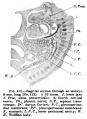
412
413
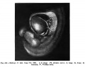
Fig. 414. Embryo 11 mm long
415-416
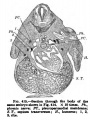
Fig. 415. Section through the body of the same embryo shown in Fig 414
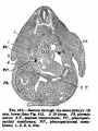
Fig. 416. Section through the same embryo 0.18 mm lower than Fig 415
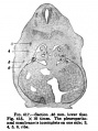
Fig. 417. Section 0.46 mm lower than Fig. 415
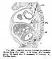
Fig. 418. Sagittal section through an embryo 14 mm long
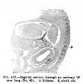
Fig. 419. Sagittal section through an embryo 16 mm long
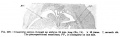
Fig. 420. Transverse section through an embryo 19 mm long
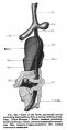
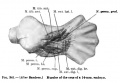
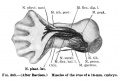
Fig. 421. Cast of the lesser peritoneal cavity encircling the intestine, from a human embryo 5 mm long
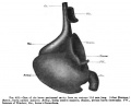
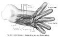
Fig. 422. Cast of the lesser peritoneal cavity from an embryo 11.5 mm long
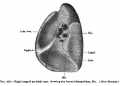
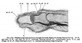
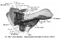
Fig. 423. Right lung of an adult man showing the bursa infracardia


| Historic Disclaimer - information about historic embryology pages |
|---|
|
Glossary Links
- Glossary: A | B | C | D | E | F | G | H | I | J | K | L | M | N | O | P | Q | R | S | T | U | V | W | X | Y | Z | Numbers | Symbols | Term Link
Cite this page: Hill, M.A. (2024, June 17) Embryology Book - Manual of Human Embryology - Figures. Retrieved from https://embryology.med.unsw.edu.au/embryology/index.php/Book_-_Manual_of_Human_Embryology_-_Figures
- © Dr Mark Hill 2024, UNSW Embryology ISBN: 978 0 7334 2609 4 - UNSW CRICOS Provider Code No. 00098G