Integumentary System - Tooth Development: Difference between revisions
| Line 20: | Line 20: | ||
|} | |} | ||
[[Talk:Integumentary_System_-_Tooth_Development|Recent References]] | [[#References|References]] | |||
== Textbooks == | == Textbooks == | ||
Revision as of 09:56, 15 January 2013
Introduction

The tooth is an extrordinary integumentary system specialization providing insights into epitheilal/mesenchymal (ectoderm of the first pharyngeal arch and neural crest, ectomesenchymal cells) interactions in development and develops with a major contribution from the neural crest. (More? Neural Crest Development)
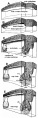
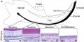
There are 4 morphological stages describing the early tooth development: bud, cap, bell, and terminal differentiation.
Some Recent Findings
|
Recent References | References
Textbooks
- Human Embryology (2nd ed.) Larson Chapter 14 p443-455
- The Developing Human: Clinically Oriented Embryology (6th ed.) Moore and Persaud Chapter 20: P513-529
- Before We Are Born (5th ed.) Moore and Persaud Chapter 21: P481-496
- Essentials of Human Embryology Larson Chapter 14: P303-315
- Human Embryology, Fitzgerald and Fitzgerald
- Color Atlas of Clinical Embryology Moore Persaud and Shiota Chapter 15: p231-236
Development Overview
- ectoderm, mesoderm and neural crest ectomesenchyme contribute
- inductive influence of neural crest with overlying ectoderm
Odontoblasts
- neural crest-derived mesenchymal cells
- differentiate under the influence of the enamel epithelium
- form predentin
- calcifies to form dentin
Ameloblasts
- produce enamal
- tooth growth occurs in ossifying jaws
- periodontal ligament holds tooth in bone socket
Tooth Stages

| Stage | Human (weeks) | Mouse (days) | |
| lamina | 
|
Week 6 | E11 |
| placode | 
|
Week 7 | E11.5 |
| bud | 
|
Week 8 | E12.5 |
| cap | 
|
Week 11 | E14.5 |
| bell | 
|
Week 14 | E15.5 |
Images: all stages | lamina | placode stage | bud stage | cap stage | bell stage
Human 2 Sets of Teeth

Deciduous Teeth
- 20 deciduous teeth
- Differential rates of growth, shed at different times over 20 year period

Permanent Teeth

- 32 permanent teeth
- Incisors - sharp cutting edge, adapted for biting the food.
- Canines - are larger and stronger than the incisors. The upper canines have also been called the "eye teeth", while the lower canines "stomach teeth".
- Premolars - or Bicuspid teeth are smaller and shorter than the canines.
- Molars - are the largest teeth adapted for grinding and pounding food.

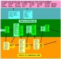
Epithelial Mesenchymal Interaction
local ectodermal thickening expresses several signaling molecules these in turn signal to the underlying mesenchyme triggering mesenchymal condensation (epithelially expressed Bmp4 induces Msx1 and Lef1 as well as itself in the underlying mesenchyme)
Four epithelial signaling molecules, Bmp2, Shh, Wnt10a, and Wnt10b, in the early inductive cascade, each signal has a distinct molecular action on the jaw mesenchyme.
Mouse (E11.0 and E12.0) - all four genes are specifically expressed in the epithelium.
Shh and Wnt10b induce general Hedgehog and Wnt targets, Ptc and Gli for Shh and Lef1 for Wnt10b,
Bmp2 is able to induce tooth-specific expression of Msx1.
(Text above modified from: Hélène R. Dassule and Andrew P. McMahon Developmental Biology, v 202, n 2, October 15, 1998, p215-227)
(More? Developmental Mechanism - Epithelial Mesenchymal Interaction)
Periodontal Ligament
The tooth is not anchored directly onto its bony socket (alveolar bone) but held in place by the periodontal ligament (PDL), a specialized connective tissue structure that surrounds the tooth root coating of cementum.
The additional roles of the PDL are to also act as; a shock absorber, transmitter of chewing forces (from tooth to bone), sensory information (heat, cold, pressure and pain).
The collagen fiber bundles within the ligament are called "Sharpey’s fibres".
Cementum (from investing layer of the dental follicle) is contiguous layer with the periodontal ligament on one surface and firmly adherent to dentine on the other surface.
Molecular Tooth Development

More than 300 genes have been associated with tooth development including: BMP4, FGF8, MSX1, MSX2, PAX9, PITX2, SHOX2, Delta/Notch, Hox-8, Runx2
Most recent review in Developmental Dynamics by Lin D, Huang Y, He F, Gu S, Zhang G, Chen Y, Zhang Y. Expression survey of genes critical for tooth development in the human embryonic tooth germ. Dev Dyn. 2007 Mar 29.
Amelogenin - abundant protein secreted by ameloblasts which is a major component of tooth enamel.
The papers below are from UNSW Embryology (version 3), information requires updating.
Bone Morphogenic Protein (BMP) / Fibroblast Growth Factor (FGF)
Growth factors in the BMP- and FGF-families are expressed in dental epithelium during initiation of tooth development and their effects on the underlying mesenchyme mimic those of the epithelium. They upregulate the expression of many genes, including the homeobox-containing Msx-1 and Msx-2, and stimulate cell proliferation suggesting that they may act as epithelial signals transmitting epithelial-mesenchymal interactions. During subsequent morphogenesis, when the characteristic shapes of individual teeth develop as a result from folding of the dental epithelium, several signal molecules including Sonic hedgehog, Bmps-2, 4, 7 and Fgf-4 are expressed specifically in restricted and transient epithelial cell clusters, called enamel knots.
(Text: Irma Thesleff and Carin Sahlberg Seminars in Cell & Developmental Biology, v 7, n 2, April, 1996, p185-193)
Delta/Notch
The expression pattern of Delta 1 in ameloblasts and odontoblasts is complementary to Notch1, Notch2, and Notch3 expression in adjacent epithelial and mesenchymal cells. Notch1 and Notch2 are upregulated in explants of dental mesenchyme adjacent to implanted cells expressing Delta1, suggesting that feedback regulation by Delta-Notch signaling ensures the spatial segregation of Notch receptors and ligands. TGF1 and BMPs induce Delta1 expression in dental mesenchyme explants at the stage at which Delta1 is upregulated in vivo, but not at earlier stages. In contrast to the Notch family receptors and their ligand Jagged1, expression of Delta1 in the tooth germ is not affected by epithelial-mesenchymal interactions, showing that the Notch receptors and their two ligands Jagged1 and Delta1 are subject to different regulations.
Text: Mitsiadis etal Developmental Biology,v 204, n 2, December 15, 1998, p420-431
Abnormalities

Adontia
A total lack of tooth development.
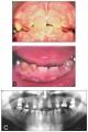
Dentinogenesis imperfecta
The teeth are translucent and often roughened with severe amber discolouration. Discoloured teeth with an opalescent sheen, dentin does not support enamel (dentin sialophosphoprotein mutation)
Dentine dysplasia
The primary teeth are translucent and amber in colour whereas the erupting secondary central incisors are of normal appearance.
Amelogenesis Imperfecta
Abnormal tooth enamel formation (AMELX, ENAM, KLK4, MMP20).
Dens Evaginatus
Dental anomaly mainly affecting premolars in people of Mongolian origin.
Hypodontia
Lack of development of one or more teeth.
Hypohidrotic Ectodermal Dysplasia
Maldevelopment of one or more ectodermal-derived tissues.
Microdontia
Small teeth.
References
Journals
Reviews
<pubmed>19875280</pubmed> <pubmed>17209531</pubmed> <pubmed>16838332</pubmed> <pubmed>16753804</pubmed> <pubmed>12615136</pubmed> <pubmed>12640730</pubmed>
Articles
<pubmed>17394220</pubmed> <pubmed>16632755</pubmed> <pubmed>16651263</pubmed> <pubmed>9520113</pubmed>
Search PubMed
Search Pubmed: Tooth Development | odontogenesis | tooth morphogenesis | adontia | amelogenesis imperfecta | dens evaginatus | hypodontia
Additional Images
Tooth development stage
Tooth molecular development
Inherited dentine disorders
Mouse - lower incisor teeth
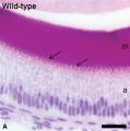
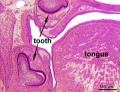
Mouse - tooth histology
Rat - neonatal teeth


Terms
External Links
External Links Notice - The dynamic nature of the internet may mean that some of these listed links may no longer function. If the link no longer works search the web with the link text or name. Links to any external commercial sites are provided for information purposes only and should never be considered an endorsement. UNSW Embryology is provided as an educational resource with no clinical information or commercial affiliation.
- StemBook - Tooth organogenesis and regeneration
- University of Helsinki Gene Expression in Tooth
- American Dental Association Overview - Tooth
- Columbia University Medical Centre Illustrations: How a Tooth Decays
- Merck Tooth disorders
- Nemours Foundation Teething Tots
Glossary Links
- Glossary: A | B | C | D | E | F | G | H | I | J | K | L | M | N | O | P | Q | R | S | T | U | V | W | X | Y | Z | Numbers | Symbols | Term Link
Cite this page: Hill, M.A. (2024, June 17) Embryology Integumentary System - Tooth Development. Retrieved from https://embryology.med.unsw.edu.au/embryology/index.php/Integumentary_System_-_Tooth_Development
- © Dr Mark Hill 2024, UNSW Embryology ISBN: 978 0 7334 2609 4 - UNSW CRICOS Provider Code No. 00098G