User:Z3418698
Welcome to the 2014 Embryology Course!
- Links: Timetable | How to work online | One page Wiki Reference Card | Moodle
- Each week the individual assessment questions will be displayed in the practical class pages and also added here.
- Copy the assessment items to your own page and provide your answer.
- Note - Some guest assessments may require completion of a worksheet that will be handed in in class with your student name and ID.
| Individual Lab Assessment |
|---|
|
| Lab 12 - Stem Cell Presentation Assessment | More Info | |
|---|---|---|
| Group | Comment | Mark (10) |
| 1/8 |
|
7 |
| 2 |
|
7.5 |
| 3 |
|
7.5 |
| 4 |
|
8.5 |
| 5 |
|
8.5 |
| 6 |
|
8.5 |
| 7 |
|
7.5 |
Lab Attendance
Lab 1--Z3418698 (talk) 14:26, 12 August 2014 (EST)
Lab 2--Z3418698 (talk) 11:14, 13 August 2014 (EST)
Lab 4--Z3418698 (talk) 11:55, 27 August 2014 (EST)
Lab 5--Z3418698 (talk) 11:38, 3 September 2014 (EST)
Lab 6--Z3418698 (talk) 11:21, 10 September 2014 (EST)
Lab 7--Z3418698 (talk) 11:21, 17 September 2014 (EST)
Lab 9--Z3418698 (talk) 12:35, 8 October 2014 (EST)
Lab 10--Z3418698 (talk) 11:26, 15 October 2014 (EST)
Lab 11--Z3418698 (talk) 12:39, 22 October 2014 (EST)
Online Assessments
Lab 1
Article 1
<pubmed>25100710</pubmed>
Vascular dysfunctions including high blood pressure and vascular remodeling seem to have a higher appearance in children who have been conceived by in-vitro fertilisation (IVF). Numerous tests have shown that IVF children between the ages of 3-13 have higher blood pressures of considerable statistical significance. The research paper aims to investigate any particular trends in the expression of a number of protein compounds in trying to deduce or narrow down a causative agent.
The study focused on children aged 3-13 years with a sample of 704 IVF and an equal number of natural birth as well as ‘control’ children from a similar area in china. Umbilical vessels and cord blood was collected post-caesarean delivery and protein digestion samples were prepared in the lab for analysis. iTRAQ (isobaric tags for relative and absolute quantitation) was used as the primary method to tag a number of proteins which were then identified using the MASCOT search engine and Proteomics tools. Western Blotting Analysis was used to calculate relative ratios of the identified peptides in IVF, natural-birth and control samples and concentrations were compared.
Results showed the expression of forty-seven proteins in the IVF veins that are typically not present in children conceived by natural birth . Further analysis using the Ingenuity Pathway Analysis was then conducted to examine signaling pathways of the differentially expressed proteins revealing that these proteins played an important role in vascular development and carbon metabolism. A reduction in rate of carbon metabolism is linked to a subsequent decrease in energy supply hence also possibly directly influencing the vascular development. Results also showed a significantly higher concentration of serum estradiol (E2) in cord blood of IVF newborns compared to newborns. Previous studies have shown that a higher concentration of E2 induces alteration of lumican and vimentin mRNA expression in human umbilical vein endothelial cells which are involved in the process of vascular modification. The results of the investigation suggest that the abnormal expression of these proteins in umbilical veins may have a potentially causative relationship or at least, be a factor involved in cardiovascular dysfunction and increased blood pressure in IVF children.
Article 2
<pubmed>25100106</pubmed>
The luteal phase during the menstrual cycle has been suggested in a number of studies to significantly improve pregnancy rates in women undergoing IVF. As the risk of ovarian hyperstimulation syndrome is higher associated with the use of hCG itself, progesterone was used as the treatment choice for the experiment. This study focuses on comparing the efficacy and safety of two different forms administration of progesterone; aqueous subcutaneous progesterone and vaginal progesterone for luteal phase support of in vitro fertilization.
A randomized controlled trial was performed at eight different fertility centres with 800 female participants aged between 18-42 years undergoing IVF treatment. A serum pregnancy test was performed at 15 ± 2 days after oocyte retrieval and, if positive, was repeated 2–3 days later. For patients with a rising β-hCG level, an ultrasound was performed at 6–7 weeks of gestation. After retrieval of at least three oocytes to an aqueous preparation of progesterone administered subcutaneously or vaginal progesterone, if a viable pregnancy occurred, progesterone treatment was continued up to 12 weeks of gestation.The primary efficacy variable was the proportion of patients who had an ongoing viable pregnancy of 10 weeks after the start of progesterone treatment.
Using a per-protocol analysis, which included all patients who received an embryo transfer the result obtained from the experiment was 41.6% for subcutaneous progesterone versus 44.4% for vaginal progesterone whereas live births were 41.1% and 43.1% respectively. Both formulations were well-tolerated however the differences in ratios were not statistically significant to provide any solid evidence for the efficacy of one method of administration of progesterone over another in IVF treatment.
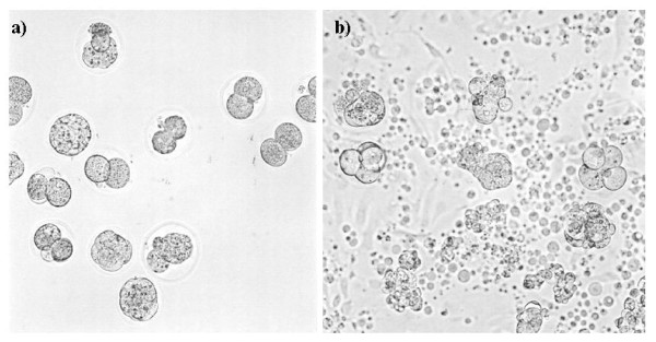
Morphology of mouse embryos degenerated by endocrine disruptors in the medium (a) or the coculture (b)[1]
1. <pubmed>3480945</pubmed>
Pineal gland
<pubmed>9852259</pubmed>
M. Hulsemann, 1971, Development of the Innervation in the Human Pineal Organ, Light and Electron Microscopic Investigations, 115: 396-415
<pubmed>16021838</pubmed>
Hypothalamus
<pubmed>11954031</pubmed>
<pubmed>7643957</pubmed>
Y. Koutcherov, J.K, Mai, G. Paxinos Hypothalamus of the human fetus, Journal of Chemical Neuroanatomy, 26:4, pp 253–270
Pulmonary surfactant metabolism dysfunctions
Pulmonary surfactant is a mixture of lipids (90%w/w) and protein (10%w/w) that coats the alveolar surface of the lungs and functions to reduce surface tension at the air-liquid interface and is essential for proper inflation and function of the lung. Surfactant is produced by alveolar type II cells, which differentiate during the last third of gestation. The substance is stored intracellularly by surfactant protein B (SP-B) and ATP-binding cassette transporter A3 (ABCA3) in specialized secretory organelles known as lamellar bodies, and is then secreted by exocytosis.
Expression of these proteins and lipids in surfactant is regulated during fetal development and is critical for pulmonary surfactant function at birth. Mutations in the genes encoding the surfactant proteins B and C (SP-C), and the phospholipid transporter, ABCA3, or dysfunction of Type II alveolar cells during the developmental period can significantly affect surfactant metabolism and result in respiratory distress and interstitial lung disease.
SP-B and SP-C have an important role in the adsorption of secreted surfactant phospholipids to the alveolar surface whereas SP-B and ABCA3 are required for the normal organization and packaging of surfactant phospholipids into lamellar bodies. In general, mutations in the SP-B gene (SFTPB) are associated with fatal respiratory distress in the neonatal period. SP-B deficiency is inherited as an autosomal recessive disorder with mutations in the SFTPD gene resulting in the complete absence or loss of function of SP-B, causing acute respiratory distress in full-term infants at birth, which is progressive and usually fatal by 3 to 6 months of age. Mutations in the SP-C gene (SFTPC) are more commonly associated with interstitial lung disease in older infants and are also likely to lead to respiratory. Mutations in the ABCA3 gene are associated with both phenotypes however more commonly in newborns.
<pubmed>2987676</pubmed>
<pubmed>15044640</pubmed>
'Lab 8 Individual Assessment'
Human ovary development
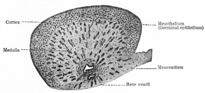
The human ovary is derived from three sources, the mesothelium which lines the posterior abdominal wall, the underlying mesenchyme and the primary undifferentiated germ cells. In the initial stages of gonad development in the embryo, the gonad structures in males and females are indifferent and develop identically till week 7. A layer of mesothelium develops on the medial side of the mesonephros, (a primitive kidney) and ongoing proliferation of this epithelium and the underlying mesenchyme forms the gonadal ridge on the medial side of the mesonephros. Epithelial gonadal cords then grow into the underlying mesenchyme. The indifferent gonad consists of an external cortex and an internal medulla.
Ovary development is only histologically visible during approximately week 10 of gestation. The XX chromosome contains genes encoding development of the ovary. Cortical cords arising from the mesothelium of periosteum extend from the surface epithelium of the developing ovary into the underlying mesenchyme during the early fetal period. Primordial germ cells are incorporated in these cortical cords. At 16 weeks, these cords begin to break up into isolated cell clusters called primordial follicles, each containing an oogonium that is derived from a primordial germ cell. Each follicle is lined by a single layer of follicular cells derived from the surface epithelium. Mitosis of oogonia occurs throughout fetal life and produce almost 2 million that enlarge to become primary oocytes and deplete gradually during ovulation.
Moore: The Developing Human, 9th ed.
'Lab 10 Individual Assessment - Peer Reviews'
Group 1
The wiki-page is very thorough and informative and addresses the majority of the marking criteria well. However there are some points I’d like to highlight for further editing. The second and third sentences in the introduction paragraph are confusing. It would be better to clarify which parts of the respiratory system are derived from endoderm and mesoderm. I particularly liked that the embryonic and fetal stages were quantified by week of development early on in the introduction to indicate what weeks of development the project was focusing on. It would also be extremely helpful to students who are using this as a learning resource if subheadings or brief descriptions were used underneath the images. What I really liked was the use of the ‘lung development table’. The layout made it easy to read and the explanations were not overly long-winded or complicated. At times there was a bit of repetition of information under different subheadings, for example regarding the two components of the respiratory system.
It would be a good idea to read through the entire project as a whole rather than one subheading at a time, and then restructure the content to minimize repetition. There were also a few minor spelling and grammatical errors in the first paragraph which can be fixed up post-editing. The current and historic findings were divided into separate headings and referencing of sources used was done extremely well. I noticed however that the schematic on lung disease doesn’t seem to really flow with the text in its current position. I would suggest to move it down to the abnormalities section.
Overall the group has done an excellent job at referencing and has derived information from a variety of mediums including video clips and animations. What I would suggest however is to keep all the references at the end of the page. I think this would make the project appear much more organized and easier to read. Other than that, I think the group has definitely produced a high quality wiki page with useful information on the fetal development of the respiratory system. I think it was pitched at an appropriate level for university students and included helpful diagrams and illustrations.
Group 2:
Overall the group has done a good job at referencing and has derived information from a variety of mediums including video clips and animations. What I would suggest however is to keep all the references at the end of the page under an exclusive references heading. I think this would make the project appear much more organized and easier to read. I think the use of quotation marks should be avoided in the introduction. It would be a better idea to summarise the sentence in your own words and reference it. Though the introduction is informative, I think it could be structured better so as to flow on from each paragraph, particularly with the last two paragraphs.
With the developmental timeline, it would be a great idea to put that into a table and expand a little more on each stage you have listed to make it easier to understand the process. Current research models mentioned were explained well and provided context to the information given however I’d suggest to use two subheadings for each of the different research models used (mice and lambs) so that the text isn’t so chunky.
I think the kidney development, urethra, bladder and abnormality section were written exceptionally well and the text was supported with relevant images. The referencing here however was a little inconsistent with the rest of the format but that can easily be fixed during the editing process. Overall however, the information was presented well in an interesting and meaningful way, perfectly suited as an educational resource for university students. Great job!
Group 3:
This group has done exceptionally well at referencing their information and has a clear references section at the end of the page, (with the exception of the recent findings article which can be fixed up during the editing process). I particularly liked the structure and layout of the page and found it extremely organized and easy to navigate. A point for improvement I’d like to suggest in the introduction is to focus on introducing the process of fetal GIT development rather than on the post-natal structure as that way you can set the scene of what the project is really about.
It would be a good idea to summarise the purpose of the page and it’s contents. With the developmental timeline, it would be a great idea to put that into a table and expand a little more on each stage that has been listed, for example include the implications of development of Cajal cells in the small intestine. Additionally, it would be a good idea to include images of the development process to help illustrate rotations and break up the text. I would also suggest to find and briefly elaborate another one or two articles for the recent findings section.
There are some links to references under the foregut heading that don’t seem to be referring to any text and should be editing out or moved to the relevant position in the text. Another suggestion is to shift the developmental problems subheading from the hindgut section to under the deformities section as I feel that would be more relevant there. The midgut and hindgut sections were written very well with relevant diagrams to support the information given. The use of original hand-drawn diagrams with colour helps to make the page more visually appealing and interesting to read. Overall the project was coherent and consistent over the different headings. I found it to be well structured and definitely informative. Good job!
Group 4:
There is definitely plenty of useful information and your group has clearly put in a lot of effort to do extensive research on the topic. However there are some inconsistencies with formatting and references, which you can easily iron out once you have time for a final edit. There’s a lot of content on the page and understandably its difficult to organize it in a way that’s meaningful and easy to read. I think there are a few too many subheadings and it becomes a little confusing to follow, for example under the current research, models and findings heading, the female subheading was hard to follow so I think that just needs a brush up. I think it would be a good idea to avoid presenting all the information under this heading just as bullet points.
Try to have at least 2-3 current research articles and under them elaborate on what the findings were and what they may imply. I think it would be much more interesting if it was presented that way rather than spread over so many bullet points. I particularly liked the use of original hand-drawn diagrams with colour helps to make the page more visually appealing and interesting to read. I see there is still work to be done under the current findings section. It would be a good idea to summarise the findings and state their implications on current knowledge under each article.
The historical findings section is definitely extremely elaborate but I feel it may be a little too much. I think it’s important to keep in mind the purpose of the assignment and focus more on the actual fetal development and keep information succinct and relevant rather than overloading with information. The Abnormalities section was done extremely well and had a lot of useful information on many different diseases. Overall a great effort by the group, definitely can see how much effort you have all put in.
Group 5:
This group has made an outstanding effort in their efforts. I particularly liked the fact that they summarized the actual purpose of the page and what it would contain within the introduction itself, something the other groups have not really done. They have structured and organized the page extremely well and it is consistent and flows between each of the headings. I would suggest to tabulate the three types of cells in skin and have a column describing their origin and then their function to make it easier to read. The table for development of dermal layers and the table for teeth development should also ideally have a title and table numbers.
In general the content is very well written and informative, supported with relevant images and diagrams that illustrate the actual development process. The recent findings heading is also well written however there are a few formatting issues with the text box sizes that need to be fixed up. I think it would be a good idea to list the sources of historic findings and then elaborate on what contribution they may have made to our current understanding. The referencing just seems to be inconsistent in this section. Additionally it would be a good idea to move all the references for the other sections to the end of the page under the actual references heading rather than having them scattered over the wiki page. The abnormalities section is highly commendable and written extremely well.
Great job on the excellent and informative wiki page you have produced.
Group 7:
Group 7 has definitely put in a lot of effort into the project however there are some points for improvement I’d like to suggest. The introduction is quite succinct and short which is good however I felt some more could be detailed about what the project was about and what aspects of development were being focused on. I would also suggest having a timeline either in dot-point form or as a table to summarise the changes that occur during fetal development of each of the organs. Though this was done with the visible anatomical details table, it would be a good idea to include some more information on function or implications of the stages of development.
I think overall a good variety of images and diagrams have been used to support the text however the structure and format of the text needs to be edited and made more consistent between the headings. With the current research findings heading, the layout is a bit confusing and hard to follow and the references listed here seem out of place. I would suggest finding at least 2-3 recent research papers and under each one, summarise the purpose of the study, the out come and then the implications. This will give a lot more meaning and purpose to the text and be an interesting read.
I am not really sure if the future research subheading is necessary, but if you have found good sources of proposed research plans then it would be a good idea to include it. The abnormalities section still needs to be completed, but from whatever work has been done, I think the information was informative and well written. There is an issue with the ‘facial expressions associated with fetal alcohol syndrome’ picture which can be sorted out by reformatting. Overall, the referencing was done well and most were listed under an exclusive references heading which is great. Bit of work still needs to be done but other than that, great job!
Group 8:
I think the group has found some useful and relevant sources of information however there still needs to be work done in writing up content under some headings. The structure of the wiki page has been laid out and I think the idea of splitting up the developmental process into three trimesters is a good idea to avoid lengthy paragraphs or an overly lengthy timeline that may be difficult to absorb. I would suggest using a table to write up the timeline with a brief description of what exactly the process occurring involves. There are some references that are missing in the tendon development section.
I think the ‘Molecular and Cellular regulation of fetal myogenesis’ section was the most well written section with a thorough description of the process involved. Try to find relevant pictures and diagrams to accompany this text, they will make the explanation much more beneficial and easier to understand. Overall a good structure has been laid out for the wiki page but more content still needs to be added.
Lab 10
Sensory Development
<pubmed>20014102</pubmed>
Differentiation of neural retinal precursor (NRP) cells in vertebrates follows an established order of cell-fate determination associated with exit from the cell cycle. Wnt signaling regulates cell cycle in colon carcinoma cells and has been implicated in different aspects of retinal development in a number of species. This research paper aims to understand the biological roles of Wnt in the developing retina through the use of transgenic and pharmacological techniques.
Through the manipulation of the Wnt signaling pathway during retinal development in medaka embryos, they were able to observe that during the early phase of retinal development Wnt signaling regulated cell cycle progression, proliferation, apoptosis, and differentiation of NRP cells.To understand the role of Wnt signaling during retinal development, a HS promoter was used to induce expression of genes so that Wnt signalling could be manipulated. Two constructs of the heat shock promoter were utilized to regulate expression of the genes Wnt8 or the C-terminal truncated form DNWnt8, which is another (specific dominant-negative) form for the Wnt pathway. Both constructs were injected into CAB medaka embryos to produce germline transgenic animal lines. To speed up the process of transgenesis, the constructs contained flanking I-SceI meganuclease sites and to facilitate identification of transgenic embryos, the constructs also included Green Fluorescent Protein (GFP). This permitted the manipulation of the Wnt signaling pathway at precise stages of retinal development without interfereing with early gene functions such as for example, anterior-posterior patterning or eye field establishment which could affect retinal development indirectly and invalidate the experiment.
Using these two constructs, the results were able to show that during the early phase of retinal development Wnt signaling did in fact regulate cell cycle progression, proliferation, apoptosis, and differentiation of NRP cells. However, during later phases of retinal development, proliferation and apoptosis process remained unaffected by the manipulation of Wnt signaling. Thus, the conclusion made by the research paper was that the response of NRP cells to Wnt signaling is actually stage-dependent.

