Review - Normal Plates of the Development of Vertebrates
| Embryology - 1 Mar 2026 |
|---|
| Google Translate - select your language from the list shown below (this will open a new external page) |
|
العربية | català | 中文 | 中國傳統的 | français | Deutsche | עִברִית | हिंदी | bahasa Indonesia | italiano | 日本語 | 한국어 | မြန်မာ | Pilipino | Polskie | português | ਪੰਜਾਬੀ ਦੇ | Română | русский | Español | Swahili | Svensk | ไทย | Türkçe | اردو | ייִדיש | Tiếng Việt These external translations are automated and may not be accurate. (More? About Translations) |
Mall FP. Normal plates of the development of vertebrates. Anat. Rec, (1908).
| Online Editor |
|---|
| Note this review of the series Normal Plates of the Development of Vertebrates perhaps indicated Mall's own intention of establishing a developmental series for human embryos.
|
| Historic Disclaimer - information about historic embryology pages |
|---|
| Pages where the terms "Historic" (textbooks, papers, people, recommendations) appear on this site, and sections within pages where this disclaimer appears, indicate that the content and scientific understanding are specific to the time of publication. This means that while some scientific descriptions are still accurate, the terminology and interpretation of the developmental mechanisms reflect the understanding at the time of original publication and those of the preceding periods, these terms, interpretations and recommendations may not reflect our current scientific understanding. (More? Embryology History | Historic Embryology Papers) |
Review - Normal Plates of the Development of Vertebrates
Normentafeln zur Entwicklungsgeschichte der Wirbelthiere
By
Herausgegeben von Prof. Dr. F. Keibel, LL.D. (Harvard). Achtes Heft. Normentafel zur Entwickelungsgeschichte des Mensehen. Von Franz Keibel und Curt Elze. Mit 6 Tafeln und 44 figuren im Text. Jena. Verlag von Gustav fischer, 1908.
Since the appearance of the first part of the Normentafeln in 1897, the great work edited by Professor Keibel has become familiar to all anatomists, and it is natural that they have looked forward with especial interest to this, the eighth part, which is devoted to a consideration of the human embryo. The monumental work of His on the anatomy of the human embryo in a way brought into existence Keibel’s Normentafeln, for in it a standard plate is given, and it was to extend this plate to include various vertebrates that Keibel assumed his task.
Nearly a quarter of a century has elapsed since the completion of His’s “Anatomic Menschlicher Embryonen”, and we see in Keibel and Elze’s “Normentafel zur Entwicklungsgeschichte des Menschen” what progress has taken place in human embryology in this period. The work gives a detailed account of the degree of development of organs and tissues of eighty-four human embryos from .5 to 26 mm. long, all of which came under the authors’ observation. The earlier ova are given in great detail and are compared with the other young ova which have been described by various authors. There is also an excellent chapter on the comparison of young human embryos with those of the monkey, and finally there is a summary of the time of the appearance and transformation of some of the organ anlages of the human embryo, which are compared with the same in other classes of vertebrates given in the first seven parts of the “Normentafeln.”
The whole work, which fills 314 large quarto pages, is concluded with the bibliography relating to human embryology since 1880. This is practically complete and includes about 5,000 titles.
The work of Keibel and Elze is intended primarily to standardize various stages of the human embryo. Naturally, the external form of the embryos must be considered first and then the degree of development of the various organs to correspond with the various stages. Not only is this expressed in carefully planned tables, but also in a series of excellent illustrations on plates which are further elaborated by many figures of sections. The descriptions thus given are graphic and equal almost, on account of their excellence and necessary analysis, to the specimens themselves. The specimens described are nearly all new ones, many of which were obtained from operations, and, therefore, are probably normal. This mine of information is analyzed most carefully, and all statements regarding the sequence of development are given in a most conservative and trustworthy way. No rash statements are found in this excellent work, and I believe that it will withstand the test of time and prove to be a most valuable aid to all investigations of human embryology. As a whole it marks the first great “next step” as a. standard since the appearance of His’s monograph in 1880. The summary found in it will prove to be of the greatest use to all interested in human embryology and of great value to investigators in this science.
Keibel ventures to construct several diagrams, based partly upon observations and partly upon hypothesis, regarding the earliest stages of development. He starts with the assumption that fertilization takes place immediately after ovulation and that segmentation is more or less complete by the time the ovum reaches the uterus. About four or five days are required for this process to take place, and the ovum as a whole does not increase in size until segmentation is completed. It is also probable that the ovum eats itself into the mucous membrane of the uterus much as is the case in the guinea pig, and, judging by the size of the opening left, it cannot have been over .5 mm. in diameter when it became implanted. At this time the mesoblast is formed. Shortly after this the coelom, the cavity amnion and that of the yolk sac must develop. I{e believes that the exocoelom is formed within a solid mesoderm on account of the isolated strands of cells which cross it in early stages, and recently the observations of Bryce and Teacher have confirmed this view. Both the amniotic cavity and the hollow yolk sac are formed from solid cell masses, the former not being the result of an infolding, as has generally been believed. These views are based upon comparative evidence as well as by observations upon the human ovum, and are illustrated beautifully by a series of diagrams which will no doubt displace those given in various text-books. The peculiarity of the human ovum is due to the very early formation of the amnion and of the exocoelom. There is no inversion of the membranes, and what appears to be an inversion is due to a confusion of the very early amniotic cavity with this process. He does not venture to give an opinion regarding the sequence of the formation of these three cavities, but the very young ovum recently described by Bryce and Teacher shows that the exocoelom is the last to be formed, for in this ovum the cavities of the amnion and yolk sac are present, while the rest of the ovum is filled with an even mesoderm, there being no exocoelom. After it is once formed the exocoelom extends very rapidly, and the medullary plate, primitive streak, allantois canal and neurenteric canal follow in rapid succession. From now on in the monograph the formation of the body of the embryo is followed in successive stages. For instance, in a discussion of the anterior neuropore, he says that in man it is just closed in an embryo with 23 somites, in Macacus with 19, in Tarsius with 18 to 20, in the pig with 20, in Lacerta with 20, in the chick with 12 to 13, and in the rabbit with from 9 to 11 somites. Here we have a comparison of various vertebrates, not dependent upon stages, size nor age, but upon the degree of development, and as far as I can determine we shall be compelled in the future to designate young embryos by their number of somites and other anatomical conditions and not by their length in millimeters nor their age in days. The latter, on account of the very probable errors, and on account of the unequal degrees of development within a given time is probably the worst of standards in human embryology and has caused endless confusion. His makes his stages dependent upon their length and their general external form, while Keibel improves His’s standards by including the anatomy of the embryo. In order to do this it has been necessary for him to study all of the details of the embryos under consideration with great care, and by the comparison of stages of the same size, which resulted in the observation that the sequence is not always the same, he has laid a. firm foundation for the scientific study of variations in the adult.
Among the youngest embryos described by His, that is, those under 4 mm. long, a dorsal kinking is observed, and His is of the opinion that this bend is to be considered normal. However, it is not present in all embryos of this stage; the form of the curve appears to be artificial ; it is rarely present in embryos that have been hardened within the ovum and it can easily be produced artificially in the fresh chick and pig embryos. Therefore, it is highly probable that this dorsal kink is an antifact. Keibel is also strongly of this opinion, and he described normal embryos of this stage as being somewhat spiral and curved ventralwards, that is. the dorsal border of the embryo is always convex. The stages around 3 mm. long (from 13 to 30 segments) given by Keibel bear upon this point, but are too few in number to give an entirely satisfactory conclusion. To fill out the series it has been necessary to include two embryos (Nos. 6 and 7) with heads that appear atrophic, much as is the case in an embryo described on several occasions by the reviewer (Mall, No. 12). Before this gap is filled out in an entirely satisfactory manner more embryos of this stage will have to be studied. However, the point made by Keibel against the dorsal kinking is sound and the distorted figures of embryos which have gradually crept into the literature will no doubt be replaced by those of normal ones with a delicate and slightly spiral dorsal convexity.
The material brought together by Keibel and Elze shows that a “Normentafel” of the human embryo can be constructed about as well as for any of the other vertebrates. Gaps do exist, but it will be quite easy to fill them in since the way to follow has been pointed out. This can be done by individual workers if they will take the trouble to tabulate their specimens as Keibel and Elze have done and not forget to give careful drawings of the specimens. No doubt a number of such unpublished drawings now exist, as they do in my own collection, which if published would be of value in the study of human embryology. It might be well for the investigators in this science to analyze still further the various stages given by His and by Keibel and Elze, making, for example, the "embryo Bulle with 14 myotomes “stage D” instead of a “14 day embryo.” For the present we must designate young human embryos after the myotomes appear by their number, and if we add to this the appearance and disappearance of organs and cavities many workers can contribute to the standardization of human embryos. They will thus help to elaborate Keibel’s “Normentafeln,” which are intended in the first place to establish the norm for various stages of development in different vertebrates.
The “Heft” by Keibel and Elze upon human embryology marks a distinct step in advance, for it arranges and compares various stages, with the control of comparative embryology The embryos included have been carefully selected and the statements regarding them are sound and conservative. This is especialy necessary in the study of human embryology, for many of the specimens obtained are pathological, and on account of the wide interest in this subject are often extravagantly and poorly described. The great work of Keibel and Elze is to be recommended equally as much for what has been omitted from it as for what has been included, and for these two reasons it will prove to be of the greatest value to all scientists and physicians who are interested in human embryology.
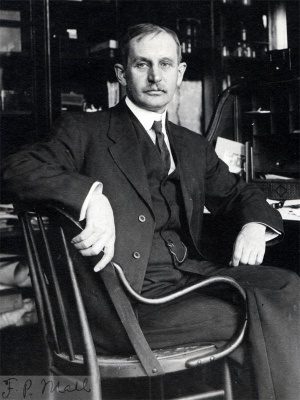
Franklin P. Mall.
Cite this page: Hill, M.A. (2026, March 1) Embryology Review - Normal Plates of the Development of Vertebrates. Retrieved from https://embryology.med.unsw.edu.au/embryology/index.php/Review_-_Normal_Plates_of_the_Development_of_Vertebrates
- © Dr Mark Hill 2026, UNSW Embryology ISBN: 978 0 7334 2609 4 - UNSW CRICOS Provider Code No. 00098G


