Paper - The distal projection of the endolymphatic sac in human embryos
| Embryology - 27 Apr 2024 |
|---|
| Google Translate - select your language from the list shown below (this will open a new external page) |
|
العربية | català | 中文 | 中國傳統的 | français | Deutsche | עִברִית | हिंदी | bahasa Indonesia | italiano | 日本語 | 한국어 | မြန်မာ | Pilipino | Polskie | português | ਪੰਜਾਬੀ ਦੇ | Română | русский | Español | Swahili | Svensk | ไทย | Türkçe | اردو | ייִדיש | Tiếng Việt These external translations are automated and may not be accurate. (More? About Translations) |
Anson BJ. The distal projection of the endolymphatic sac in human embryos. (1933) Anat. Rec. 701. 57, 110. 1, pp. 55-58.
| Historic Disclaimer - information about historic embryology pages |
|---|
| Pages where the terms "Historic" (textbooks, papers, people, recommendations) appear on this site, and sections within pages where this disclaimer appears, indicate that the content and scientific understanding are specific to the time of publication. This means that while some scientific descriptions are still accurate, the terminology and interpretation of the developmental mechanisms reflect the understanding at the time of original publication and those of the preceding periods, these terms, interpretations and recommendations may not reflect our current scientific understanding. (More? Embryology History | Historic Embryology Papers) |
The Distal Projection of the Endolymphatic Sac in Human Embryos
Department of Anatomy, Northwestern University Medical School, Chicago, Illinois
Twenty Five Figures
Contribution no. 190 from the anatomical laboratory, Northwestern University Medcal School.
Paper read at the New York City meetings of the American Association of Anatomists, February, 1932; abstract, Anatomical Record, vol. 52, no. 1, pp. 2-3, February 25, 1932.
Introduction
The endolymphatic duct in the mammalian embryo is described conventionally as ending in a simple dilated extremity, the endolymphatic sac; and so far as the Writer knows, it is not stated in any textbook that from the distal aspect of the endolymphatic sac in young human embryos there regularly extends a considerable projection the length of which may actually exceed that of the endolymphatic duct.
The projection has, however, been figured by several investigators as it appeared in models of the human membranous labyrinth. Thus in Streeter’s (’07) figure of the labyrinth in the embryo of 30 mm., there is a knob—like protuberance on the dorsum of the endolymphatic sac; in Macklin’s (’21) models of the 43—mm. (CH) embryo, the projection is long and thin; and in those by Bast ( ’30) of the 135-mm. and the 147—mm. embryos it is short, and of triangular outline. Doctor Macklin described what he observed, recording briefly that the dorsal extremity of the broad flattened saccus “is a very slender filamentous process containing an almost imperceptible lumen.”
The present study is concerned with the appearance of this process in a graded series of human embryos.
Material and Methods
The observations recorded herein are based upon a study of the mammalian series available in the Harvard Embryological Collection, and constitute part of a more comprehensive comparative study on the development of the ear in vertebrates.
For the study of the endolymphatic sac and its projection, 141 embryological series were examined, distributed among eleven species of mammals as follows: forty-eight series of human embryos, from 10 to 74 mm. in length (CR)2; twentytwo of the cat, from 6 to 39 mm.; five of the dog, from 12.5 to 17 mm.; seventeen of the rabbit, from 5 to 33.8 mm.; six of the guinea—pig, from 6.1 to 30 mm.; nine of the rat, from 6 to 24.8 mm.; fourteen of the pig, from 7.8 to 48 mm.; three of the calf, 14.2 to 25.2 mm.; ten of the sheep, from 6.5 to 48.4 mm.; two of the deer, 9.8 and 18.6 mm.; seven of the opossum, from 13 to 26 mm.
Two series (’29 mm. and 40 mm.) were modeled by the Born wax-plate method, at a magnification of 250 diameters.
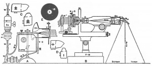
Twenty-five of the series of human embryos were reconstructed graphically, from tracings of each fifth section prepared with an Edinger apparatus at a magnification of 550 diameters. The reconstructions” were stylized to the extent of being built along a straight central axis without regard to the slightly curving course of the duct (compare, for example, figs. 1C and 25).
- Online Editor - The |Edinger projection apparatus was developed in 1907 by Dr. L. Edinger (director of the Neurologic Institute at Frankfurt on Main). He replaced his drawing and photographing apparatus using either oil or gas light, with one using a small arc lamp. This small arc lamp was worked out and perfected by the optical works of Leitz at Wetzlar.
Observations and Discussion
The entire endolymphatic appendage is, in its early development, at first a digitiform (figs. 2 to 5), and then a lanceo— late (figs. 6 to 10) diverticular process of the otocyst. Very soon after the Whole appendage becomes differentiated into the commonly recognized subdivisions, ductus and saccus, a third segment appears, in the form of a narrow, distally placed, prolongation. This third subdivision, the distal projection, is unmistakably present in the embryo of 19 mm. (fig. 12; also 11 and 13), and seems to owe its formation not only to elongation of the terminal third and the primitive appendage, but also to accompanying constriction of this part. Then, while retaining its typical shape, further extension occurs through the 27 mm stage (figs. 15 to 18) — the length of the entire endolymphatic appendage being tripled or quadrupled while its greatest width is but doubled. Thereafter, as shown by the most advanced stages included in our reconstructions (31—, 36-, 37-, and 45—mm. embryos; figs. 21, 22, 23, and 25), the distal projection retains its long thin form, while the saccus becomes more bulbous; the projection is still present in most advanced series available, the 74—mm. embryo. Concurrently, With the alterations described above, the ductus becomes, in general, progressively more elongate.
3 Only one series, the 74 mm, was more than 45 mm. in length, and was alone exceptional to the graduated arrangement of series.
3 These are reproduced in figures 2 to 25, and are the following: fig. 2, H.E.C., 1000; fig. 3,‘2247; fig. 4, 2284; fig. 5, 2036; fig. 6, 2161; fig. 7, 2000; fig. 8, 839; fig. 9, 2051; fig. 10, 2155; fig. 11, 2246; fig. 12, 819; fig. 13, 1597; fig. 14, 871; fig. 15, 2048; fig. 16, 2046; fig. 17, 2045; fig. 18, 2042; fig. 19, 2248; fig. 20, 913; fig. 21, 2043; fig. 22, 2050; fig. 23, 820; fig. 24, 841; fig. 25, 2128.
Fig.1 Endolyniphatic duct, sac, and distal prolongation of the latter, from human embryos in the Harvard Embryological Collection.
A. Tracing from a frontal section of a 29-mm. embryo (H. E. C., 914, sect. 392). X 30. B. Model prepared from the same series. The level of figure A is indicated by broken line. X 30. C. Model from a 40-mm. embryo (H. E. C., 1917). X 30. D. Reconstruction, from a. 19-mm. embryo (H. E. C., 819; see also fig. 12). Levels of sections shown in the two succeeding figures are indicated. X 67. E and F. Drawings (camera-lucida) at two levels (sections 87, 103) of the distal projection seen in figure D. X 760. 56
Figs.2 to 25 Graphic reconstructions of the right endolymphatic sac and duct from transverse series of human embryos. (Harvard Embryological Collection.) Lateral views. X
The development of the projection is not, in the successive embryos, invariably progressive—there occur exceptional forms and sizes. Thus in the 19-mm. embryo the projection (fig. 12) is in an advanced stage of development (a condition which is duplicated in the other labyrinth of the same specimen); in the 25—mm. embryo (fig. 18) there is a striking development of the appendage, this structure attaining a length one and a half times that of the endolymphatic duct.‘ Contrariwise, the outgrowth is occasionally poorly developed; in the 22.8-mm. embryo (fig. 14) it is merely nodular, and in an older embryo, one of 42 mm. (fig. 24), its basal portion forms a relatively wide bulging.
When seen in section (fig. 1 A), or When modeled (figs. 1 B and 1 C), the configuration of the projection is in substantial agreement with that displayed by the reconstructions.
Histologically, the wall of the distal projection, like that of the saccus and of the ductus, consists of a single layer of cuboidal epithelial cells. In the region where the sac definitely narrows to become the dorsal extension, it is approximately ten cells in circumference (fig. 1F); the number of cells gradually decreases to four or five in the terminal fourth, and the lumen becomes progressively smaller (fig. 1 E); the latter usually disappears in the distal reaches, so that the tip of the process is a solid cord, composed, in transverse section, of four, three, or even fewer cells.
- In this series the writer first encountered the structure which he then be» lieved might represent the original stalk of communication between the outer ectoderm and the endolymphatic sac drawn out into attenuate form. Further study of series, as herein figured, proved that such was not the case. The history of the rupture of the connection with the ectoderm will be discussed in a subsequent publication.
The distal prolongation of the endolymphatic sac is, so far as our observations indicate, restricted in its occurrence to the embryo of man; it was not seen in the embryos of the other ten mammalian species included in this study.
A further study of this striking prolongation of the saccus endolymphaticus is now in progress. Although its fate in the fetal or the adult ear is as yet undetermined, it may be suggested that the structure is perhaps represented by the very irregular, or occasionally even contorted, termination of the endolymphatic duct observed in serial section of ears from some postnatal specimens.
Literature Cited
Streeter GL. On the development of the membranous labyrinth and the acoustic and facial nerves in the human embryo. (1906) Amer. J Anat. 6:139-165.
Macklin CC. the skull of a human fetus of 43 millimeters greatest length. (1921) Contrib. Embryol., Carnegie Inst. Wash. Publ., 48, 10:59-102.
Bast TH. Ossification of the otic capsule in human fetuses. (1930) Contrib. Embryol., Carnegie Inst. Wash. 121, Publ. 407, 53-82.
Cite this page: Hill, M.A. (2024, April 27) Embryology Paper - The distal projection of the endolymphatic sac in human embryos. Retrieved from https://embryology.med.unsw.edu.au/embryology/index.php/Paper_-_The_distal_projection_of_the_endolymphatic_sac_in_human_embryos
- © Dr Mark Hill 2024, UNSW Embryology ISBN: 978 0 7334 2609 4 - UNSW CRICOS Provider Code No. 00098G


