Paper - Stapes, fissula ante fenestram and associated structures in man 2
| Embryology - 28 Apr 2024 |
|---|
| Google Translate - select your language from the list shown below (this will open a new external page) |
|
العربية | català | 中文 | 中國傳統的 | français | Deutsche | עִברִית | हिंदी | bahasa Indonesia | italiano | 日本語 | 한국어 | မြန်မာ | Pilipino | Polskie | português | ਪੰਜਾਬੀ ਦੇ | Română | русский | Español | Swahili | Svensk | ไทย | Türkçe | اردو | ייִדיש | Tiếng Việt These external translations are automated and may not be accurate. (More? About Translations) |
Anson BJ. Karabin JE. and Martin J. Stapes, fissula ante fenestram and associated structures in man: II. From Fetus at Term to Adult of Seventy (1938) Arch. Otolaryng. 28: 676-697.
| Historic Disclaimer - information about historic embryology pages |
|---|
| Pages where the terms "Historic" (textbooks, papers, people, recommendations) appear on this site, and sections within pages where this disclaimer appears, indicate that the content and scientific understanding are specific to the time of publication. This means that while some scientific descriptions are still accurate, the terminology and interpretation of the developmental mechanisms reflect the understanding at the time of original publication and those of the preceding periods, these terms, interpretations and recommendations may not reflect our current scientific understanding. (More? Embryology History | Historic Embryology Papers) |
Stapes, fissula ante fenestram and associated structures in man: II. From Fetus at Term to Adult of Seventy
Barry J. Anson Ph.D. (Mm Sci.) John E. Karabin, M.D. and John Martin, M.D. Chicago
From the Department of Anatomy, Otolaryngology and Surgery, Northwestern University Medical School. Contribution 268 from the Anatomical Laboratory.
Read at the fifty-fifth Annual Session of the American Association of Anatomists, Boston, April 8, 1939 (abstracted, Anat. Rec. [supp. 2] 73:37, 1939). The investigation was conducted under the auspices of the Central Bureau of Research of the American Otological Society, with the general superintendence of Dr. J. Gordon Wilson.
Introduction
As mentioned in an earlier article[1] on the structure of the stapes, more precise information has been needed on the development and the adult form of this ossicle; until such information was made available, pathologic structure of the stapes could not, with certainty, be distinguished from normal. Equally important is information concerning the histologic nature of the vestibular window, the nearby fissular tracts through the otic capsule and the islands of chondral tissue situated in or near the latter. The current paper represents a second phase of this study and includes the description of six stages from the fetus at term to the adult of 70 years. A third article will trace the developmental changes which occur between midfetal life and term.
Curiously, while structures in the general locality of the stapes have been described fully in recent publications, the ossicle itself has received little attention. Thus, the fissula ante fenestram and the fossula post fenestram — channels through the capsule to the two sides of the vestibular (oval) window — have been discussed in some detail. The formative stages of the fissula have been fully discussed by Bast (1930)[2] the variety in form of fissulae in postnatal stages and the relation of constituent fissular tissues to sclerotic bone have been studied by Bast (1933)[3] Anson and Martin (1935),[4] Wilson (1935)[5] and Bast (1936)[6]; the fossula post fenestram has been more recently described by Bast (1938)[7] Since these reports have been reviewed in our earlier paper,[1] consideration of their results need not be repeated here.
Although the extrafenestral channels have thus received considerable attention, a full account of the stapes and the vestibular Window, with consideration of the changes which occur during infancy, childhood and adult life, has never been presented. It is our object in the present group of articles to consider the morphologic and histologic changes in these several structures—treating them as interrelated parts of a general stapedial region. An earlier publication described the capsule and the stapes in the 29 mm. embryo[8]
Materials and Methods
For this investigation reconstructions of the stapes, together with the surrounding area, were prepared by the wax plate method.[9] Representative developmental stages were selected from the series of sections in the department of anatomy at the University of Wisconsin and in the department of otolaryngology at the Northwestern University Medical School. These series were lent by Profs. Theodore H. Bast and J. Gordon Wilson. All series in the otologic collection at Northwestern University were prepared by Miss Helen Banks, R.N.
The stapes and the surrounding portion of the otic capsule in the fetal series have been described and illustrated in our earlier paper[1]; only the details of fissular structure, then, will be presented in the current account.
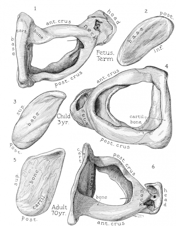
Fig. 1 to 6. Reconstructions of the stapes; superior and medial views; X 21. The arrows in these and the next group of figures pass through the foramens.
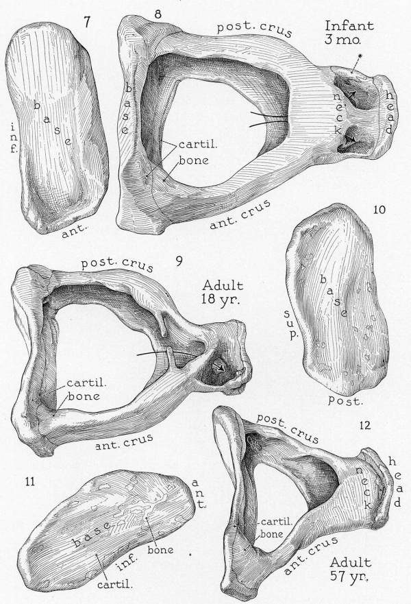
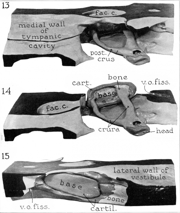
In these and in the succeeding figures ant. crus. indicates anterior crus of stapes; cart. or cartil., cartilage; far. c., facial canal ; fen. cart, fenestral cartilage; fiss. a. f., fissula ante fenestram; fiss. cart, fissular cartilage; foss. p. f., fossula post fenestram; hav. sp., haversian spaces; mm. membn, mucous membrane; obt. membr., obturator membrane; perich., perichondrium; post. crus, posterior crus ; pryr, pyramid; sc. tymp, scala tympani; semis. t. t. m., semicanal for tensor tympani muscle; st. lig., stapedial ligament; t. c. or tymp. cavity, tympanic cavity; t. o. foss., tympanic orifice of fossula; v. or vest, vestibule; vestib. orif. or v. o. fiss, vestibular orifice of fissula, and 'v. o. foss., vestibular orifice of fossula.
Figs. 7 to 12. Reconstructions of the stapes; superior and medial views; X 21. In figure 8 the asterisk marks the point of attachment of the tendon of the stapedius muscle.
Reconstructions were prepared from the following series:
| Reconstructions | |||
|---|---|---|---|
| Age and Length | Series | Side | Figures |
| 17 fetal weeks (135 mm) | 5 | Left | 60 |
| 19.5 fetal weeks (161 mm) | 13 | Left | 61 |
| 21.5 fetal weeks (183 mm) | 21 | Left | 62 |
| Fetus at term | Apr. 21 | Right | 1, 2, 13 to 15 |
| 3 mo. | 2/ 1/32 | Left | 7, 8, 16 to 19, 63 |
| 3 yr. | 6/18/29 | Left | 3, 4, 22, 23 |
| 18 yr. | 1/17/31 | Left | 9, 10, 24, 25 |
| 57 yr. | 1/14/33 | Left | 11, 12, 26 to 28 |
| 70 yr. | 3/ 3/34 | Left | 5, 6, 20, 21, 29, 30, 64 A |
Sections from the following series are illustrated:
| Sections Illustrated | ||
|---|---|---|
| Age | Series | Figures |
| Fetus at term | March, ’27 | 31 |
| Fetus at term | 4/18/30 | 38 |
| 7 days | 5/ 8/36 | 32, 32a |
| 7 wk. | 9/ 7/31 | 41 |
| 2 mo. | 9/ 9/31 | 40 |
| 3 mo. | 2/ 1/32 | 33, 34 |
| 4 mo. | 5/24/32 | 54 |
| 1.5 yr. | 9/28/32 | 35, 55 |
| 2 yr. | 3/30/28 | 36 |
| 3 yr. | 2/ 4/32 | 52 |
| 3 yr. | 6/17/29 | 56 |
| 3.5 yr. | 2/21/33 | 37 |
| 4.5 yr. | 6/23/33 | 43 to 47 |
| 13 yr. | 3/ 8/35 | 39, 39a |
| 50 yr. | 4/ 8/35 | 57 |
| 57 yr. | 1/14/33 | 48 to 51, 53, 58 |
| 70 yr. | 3/ 3/34 | 42, 42 a |
| 74 yr. | 4/ 4/34 | 59 |
I. Reconstructions
(a) Fetus of 135 mm Crown-Rump Length
(17 Weeks): The stapedial and fenestral structures of the specimen have already been described; 1 the fissula ante fenestram remains to be discussed.
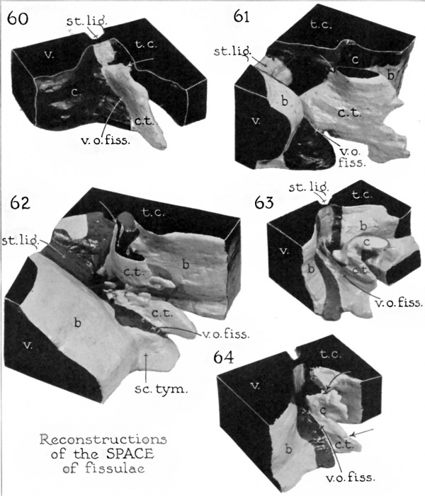
The entire capsular wall in the fissular area is cartilaginous; it encloses the strip of connective tissue which fills the fissula ante fenestram. The fissula is of simple form. Its appearance when reconstructed as a solid is that of an invagination of the vestibular cavity, but it has a small connection with the tympanic submucosal tissue (fig. 60). The former is its vestibular, and the latter its tympanic, extremity. The vestibular extremity, or orifice, is elongate, measuring 1.04 mm. in length and from 0.08 mm. to 0.12 mm. in width. Coinciding with the contour of the vestibular wall on which it opens, the inclination of the orifice is sharply away from the vertical. Inferiorly, the communication is continued along the wall of the scala vestibuli. The fissula extends into the capsule for a maximum distance of 0.8 mm. measured from the vestibular orifice; turning sharply at an angle of 90 degrees, it opens into the tympanic cavity through an orifice which is 0.32 mm. long and 0.08 mm. wide. So close together are the two orifices that the merest strip of cartilage separates them. Within the capsule, the fissula broadens somewhat, attaining a maximum breadth of 0.2 mm.
(b) Fetus of 161 mm Crown-Rump Length
(19.5, Weeks): The stapedial and fenestral structures have been discussed in the initial paper in the series.[1]
The fissula in this specimen is larger than that in the preceding stage (fig. 61); the surrounding tissue is still cartilaginous. The vestibular extremity is shorter by one third than that in the 135 mm. embryo (0.64 mm. in length); it is also wider (0.24 mm.) and communicates mainly with the scala. The fissula extends well above the level of the tympanic orifice, attaining its maximum thickness (0.36 mm.) in this superior position. The tympanic orifice is round and small (0.16 mm. in diameter), appearing to be merely a stemlike extension of the main space. The fissula invades the bone for a distance of 1.84 mm.; its distal margin is irregular, not smooth as in the preceding stage. As a whole it is relatively large, more than a mere chink in the cartilage of the capsule.
(c) Fetus of 183 mm Crown-Rump Length
(21 Weeks): The stapes, the vestibular window and the surrounding capsular wall have been described in our earlier publication.[1]
Bone has now replaced cartilage to such an extent that the former tissue remains only in the areas of the fissular orifices and the vestibular window. Remarkably, the fissula is smaller than it is in the 161 mm. embryo (fig. 62). The vestibular orifice is 0.88 mm. in length; the tympanic, approximately 0.8 mm. The fissula is divided into two portions, separated by cartilage; the upper subdivision is roughly quadrangular, its margin irregular. The adjacent margin of the lower piece is similarly irregular, at one point widening into a finger-like projection (0.24 mm. in width). The upper piece curves on itself, the concavity being on the medial side; the lower piece is not curved, and it extends anteriorly toward the cochlea.
(d) Fetus at Term
The stapes has attained its adult dimensions in the embryo of 135 mm., i. e., before the embryo itself has reached the midfetal stage} In the fetus at term the adult configuration of the constituent parts of the stapes is evidenced (figs. 1, 2, 13 to 15); between the stage of the fetus at term and that of the oldest adult only relatively moderate alterations occur — moderate, at least, in comparison with those which the stapes undergoes in the interval between the midfetal stage and birth.
Figs. 13 to 15. Reconstruction of the stapedial region in a fetus at term; ) x 18. figures 13 and 14 are tympanic views; figure 15, a vestibular (general descriptions[9]).
The features which distinguish the stapes of the fetus at term from that of the 183 mm. embryo and those which are characteristic of the adult ossicle may now be discussed. Outstanding among them is the new structure of the base (footplate) and the crura: These parts are no longer hollowed bars perforate ‘On one surface but are channeled members with the excavations mutually facing each other, that is, directed toward the intercrural space (figs. 1, 2, 13 and 14). Structurally the crura differ from the base, however, in being evenly grooved to form a semicircle in cross section and in consisting entirely of bone.
The base, although also grooved, possesses a different form, being flattish where it faces the vestibular cavity. The base projects backward marginally in the form of lips which are continuous with the free edges of the crura (fig. 14). The base is a bulkier structure than either crus and consists of both cartilage and bone, arranged in two laminas (figs. 1 and 14) ; the cartilage forms a complete layer on the vestibular surface of the base (fig. 15) ; the bone, an almost unbroken stratum on the tympanic surface (fig. 14); in only occasional small areas on the latter aspect of the footplate does cartilage persist. The explanation of the structure just described is not far to seek; the process of perforation, seen in its initial stages in the embryo of 183 mm.,1 has ultimately destroyed the inner wall of each crus and the tympanic wall of the base of the stapes. Thus, while cartilage has been wholly replaced by bone along the crura, it has not supplanted — merely formed a coating for — the cartilaginous lamina of the base; in the course of this process marrow has been succeeded by connective tissue, now submucosal in position. Were a long bone halved through its length and its marrow replaced by p-eriosteal tissue, the morphologic result would be similar.
The crura arise from the inferior portion of the base, especially the posterior crus (fig. 14). They are directed laterally, not in an exactly horizontal plane but at an acute angle with the base; they therefore lie close to the infrafenestral wall of the otic capsule, a proximity increased by the jutting character of the wall itself. The anterior crus is the shorter and less curved of the two; it is also less deeply grooved; in section, it has the form of a letter “C” with a short inferior arm, while the posterior crus resembles the same letter with a long inferior arm.
The neck of the stapes, like the crura, is entirely osseous (figs. 13 and 14). Like the crura, it is excavated (figs. 1 and 14), but in the form of an incomplete cylinder, entirely open medially (i. e., toward the base) and partially so superiorly; the space is continuous with that enclosed by the head. In the head, cartilage is limited to the articular surface (fig. 14), the latter being cupped for reception of the incus. The head resembles the base in consisting of two laminas, the external one, just described, which is formed of cartilage, and an internal layer, formed of bone.
The facial canal at this stage is a complete bony channel, the jutting character of which adds considerably to the depth of the fossa in which the stapes lies.
The stapedial ligament exhibits a structure which is not distinguishable from that in the adult.
The fissula ante fenestram of the specimen has been described in earlier papers.[10] It consists of a small round tympanic extremity (diameter, 0.09 mm.) situated on the floor of a depression in the otic capsule (figs. 13 and 14). From this extremity, situated 0.7 mm. in front of the vestibular window, it widens rapidly, extending medially, inferiorly and then, Without interruption, posteriorly, to communicate with the vestibular window by an auxiliary orifice (0.12 mm. wide and 0.15 mm. long). The true vestibular extremity opens into a groove at the junction of the vestibule and the scala vestibuli as an oval orifice (0.12 mm. wide and 0.3 mm. long), placed 0.4 mm. from the vestibular window (fig. 15). Its connective tissue is embedded in cartilage which is a continuation of the chondral rim of the vestibular window.
(e) Infant of 3 Months
The stapes in the infant is slightly larger than is that of the fetus at term (figs. 7 and 8 and table), but the increase is so slight that it might reasonably be regarded as due to individual variation.
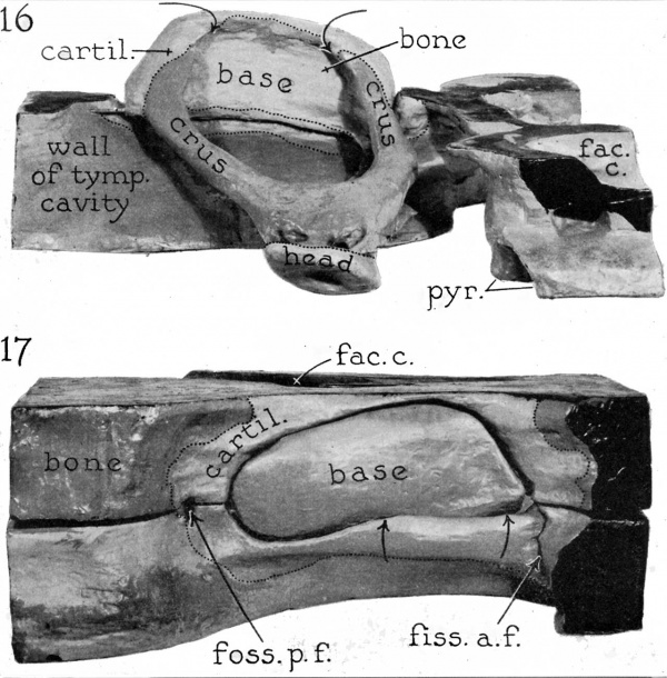
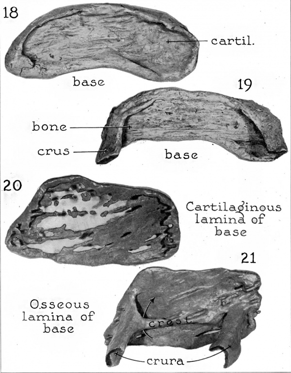
The base consists of two layers, as does the stapes in the fetus; the vestibular lamina is entirely cartilaginous; the tympanic, almost completely osseous (figs. 18 and 19). The ratio of cartilage to bone in the base is approximately 2:1. The cartilage of the vestibular lamina extends well over the margin to the tympanic surface, forming a rim at the periphery of the base (figs. 16 and 17 ) . Anteroinferiorly the base narrows into a blunt point; its curvature imparts a reniforin outline. The base is not seated in the window plunger fashion, but at its anterior extremity lies against the tympanic surface of the window’s shelving margin 17, between arrows).
Figs. 16 and 17. Reconstruction of the stapedial region in an infant 3 months old; X 21. figure 16 is a tympanic view; figure 17, a vestibular. In figure 16 the unlabeled arrows point into the space between the base and the crura; in figure 17 they mark the area in which the stapedial base is overlapped by the fenestral margin.
The crura are channeled osseous columns which arise from the inferior aspect of the base and as they extend laterally incline inferiorly in close proximity to the labyrinthine capsule (figs. 8 and 16). The anterior crus is straighter, smaller in circumference and less deeply channeled than the posterior. A cross section through any part of either crus would present a C-shaped figure with an exaggerated lower arm; that is to say, as seen from above the inferior margins of the two crura extend into the intercrural space for a greater distance than the superior, the circumference of the intercrural space along the inferior margin therefore being less than it is along the superior.
The head and neck are hollowed rather completely, except at the articular surface of the head (fig. 8, arrows, and fig. 16). The neck is short, and its exact limits are difficult to determine; such is regularly the case, because the change from crura to neck and then to head is gradual and not marked by incisures. On the posterior margin of the neck of the stapes occurs the inconstant tubercle of bone which served for insertion of the ligament of the stapedial muscle; it is covered by a thin layer of cartilage. The articular surface of the head points somewhat inferiorly; it is invested by cartilage (fig. 16).
The fistula ante fenestram presents three extremities, as does that of the fetus at term and that of the 135 mm. embryo. The opening into the vestibular window (fig. 63), in direct contact with the annular ligament, is a feature not frequently encountered. The vestibular extremity is elongate (1.1 mm. long and 0.1 mm. wide), as is usually the case. The tympanic extremity is a mere depression. The cartilage surrounding the vestibular window still forms a thick fenestral rim. From its anterior aspect the cartilaginous mass extends to incorporate the fissula ante fenestram, most prominently at the vestibular extremity.
A fossula post fenestram occurs in the specimen. It possesses a distinct vestibular extremity (fig. 17) and a less prominent tympanic opening. The fossula post fenestram, like the fissula, is lodged in cartilage which is directly continuous with that which surrounds the vestibular window.
The recess in the otic capsule occupied by the stapes is a deep excavation; its roof is the prominence formed by the facial canal (fig. 17) ; its floor, the inclined wall of the tympanic cavity below the crura (fig. 16). The space intervening between the stapes and these bony walls is of course considerably reduced by the mucosal lining of the tympanic cavity?
(f) Child of 3 Years
The stapes of the child has not undergone marked change in shape; surprisingly, it is smaller than that of the infant (table).
The vestibular portion of the base of the stapes is still entirely cartilaginous (figs. 3 and 23). The vestibular surface is not smooth, being marked by a concavity near its anterior, and a convex portion near its posterior extremity. The cartilaginous lamina of the base is turned laterally along its periphery in such a way as to produce a deep, elongate fovea on the tympanic aspect (figs. 4 and 22) ; this hollowing is continuous with that which is internal to each crus. The tympanic surface, except peripherally, Where it is covered by cartilage, is made up almost entirely of bone; a few small areas of cartilage remain on this lateral surface (fig. 4). The two laminas of which the fetal stapes is composed thus remain distinct in that of the child; the ratio of cartilage to bone is approximately 2: 1.
Figs. 18 to 21. Reconstructions of the osseous and the cartilaginous portion of the base of the stapes; tympanic aspect; X 28. figures 18 and 19, infant 3 months old, and figures 20 and 21, adult 70 years old.
File:AnsonKarabinMartin1939 fig22-23.jpg
Figs. 22 and 23. Reconstruction of the stapes and the stapedial area in the child 3 years old; X 25. figure 22 is a lateral (tympanic) view; figure 23, a medial (vestibular) view.
The crura are composed entirely of bone. They arise from the base at such an acute angle that inferiorly they lie in close proximity to the wall of the labyrinthine capsule (fig. 22).
The head and neck are even more deeply hollowed than these parts of the infantile stapes. Only a small amount of cartilage remains to cover the articular surface and the bony prominence for the insertion of the stapedial muscle. The articular surface of the head is directed inferiorly. The flanged margins of the crura join the capital and basal extremities in such even curves that the intercrural space is oval rather than triangular in outline. The inferior margin of the intercrural space is smaller in circumference than the superior.
There is no appreciable change in the structure of the stapedial (annular) ligament.
The wall of the facial canal has increased somewhat in thickness.
The fissula ante fenestram islfilled with a slightly vascular connective tissue, which is continuous laterally with the submucosa and medially with the periosteal lining of the vestibule.[11] The vestibular extremity of the fissula is oval in outline (0.05 mm. long and 0.04 mm. wide) ; it is situated 0.5 mm. from the margin of the oval window and extends for a distance of 0.9 mm. into the otic capsule (fig. 23). The tympanic extremity is round (0.06 mm. in diameter) ; it is removed 0.3 mm. from the margin of the vestibular window. The cartilaginous shell of the fissula is continuous with the fenestral rim.
In this specimen a shallow depression occurs on the vestibular wall, representing an incomplete fossula post fenestram.
(g) Adult of 18 Years
The stapes in the adult of 18 years is a less bulky structure than it is in the child 3 years old.
The vestibular surface of the base is smooth and composed of cartilage except for small, scattered areas of bone which have spread from the osseous lamina on the opposite surface (figs. 10 and 25). At this stage, therefore, the slow process of replacement of cartilage by bone has progressed to the point at which islets of bone make up part of the vestibular surface of the base. The cartilage around the margin of the base, though somewhat reduced in extent, still forms an unbroken layer; at no point has bone extended through the cartilaginous rim to reach its free surface. On the tympanic surface of the base cartilage remains 0111}? in small patches 24). In the base as a wliole the ratio of cartilage to bone is now approximately 1:1.
The crura are thinner and shallower than they are in the child (fig. 9) ; they take origin at approximately the center of each extremity of the base. As they extend laterally they exhibit but slight inferior inclination.
File:AnsonKarabinMartin1939 fig24-25.jpg
Figs. 24 and 25. Reconstruction of the stapedial region in an adult 18 years old; lateral and medial views, respectively; X 20.
The walls of the head and neck are thinner. The bone of the superior surface of the head and neck remains only as two thin projections and one unbroken bridge; the projections extend toward each other, one from the capital end of each crus; the bridge of bone appears as a prolongation of the anterior crus (figs. 9 and 24). The articular surface of the head is markedly excavated, with definite lipping of the edges; it consists of a thin cartilaginous layer applied to equally thin bone. On the posterior surface of the neck is a slight elevation serving for the insertion of the stapedial muscle.
The fissula ante fenestram is inclosed in cartilage which is continuous with that surrounding the vestibular window; its orifices terminate on the surface of the cartilage (fig. 25, vestibular opening). The tympanic extremity is small (0.08 mm. in diameter), situated 0.06 mm. from the margin of the vestibular window. In this specimen, bone of sclerotic character almost completely replaces the normal connective tissue of the fissular space; at the tympanic extremity it is in actual contact with the mucous membrane.[12]
A fossula post fenestram is present but is an incomplete channel. Its vestibular extremity is an elongate slit situated within, but near the posterior margin of, the fossular prolongation of the fenestral cartilage (fig. 25) ; a small tympanic depression occurs.
(h) Adult of 57 Years
At the age of 57 there is no appreciable change in the size of the stapes (figs; 12 and 27). The reduction in bulk and the continued replacement of cartilage by bone are the only changes which make the ossicle of the old adult different from that of the young one.
The base of the stapes is much thinner; it is one third as thick as that of the fetus at term and one half as thick as that of the young adult (18 years). On the vestibular surface the replacement of cartilage by bone has progressed to such a point that approximately one eighth of the surface is made up of cartilage (figs. 11 and 28). The tympanic surface is now composed entirely of bone. The ratio of cartilage to bone in the base as a whole is approximately 1:2. The cartilaginous rim of the Window has decreased considerably in thickness.
The crura are thin osseous structures; each has the form of a hollow column halved through its long axis. The crura extend laterally with a marked downward inclination and thus are closely approximated to the wall of the labyrinthine capsule. The anterior crus, in cross section, has the form of a “C”; the same letter with a lengthened lower arm would represent the cross sectional form of the posterior crus (figs. 12, 26 and 27 Correspondingly the circumference of the intercrural space in the plane of the superior surface of the stapes is greater than the circumference at its inferior margin. Neither crus arises at its basal extremity simply from the anterior or the posterior margin of the base; rather, its edges are continued into the elevated lips of the base so that it meets the base not at a sharp angle but in a curve which is concave inward (figs. 12, 26 and 27 The liplike elevation is lost over the middle third of the base. Joining again at their lateral ends, in curving fashion, the crura form the neck of the ossicle.
File:AnsonKarabinMartin1939 fig26-28.jpg
Figs. 26 to 28. Reconstruction of the stapes and neighboring structures in an adult 57 years Old; X 20. figures 26 and 27 are lateral views; figure 28, a medial. In figure 26 an arrow points into the pyramid.
The neck is short and hollowed and contains on its posterior surface a low tubercle for attachment of the stapedial muscle. The head is likewise hollow internally. Unlike that of the stapes in the fetus, infant, child and young adult, the neck is not perforated with irregular openings ; it is smooth and marked off from the head by the sharply elevated margin of the latter (fig. 12) ; the presence of this marginal ridge and the occurrence of an unbroken cervical area render the head more clearly distinguishable from the neck than in any of the other stages reconstructed for the current article. The articular surface is no longer completely cartilaginous; bone, assumedly extending through from the internal surface of the head, appears as islands; a similar process, operative earlier, resulted in the presence of bone on the vestibular surface of the stapedial base.
The anterior attachment of the stapedial ligament, which has not changed appreciably, is much wider than the posterior.
The fissula ante fenestram is prominent, and there is a definite increase in resorption of the cartilage which surrounds the structure — especially at the tympanic extremity. The tympanic extremity is 0.12 mm. wide and 0.2 mm. long; it is situated 0.5 mm. from the margin of the vestibular window. The vestibular extremity (fig. 28) lies in a deep groove, into which there are two direct orifices, the upper oval (0.3 mm. by 0.07 mm.) and the lower circular (0.04 mm. in diameter). The common ampulla for the two openings is 1.6 mm. from the margin of the vestibular window.
The cartilage which forms the rim of the vestibular window has decreased, but not in a uniform manner, with the result that the limits of cartilage and bone form an irregular line (fig, 28). In spite of this resorption of the cartilaginous shell about the window, the internal, or articular, surface is free from bony spicules or islands.
The facial canal in its course over the vestibular window forms a prominent ledge, deepening the recess in which the stapes rests (figs. 26 and 28).
(i) Adult of 70 Years
The stapes is much thinner than in any of the less advanced stages (figs. 6 and 29). The crura are slightly longer and the base somewhat shorter but wider than are the corresponding portions of the stapes in the adult of 57 ; the entire stapes is shorter and the base wider than in the adult of 18 (table).
The base is as a whole a thin plate, only slightly thickened at its margins (fig. 6). The vestibular surface is curved sinuously, with a concavity posteriorly and a convexity anteriorly. The periphery of the base is cartilaginous, while the center, on the vestibular surface, is studded with islands of bone (figs. 6 and 30). On the tympanic surface cartilage remains only at the periphery of the base and as small areas more centrally placed; otherwise, this surface is osseous. When viewed separately (i. e., in reconstruction) the cartilaginous lamina appears as a fenestrated plate through which bone of the opposite lamina would be exposed (fig. 20; compare fig. 21). The two tissues remain distinct when seen microscopically (figs. 42 and 42 a.) ; their apposed surfaces are not smooth, being marked by interlocking irregularities. The ratio of cartilage to bone (estimated) in the base as a whole is 2: 3. The specimen possesses an inconstant feature, the crista stapedis, a ridge of bone on the tympanic surface of the base; the long axis of the crest coincides with that of the base itself (figs. 6 and 29).
File:AnsonKarabinMartin1939 fig26-28.jpg
Figs. 29 and 30. Rcconstruction of the stapedial region in an adult 70 years old; lateral and medial views, respectively; X 20.
The crura are thin walled and bowed outward; they are deeply channeled and less broadened at their basal extremity than are the crura in the adult of 57 years (figs. 6 and 29; contrast fig. 27). They originate from the inferior aspect of the base of the stapes and incline inferiorly as they extend laterally (fig. 29). The circumference of the intercrural space is greater in the plane of the superior margin of the stapes than it is in that of the inferior because the crura at their capital ends join earlier along their inferior margins, thus encroaching to a greater extent on the space which they bound. The stapedial crest bounds the intercrural space medially (fig. 21). At each end the elevation divides, each part of the bifid arm extending toward the corresponding free edge of a crus; on the anterior end they reach the margins of the crus, covering in a hiatus as they do so (fig. 21, arrow) ; on the posterior end of the base, the lower member is deficient; the upper one, however, reaches the superior margin of the posterior crus—but as a very low ridge. The deficiency in parts of this ridge, the absence of a pronounced labial elevation on the periphery of the base, the reduced size of the basal ends of the crura and the canalization of the neck all appear to be the result of a general process of senile erosion.
The excavation of the neck and the marginal lipping of the articular portion are more pronounced than in the previous stages. Islands of bone have penetrated the articular surface. There is a slight tubercle covered by cartilage for the insertion of the stapedial muscle.
The fissula ante fenestram opens at a small round tympanic extremity (0.03 mm. in diameter), situated 0.3 mm. from the margin of the vestibular window (fig. 29) ; the vestibular extremity is as usual larger and oval (0.7 mm. long and 0.2 mm. wide) ; it is removed by a distance of 0.2 mm. from the margin of the oval window (fig. 30). At the lateral extremity the space of the tympanic cavity is deepened into an infundibular depression, and from the latter the fissula arises by a small neck (reconstruction of space, fig. 64). Extending inward this short communication meets a dilated portion (body) at an angle of 90 degrees.[13] The body of the fissula is cleftlike and constricted in midcourse. As is commonly the case, the vestibular orifice is relatively capacious and meets the space of the bony labyrinth in the area of junction of the vestibule and the scala vestibuli,[14]
A fossula post fenestram is present; the channel possesses a minute vestibular orifice (fig. 30). Unlike the fissula ante fenestram, the fossula opens on the flattish wall of the vestibule and is not overarched by a projecting ridge. It is incomplete, there being no tympanic portion. The thin shell of cartilage immediately surrounding the vestibular end is isolated, that is, not continuous with the rim of cartilage which lines the window.
The fenestral shell of cartilage has been reduced to a thin rim. In the fetus of 183 mm. the cartilage which encloses the fissula is massive,[1] but in the specimen from an adult 70 years of age only a small portion remains, bone having replaced all but an incomplete fissular shell, still continuous with the small fenestral rim of cartilage.[15] This alteration is reflected in the progressive change in proportion of cartilage and bone in the base of the stapes; in the infant it is 2: l ; in the child, 3:2, and in the 70 year old adult, 2:3.
Change in Measurements with Age
| Change in Measurements with Age | ||||||
|---|---|---|---|---|---|---|
| Age | Length of Stapes mm | Length of Base mm | Length of Anterior Crus mm | Length of Posterior Crus mm | Width of Base (anterior) mm | Width of Base (posterior) mm |
| Fetus at term | 3.048 | 2.488 | 2.304 | 2.523 | 0.840 | 0.936 |
| 3 months | 3.424 | 2.712 | 2.952 | 2.536 | 1.008 | 1.264 |
| 3 years | 3.216 | 2.600 | 2.840 | 2.688 | 0.784 | 0.960 |
| 18 years | 3.144 | 2.544 | 2.416 | 2.136 | 1.024 | 1.130 |
| 57 years | 2.272 | 2.592 | 2.456 | 2.256 | 1.264 | 1.024 |
| 70 years | 2.696 | 2.536 | 2.496 | 2.328 | 1.376 | 1.168 |
II. Sections
In histologic structure the stapes in fetal and postnatal stages is composed of bone and cartilage. The head is cartilaginous on the articular (incudal) surface (figs. 38, 50, 51 and 54) ; otherwise the structure is osseous throughout the neck and the head; these subdivisions are not solid, but hollow and thin walled, the enclosed space being lined with mucous membrane continuous with that of the tympanic cavity generally. The crura, from their origin at the neck tb the area of articulation at the base, are composed of bone. They are thin, covered internally and externally by mucous membrane (figs. 31, 32 and 39); these features are characteristic of the crura, whether near the cervical end (fig. 49), in midcourse (figs. 52 and 53) or at the basal extremity (figs. 31 to 34, 37 to 40 and 43 to 45) ; at the area of junction of each crus and the base, the bone regularly contains haversian spaces (especially figs. 32, 37 and 41). Covering the osseous lamina of the base on its vestibular, ormedial, surface is a plate of cartilage, which, extending upward for a short distance on the basal portion of each crus, forms a caplike covering for this extremity; it is therefore more than merely articular, since it not only invests that circumferential surface which rests within the vestibular window but covers the surface which faces the space of the vestibule itself (figs. 31 to 33, 38, 39, 42 to 48). The “footplate” may thus be described as consisting of two contiguous plates of tissue, one of bone, the other of cartilage.
The proportion of cartilage to bone changes during the life of the person. In the fetus at term (fig. 31) and in the infant (figs. 32 and 33) cartilage predominates over bone. Bone forms a complete, or almost complete, stratum, excessively thin in some areas and thicker in others (fig. 32 a)—as if growing islands of osseous tissue were gradually invading and replacing the cartilage of the primordial base. In the older child, bone has come to compose approximately one half of the thickness of the base (fig. 39 a). In the adult of advanced years bone predominates over cartilage, the former tissue appearing as plaques on the vestibular aspect of the base (fig. 42 a).
File:AnsonKarabinMartin1939 fig31-37.jpg
Figs. 31 to 37. Sections through the stapedial area; figure 32a, X 70; others, X 20. figure 31, fetus at term; figures 32 and 32 0:, infant 7 days old; figures 33 and 34, infant 3 months old; figure 35, child 1% years old; figure 36, child 2 years old, and figure 37, child 3% years old. figure 32 a is a detailed representa~ tion of the area indicated by arrows in figure 32. The arrows in figures 35 and 37 mark the limits of the area in which the stapedial base is in Contact with the fenestral rim. Bone is represented in solid black; cartilage, in stipple.
File:AnsonKarabinMartin1939 fig38-42.jpg
Figs. 38 to 42. Stapedial area, continued; figures 39 a and 42 a, X 70; others, X 20. figure 38, fetus at term; figures 39 and 39a, child 13 years old; figure 40, infant 2 months old; figures 42 and 42 a, adult 70 years old, and figure 41, infant 7 weeks old. figures 390! and 42a are detailed representations of the areas indicated by arrows in figures 39 and 42, respectively. The arrows in figure 41 mark the limits of the area in which the stapedial base is in Contact with the fenestral rim.
File:AnsonKarabinMartin1939 fig43-47.jpg
Figs. 43 to 47. Stapedial area, continued; successive sections from a child 4% years old; X 20.
The relation of the basal part of the stapes to the vestibular window is a surprising one: The base fits into the fenestral opening in such a way that a considerable overlapping occurs anteriorly, with the result that a part of the base rests against the fenestral rim (figs. 35, 37, 40 and 41); in this area of contact the margin of the vestibular window is cartilaginous, as it is in that part where the stapes appears to be free to move in the plane of its own long axis (figs. 32, 33, 36, 38, 45 and 48).
Just as the cartilage in the stapes extends over nonarticular surfaces, so it does also in the fenestral territory of the otic capsule, covering the bone as it does so. It is especially prominent anteriorly, where the otic capsule is thin; in this situation, as seen in sections, it projects as a compressed cartilaginous lip containing a core of bone (figs. 34, 35, 37 and 41). Extending anteriorly on the wall of the capsule (figs. 31 to 33 and 44 to 46), it may even reach the tympanic orifice of the fissula, in continuity with the chondral lining of the latter (fig. 38).
File:AnsonKarabinMartin1939 fig48-51.jpg
Figs. 48 to 51. Stapedial area, continued; successive sections from an adult 57 yearsold; X 20. In figure 48 the middle portion of the stapedial base is omitted.
In a comparable manner, this tissue is carried anteriorly on the vestibular wall of the capsule, to become the cartilaginous wall of the vestibular orifice of the fissula ante fenestram (figs. 34, 36, 37, 41, 44 and 45). And as it passes inward, the cartilage may further extend into the supernumerary, or fenestral, orifice of the fissula (figs. 32 and 33). The degree to which the cartilage persists within the fissular tract varies with age.
File:AnsonKarabinMartin1939 fig52-59.jpg
Figs. 52 to 59. Stapedial area, continued; X 20. figure 52, child 3 years old; figure 53, adult 57 years old; figure 54, infant 4 months old; figure 55, child 112 years old; figure 56, child 3 years old; figure 57, adult 50 years old; figure 58, adult 57 years old, and figure 59, adult 74 years old.
On the posterior aspect of the window the cartilage has important relations to the fossula post fenestram, extending into either or both of its orifices (figs. 33 and 41) ; but in general, cartilage does not extend as far beyond the fenestral margins there as it does on the opposite aspect (figs. 37 and 41).
The proximity of the stapedial crura, neck and head to the tympanic wall inferior to the window is worthy of note. So close are they to the wall that frequently the mucous membrane of the head and neck (figs. 50, 51, 55, 57 and 58) and even of the crura (figs. 49, 52 and 53) is draped over them from the medial wall of the space. The membrane lines the excavation within the head and neck (figs. 50, 51 and 55 to 59). The submucosal tissue is vascular, vessels being particularly striking in that connective tissue which fills the hollowed crura (fig. 38).
The plane of the articular surface is not uniform throughout the circumference of the window. At one level, the window widens laterally (figs. 43 and 44) ; at another its form is that of a short cylinder (fig. 45), while at a still lower level in the same specimen it widens medially (figs. 46 and 47).
Comment and Conclusions
In the embryo of 40 mm. crown-rump length the otic capsule is still entirely cartilaginous;[3] the stapes is likewise composed of cartilage throughout, and its form suggests that of a stirrup. In keeping with the gradual replacement of cartilage by bone[2] in the otic capsule, a center of ossification has appeared in the base of the stapes at the 161 mm. stage[1] In the 183 mm. fetus osseous centers approach the vestibular window, leaving a fenestral shell of cartilage, from which a chondral tube for the connective tissue of the fissula ante fenestram extends as far as the cochlea;[1] in the stapes, the crura and the lateral part of the base have become bone, but they are hollowed and contain vascular tissue. Gradually excavated on their internal surface — by a mechanism which is now being studied — the base becomes a flattish plate, the crura guttered columns and the neck a hollow cylinder. The cartilage which lines the vestibular window and encloses fissular and fossular channels is slowly reduced. The outcome of these widespread changes is evident in the capsule and ossicles of the fetus at term, becoming more marked as age increases. The course of the change may now be reviewed as it affects the structures of the stapedial region, beginning with the fully developed fetus and ending with the adult of 70 years.
The stapes exhibits no regular increase in size as age advances (table). While it is longer in the infant than in the fetus at term, in the child and in 3 specimens from adults (18, 57 and 70 years) its length is actually less than it is in the 3 month old infant. Assumedly, then, once adult dimensions have been attained in the fetus of 21 weeks, considerable individual variation may be expected in all later stages. Once definitive form is established in late fetal life, the alteration proceeds at a slow pace, producing ultimately a stapes which is thinner than that of the fetus at term. This observation is to be expected when one considers that the basal dimensions are fixed through the relation of stapes to fenestra and that the crura and the neck are without epiphysial centers for growth; moreover, in later stages the stapes is affected by the generalized osteoporosis which the skeleton undergoes.
The base of the stapes, similarly, is longer in the infant than in any of the other specimens studied; the width is highly variable, bearing no constant relation to advance in years (table). In regard to form and to histologic structure, however, progressive change is more strikingly evidenced. In the specimen from the fetus at term (figs. 1 and 14), the infant (figs. 8, 16 and 18) and the child (figs. 4 and 22), the cartilage along the margin of the base is rolled backward toward the crural or tympanic aspect, most prominently on the two extremities, where these marginal lips meet the corresponding margins of the crura above and below. In the adults, this feature of peripheral lipping is less pronounced; in the adult of 18 years (figs. 9 and 24) or of 57 years (figs. 12 and 27) the middle third of the margin is virtually smooth; in the adult of 70 years (figs. 6, 20 and 29) the base is almost flat, the crura being implanted in it with but slight expansion. The base of the stapes, once definitive “adult” form is attained in the late fetal stage, remains throughout life a bilaminar plate; cartilage, however, progressively gives way to bone, the proportion of cartilage to bone changing from 3: 1 or 2: 1 in the fetus at term to 1:2 in the older adults. In the fetus at term (fig. 1), in the infant (fig. 8) and in the child (fig. 4), the base is thick; cartilage, predominating, not only covers the vestibular aspect completely (figs. 2 and 15, fetus; figs. 7 and 17, infant, and figs. 3 and 23, child) but also appears on the opposite or tympanic surface of the base in the form of small patches of chondrial tissue (fig. 14, fetus; fig. 16, infant, and fig. 22, child) ; since the cartilaginous lamina envelops the bony plate peripherally, its surface is greater (figs. 18 and 19). In adult stages ossification has advanced to a point at which osseous tissue presses through the vestibular lamina, to appear on its medial surface as islets of bone; in the adult of 18 years these are small and isolated (figs. 10 and 25); in the adult of 57 years some have coalesced to produce a larger area of bone (figs. 11 and 28), while in the adult 70 years old cartilage occupies approximately half the surface (figs. 5 and 30). The cartilaginous plate is then riddled by invasive bone (separately reconstructed, figs. 20 and 21). Concurrently, the entire base becomes thinner (compare fig. 8 with fig. 6). In individual sections this difference is often clearly exhibited ; thus in sections through the base of the stapes in the infant (fig. 32 a) and in the child (fig. 39 a) the part is relatively thick, while in those from old adults (figs. 42a and 48) it is extremely thin; both laminas alike are affected. Similarly, it is common experience to find in an excised specimen of the stapes from an aged person that the base is astonishingly fragile—as are the crura and head in the same specimen.[16]
Progressively, too, the overlapping cartilage of the lamina retreats from the basal ends of the crura; still a considerable sheath in the child (fig. 4), it covers little of either crus in the adult (figs. 6 and 12). In general, the crura become thinner as age advances, and, just as they are implanted into the base by less wide expansions in older persons, so they usually enclose less of a circle when seen in cross section. This change would indicate that resorption has acted not only to thin the crura but also to erode their free margins and to expose more of their internal surface (compare figs. 4 and 12). The channeling of the crura is especially striking in transverse or tangential sections through the stapes (figs. 40, 49, 52 and 53). The intercrural space is variable in outline; usually in specimens from the young it is an irregular oval (fig. 8, infant, and fig. 4, child), while in older specimens it tends to be triangular (fig. 9, 18 years; fig. 12, 57 years, and fig. 6, 70 years) —a change dependent on erosion of the crura at their basal and capital extremities.
The neck and head of the stapes are still continuous parts of a solid cartilaginous cylinder in the embryo of 21 weeks, at which stage the crura have become hollow columns composed entirely of bone} In the fetus at term, cartilage remains only on the articular surface of the head, the capital and the cervical portion otherwise being osseous. Not only has the neck been hollowed, but the superior wall has been made perforate (fig. 1, arrows, and figs. 13 and 14) ; such puncturing is even more prominent in the infantile stapes (figs. 8 and 16), in which two large openings occur, and in the child (figs. 4 and 22). In 2 of the 3 adult series selected for reconstruction excavation has profoundly altered the form of the neck and the head. In the adult of 18 years the greater part of the superior surface of the neck is wanting, the crura joining near the head — the capital end of the anterior crus remains as a bridge of bone; the medial margin of the head, as disjoined prongs (figs. 9 and 24). In the adult of 70 years the posterior wall of the head is deficient, the anterosuperior wall widely perforate (figs. 6 and 29) and the head less broadly connected with the neck than in any other example in the present group. Oddly, the neck of the stapes from the adult of 57 years exhibits no perforations, merely extensive hollowing and cutting away of its superior plate. The occurrence of such a simple form where complex alteration would be expected serves as another example of individual variation, commonly encountered in our anatomic study of the stapedial region.
As seen in sections, hollowing of the neck and head of the stapes is prominent and constant, occurring in the fetus, the infant (fig. 54), the child (figs. 55 and 56) and the adult (figs. 51 and 57 to 59). Like the base of the stapes, the head is bilaminar, cartilage locally covering a complete bony shell (figs. 38, 50, 51, 54 and 59). The shape of the articular surface varies considerably in specimens from persons of different ages. In those from young persons from which reconstructions were prepared, the head is oval in outline and centrallycupped to receive the incus (figs. 13, 16 and 22). In that from an adult of 18 years it is roughly triangular and foveate at the center (fig. 24) ; in that from an adult of 57 years also it is triangular, but it is broadly grooved horizontally (fig. 26). In the adult of 70 years the articular surface of the head is almost quadrilateral and, as in the preceding case, grooved rather than foveate (fig. 29) ; at the posterior margin it is pressed inward toward the neck, assumedly because the latter part of the ossicle is structurally deficient (fig. 6). The area of attachment of the tendon of the stapedius muscle is in some cases only slightly elevated and difficult to locate in the excised, dried ossicle.” When seen in sections, also, it may lack definiteness (figs. 51 and 56). The fibers of the tendon may extend to the edge of the articular cartilage (fig. 51).
In the submucosal tissue which immediately surrounds the stapes, blood vessels are frequently abundant (fig. 38) ; they are traceable into the corresponding layer on the tympanic aspect of the base, where they enter haversian spaces; the latter in some specimens render the osseous lamina strikingly cancellous (fig. 41). Haversian spaces seem to be most regularly present in the marginal part of the base at the points of attachment of the crura (figs. 31 to 33, 38 and 39).
The stapes rests within a deep fossa, of which the inferior boundary is the shelving wall of the vestibule and the superior boundary the arching facial canal (especially figs. 13, 24 and 26); so close to the inferior wall does the stapes lie that the mucous membrane is merely elevated from the adjacent Wall of the tympanic cavity to cover the crura, the neck and the head—as may be seen advantageously in sections (figs. 40, 49 to 53, 55, 57 and 58). Between the crura the mucous membrane forms the obturator membrane (figs. 43, 44 and 49). At the insertion of the tendon of the stapedius muscle the membrane is carried posteriorly along the muscle to the wall at the site of the pyramid (figs. 51 and 56). In view of these facts it is remarkable that adhesion of the stapes is not a more common pathologic condition.
The shell of hyaline cartilage which lines the vestibular window is thick in the infant and the child (figs. 17 and 23, respectively) and thin in the older adults (figs. 28 and 30). It represents the persistent portion of the cartilage of the otic capsule, which in the fetus of 21 weeks has already been greatly reduced.[2][1] In sections, the relation of the fenestral rim of cartilage to the bone of the window is clearly demonstrated: The cartilage not only covers the bone of the window proper but spreads to the neighboring tympanic and vestibular wall as a thin pellicle (figs. 31 to 38, 41 and 43 to 46). Since, regularly, the base of the stapes does not fit into the window as would a simple piston into a cylinder, but rather impinges on the lower fenestral margin, the relation between the two elements is in part like that between the gliding members of a diarthrodial joint. This area of impingement is situated on the inferior aspect of the window, near the anterior (fissular) extremity of the fenestral shell (fig. 17, between arrows, and 30). In sections which pass through the cartilaginous rim tangentially, the effect of overlapping is striking (figs. 35, 37 and 41, between arrows, and figs. 34 and 40).
In spreading beyond the confines of the window itself to invest portions of the tympanic and the vestibular wall of the osseous capsule, the hyaline cartilage extends into the territory of the fissula ante fenestram in front (figs. 15, 17, 23, 25, 28 and 30) and into that of the fossula post fenestram behind (figs. 17 and 25). The cartilage extends for a variable distance into the orifices of the fissula, forming a lining for the osseous fissure (figs. 32 to 34, 37, 38, 42 and 44 to 46) ; into the smaller fossula, when one is present, it likewise sends a partial lining (fig. 41). Usually the cartilage is gradually resorbed as age advances. Thus the fissula, considered as a channel through the capsular wall, ordinarily has on its walls both cartilage and bone, in proportions that change rather regularly as the person grows older.
In the caliber of the orifices, in location and in the form and size of the main channel, fissulae display the greatest variety. The most variable part is the tympanic opening. Considering now the specimens from which reconstructions were prepared, in the fetus of 17 weeks the tympanic orifice is small (fig. 60) ; in the fetus of 19% weeks it is smaller still (fig. 61). In the fetus of 21 weeks, however, it is a vertical slit of considerable length, being as long as the vestibular orifice in the same specimen—a condition rarely encountered (fig. 62). In the infant a true tympanic extremity is wanting (fig. 63), while in the adult of 70 years the tympanic extremity is minute (fig. 64). Since in postnatal stages generally[17] and in some of the fetal stages during the formative period this orifice is small, the condition may be regarded as usual.
Figs. 60 to 64. Reconstructions of the fissula ante fenestram; anterolateral views; X 17. figure 60, embryo of 135 mm; figure 61, embryo of 161 mm: figure 62, embryo of 183 mm; figure 63, infant 3 months old, and figure 64, adult 70 years old. The unlabeled arrows point to the tympanic orifices of the fissulae; the additional, lower, arrow in figure 64 marks the point of junction of cartilage and fibrous tissue.
The vestibular extremity is far more constant in form and size. In the fetus (figs. 60 and 62), in the infant (fig. 63) and in the adult (fig. 64), it is long and narrow, with the long axis in the vertical plane, opening on the anterior wall of the perilymphatic space in the territory of union of the vestibule with the scala vestibuli. In no specimen so far studied has a vestibular orifice been wanting.
The body of the fissula is subject to considerable variation in form and size. In the 135 mm. fetus it is extremely simple in shape, being little more than a cleftlike cul-de-sac derived from the vestibule and scala, no greater in length than its opening into the labyrinth (fig. 60). But in the 161 mm. fetus it is three times as long as its vestibular opening (being prolonged upward and downward beyond the limits of the orifices), is thick and extends relatively a great distance into the capsular substance (fig. 61). In the 183 mm. fetus it is not a continuous strip of connective tissue but is interrupted in midcourse by cartilage (fig. 62), whether because of local failure in the process of dedifferentiation of the cartilage of which the primordial otic capsule consisted or of the production of new cartilage, in a wave of growth, is not determinable. Since in postnatal stages incomplete fissulae are not infrequently encountered, it appears likely that disjunction of the sort here presented may represent the congenital basis for the anomaly in the adult ear. In the infant, a mass of new cartilage occupies part of the fissular tract (fig. 63); it is clearly distinguishable histologically from the tissue of the original chondral shell; it occupies the upper part of the tract and merges with the connective tissue near the vestibular opening. The cartilaginous mass is finger shaped and comes near but does not reach the tympanic space; the latter extends toward the fissula as an infundibular depression, which doubtless represents the site of an earlier tympanic communication. Broadening markedly in its lower portion—where the space contains the regular connective tissue———not only does the fissula open the vestibule, but on the free surface of the vestibular window its fibrous tissue becomes continuous with the perichondrium of the vestibular wall and the ligament of the stapes. In the adult of 70 years, the fissula as a whole resembles that in the 135 mm. fetus (fig. 64; compare fig. 60), but, as in the infant, its space is partially obliterated by a cartilaginous mass; the latter presses into the minute tympanic orifice and at the opposite end occupies two thirds of the vestibular opening.
It is a common experience to find such a cartilaginous mass, or nodular collection, in the fissula of the fetus or the infant, as has been reported by Anson and Martin,‘ Wilson 5 and Bast; ‘’ in an examination of otologic series we have found such a condition in 16 of 25 ears from specimens between the stage of the fetus at term and the 10 year old child;[18] it may, as reported earlier,‘ be isolated within the bone, lacking both tympanic and vestibular continuities. Since it is less frequently encountered in adults, the assumption is that it may either be resorbed or become the center for production of normal or of sclerotic bone,[19] the growth of which destroys the cartilage.
The fossula post fenestram, like the fissula, is an evagination into the bony capsule of the periotic tissue; but it is situated posterior to the vestibular window, and as described and extensively figured by Professor Bast 7 it is the more saccular and the less regular in occurrence of the two channels. In the 97 ears (of fetuses, infants and children) studied by Bast, the fossula occurred in 67 per cent; but in only 25 per cent of the bones having this fissure was the channel complete, i. e., extending unbroken from the tympanic cavity to the vestibule. In the course of the present study we found the fossula, With both extremities present, in the infant of 7 weeks (fig. 41) ; an incomplete fossula—with a vestibular orifice only— occurred in the infant (fig. 33), the young adult (fig. 25) and the old adult (fig. 30). In each it is lined at least partially by a chondral shell.[20] When it is recalled that the fissula ante fenestram and the fossula post fenestram are produced through a histologic mechanism of dedifferentiation in cartilage already formed[21] and that their contained fibrous tissue may be encroached on in later stages by cartilage derived from the lining of the space,“ or by new bone, it is not surprising that they exhibit an amazing variety in form and contents.
References
- ↑ 1.0 1.1 1.2 1.3 1.4 1.5 1.6 1.7 1.8 Anson BJ. Karabin JE. and Martin J. Stapes, fissula ante fenestram and associated structures in man: I. From embryo of seven weeks to that of twenty-one weeks (1938) Arch. Otolaryng. 28: 676-697.
- ↑ 2.0 2.1 2.2 Bast TH. Ossification of the otic capsule in human fetuses. (1930) Contrib. Embryol., Carnegie Inst. Wash. 121, Publ. 407, 53-82.
- ↑ 3.0 3.1 Bast TH. Development of the otic capsule II. The origin, development and significance of the fissula ante fenestram and its relation to otosclerotic foci. (1933) Arch. Otolaryng. 18(1):
- ↑ Anson, B. J., and Martin, J.: fissula Ante Fenestram: Its Form and Contents in Early Life, Arch. Otolaryng. 21:30.3-323 (March) 1935.
- ↑ Wilson, J. G.: fissula Ante Fenestram and the Adjacent Tissue in the Human Otic Capsule, Acta oto-laryng. 22:382-392, 1935.
- ↑ Bast TH. Development of otic capsule III. Fetal and infantile changes in fissular region and their probable relationship to formation of otosclerotic foci. (1936) Arch. Otolaryng. 23: 509-525.
- ↑ Bast TH. Development of otic capsule IV. Fossula Post Fenestram. (1938) Arch. Otolaryng. 27: 402-412.
- ↑ Martin, J., and Anson, B. J.: Otic Capsule and Membranous Labyrinth of the Twenty-Nine mm. (Crown-Rump) Human Embryo, Arch. Otolaryng. 27: 279-303 (March) 1938.
- ↑ 9.0 9.1 All reconstructions were prepared by the wax plate method from tracings made with an Edinger projection apparatus at a magnification of 125- diameters. The drawings of the reconstructions (figs. 1 to 12) were prepared at one-half the size of the reconstructions; those of the sections (figs. 31 to 59) at a magnification of 50 diameters, with the aid of an Edinger projection apparatus. sThe photographs of the reconstructions (figs. 13 to 17, 22 to 30 and 60 to 64) were taken at approximately one-fifth the size of the reconstructions; those of the cartilage and bone of the base (figs. 18 to 21) at approximately one-fourth the original dimensions. The reconstructions include sections as follows: 17 weeks (fig. 60), 35 sections; 19.5 weeks (fig. 61), 42 sections; 21 weeks (fig. 62), 60 sections; fetus at term (figs. 1, 2 and 13 to 15), 52 sections; 3 months (figs. 7, 8, 16 to 19 and 63), 155 sections; 3 years (figs. 3, 4, 22 and 23), 77 sections; 18 years (figs. 9, 10, 24 and 25), 116 sections; 57 years (figs. ll, 12 and 26 to 28), 121 sections, and 70 years (figs. 5, 6, 29, 30 and 64), I00 sections. Most of the sections are 20 to 25 microns in thickness. In magnifying the anatomic features in the reconstructions to the extent of 125 diameters all important details are brought to gross dimensions; e. g., the whole reconstruction may be 3 feet (90 cm.) in length. The reconstructions from the fetus (figs. 13 to 15), the infant (figs. 16 and 17), the child (figs. 22 and 23) and the 2 older adults (figs. 26 to 30) were made in segments in order that, by removing one portion, the form of the stapes and the course and relations of the fissula could be more effectively demonstrated; in the case of the child, the reconstruction was fabricated in three segments. (The upper piece is removed in figure 22 and the lower in figure 23.) The gray color indicates cartilage, while black marks a “cut surface” as represented by a transverse section in the series. Areas in the reconstructions marked out by white lines (figs. 24, fen. cartz'l., and fig. 29, 12.0. fiss.) are composed of cartilage in the section. Only cartilage and bone are separately indicated and not intrachondrial bone (cartilage islands). In figures 18 to 21 these two elements are reconstructed separately. Except for those of the fissula (figs. 60 and 64) the reconstructions represent solid tissues, namely, bone and cartilage. The fissular models, on the contrary, represent space (of the tympanic cavity, the vestibule and the fissular channel) ; they are essentially “casts” of the spaces enlarged to gross dimensions; they are employed as the only device by which the intraosseous space of the fissula could be adequately shown. In the sections (figs. 31 to 59) haversian bone and hyaline cartilage are shown (not the modified cartilage, or intrachondrial bone) ; merely free margins of mucous membrane and perichondrium are shown, in order to render the figures somewhat diagrammatic. In the specimens from which figures 34 and 36 were prepared the mucous membrane is edematous and the layer therefore thickened. In otitis media, it may be recorded, edema of the submucosal tissue is sufficient to obliterate the space around the stapes. That this was actually the case was shown by preparing a reconstruction (not illustrated) of the mucous membrane. In obtaining the measurements presented in the table, the length of the stapedial crus was taken as the distance (in a straight line) between the point of continuity of the crus with the tympanic surface of the base and the line of fusion of the crura to form the neck. In describing the head and neck it is difficult to find dependable limits for these subdivisions of the stapes; the authors have chosen to regard the head as substantially equivalent to the area of articulation, including not only the cartilaginous cap but the osseous lamina internal to the latter; the neck, then, is the portion of the ossicle intervening between the head and the conjoined crura. Since the crura usually meet earlier on the inferior than on the superior surface, the length of the neck will differ slightly on the two aspects. These morphologic features will be more fully discussed in a paper now in preparation (Beaton and Anson 13).
- ↑ Reconstructions are seen in Anson and Martin 4 figures 22 to 27, and Wilson, 5 figure 4.
- ↑ A reconstruction is illustrated in Anson and Martin4 figures 18 to 21.
- ↑ The significance of this tissue has been discussed and the pathologic tissue as a mass reconstructed by Anson and Martin,4 figures 61 to 65.
- ↑ The tympanic orifice is of similar form in the adult of 57, the child of 5 years and the fetus at term (Anson and Martin} figures 6, 28 and 22, respectively).
- ↑ In general, the form of the fissula in the specimen suggests that of the channel in the child of 212 years (Anson and Martin,4 figures 10 and 11).
- ↑ Both are represented by reconstructions in Anson, Karabin and Martin} figures 20 and 22, respectively.
- ↑ Beaton, L. E., and Anson, B. J.: To be published.
- ↑ 18. Anson and Martin.4 Wilson 3 Bast 6
- ↑ Anson, B. J.: Investigation in progress.
- ↑ Anson and Martin.4 Bast.6
- ↑ In the numerous series studied by Dr. Bast, the earliest indication of the fissula ante fenestram occurred in an embryo of 50 mm. crown-rump length; the fossula post fenestram, however, was first discernible in the 100 mm. embryo. It may be mentioned, incidentally, that in the preparation of reconstructions of the otic capsule in the 29 mm. embryo neither cleft has appeared, nor is either present in the 40 mm. embryo (Anson, Karabin and Martin 1).
- ↑ Bast, footnotes 2 and 7.
| Historic Disclaimer - information about historic embryology pages |
|---|
| Pages where the terms "Historic" (textbooks, papers, people, recommendations) appear on this site, and sections within pages where this disclaimer appears, indicate that the content and scientific understanding are specific to the time of publication. This means that while some scientific descriptions are still accurate, the terminology and interpretation of the developmental mechanisms reflect the understanding at the time of original publication and those of the preceding periods, these terms, interpretations and recommendations may not reflect our current scientific understanding. (More? Embryology History | Historic Embryology Papers) |
Cite this page: Hill, M.A. (2024, April 28) Embryology Paper - Stapes, fissula ante fenestram and associated structures in man 2. Retrieved from https://embryology.med.unsw.edu.au/embryology/index.php/Paper_-_Stapes,_fissula_ante_fenestram_and_associated_structures_in_man_2
- © Dr Mark Hill 2024, UNSW Embryology ISBN: 978 0 7334 2609 4 - UNSW CRICOS Provider Code No. 00098G


