Paper - On Ossification Centers in Human Embryos
| Embryology - 4 Jul 2026 |
|---|
| Google Translate - select your language from the list shown below (this will open a new external page) |
|
العربية | català | 中文 | 中國傳統的 | français | Deutsche | עִברִית | हिंदी | bahasa Indonesia | italiano | 日本語 | 한국어 | မြန်မာ | Pilipino | Polskie | português | ਪੰਜਾਬੀ ਦੇ | Română | русский | Español | Swahili | Svensk | ไทย | Türkçe | اردو | ייִדיש | Tiếng Việt These external translations are automated and may not be accurate. (More? About Translations) |
Mall FP. On ossification centers in human embryos less than one hundred days old. (1906) Amer. J Anat. 5:433-458.
| Historic Disclaimer - information about historic embryology pages |
|---|
 |
On Ossification Centers In Human Embryos Less Than One Hundred Days Old
From the Anatomical Laboratory Of The Johns Hopkins University.
With 6 Text figure and 7 Tables.
The Amer. J Anat. Vol. 5. 433-458 (1906)
As the study of the bones preceded that of the other structure of the human body, so their ossification was the first subject investigated in embryology. The early anatomists became interested in the development of bones on account of the difference between them in adults and children, and it was but a step to their study, first in the fetus and then in the embryo.
More than one hundred years ago the early ossifications were studied with vigor and in a short time the subject was closed, and we may say that our present knowledge dates mostly from before 1820. With the improve ment in embryological methods so many new fields were opened that it did not seem worth while to destroy good specimens nor to make laborious reconstructions to study a subject which seemed so unpromising in re sults. However, it is apparent that there is considerable difference of opinion regarding the time of ossification as well as the number of centers in certain bones, which frequently diminish as they are studied more carefully.
We notice in looking over the older literature that the ossification was studied by means of ordinary dissection after which the very small specimens were dried upon a glass slide. Such specimens show sharply marked bone centers, and are very useful, but unfortunately the embryos are pretty well destroyed in their preparation. Furthermore, very small centers and delicate attachments are difficult to see, and this defect in the specimens has led to numerous erroneous conclusions. The time of ossification and the order of the appearance of the centers has never been definitely settled, mainly because the specimens were not numerous and were much injured in their study, and because the various investigators did not determine correctly the age of the embryos. Thus, for instance, we read in Béclard’s article that the centers for the mandible, maxilla, clavicle, humerus, ulna, radius, femur and tibia are present in an embryo 35 days old (16 lines long) which, according to my table, must be 54 days old. Numerous other embryos are studied in this article, each time giving their age in days or weeks, and usually omitting their length. A similar criticism may be made regarding Meckel’s great paper from which is derived our main information regarding the development of the spinal column and skull.[1]
The older illustrations of primary bone centers are not especially good, for in them the finer details are obscure and the enlarging glass did not aid to make them sharper. However, some of Meckel’s pictures are still used in the anatomies,[2] but the small dried arms pictured in Bell’s Anatomy[3]and in Rambaud & Renault’s Atlas[4] have not been copied extensively. Semidiagrammatic illustrations, i. e., older bones with the earlier centers marked in them have gradually taken their place. And it is only in very recent years that suitable illustrations from X-ray pictures,[5] from transparent embryos[5] or from reconstructions[6] are taking their place.
The newer methods enable us to recognize and to follow the early ossi- fication centers with much greater precision than was possible in Meckel’s time. Instead of a dissected and dried specimen we now have sections and reconstructions, and what is still better transparent specimens made according to the method of Schultze, which enable us to detect the minutest bone (0.1 mm. in diameter) and to study it in relation to the rest of the skeleton without destroying the embryo. And last, but not least, we have a standard of measurement (the crown-rump line of His) from which we can determine the age of the embryo with an error of but a few days. It naturally follows that all that is now required is a good collection of transparent embryos to determine the time of appear- ance and order of development of the bone centers.
During the past half dozen years I have cleared from time to time human embryos which were shrivelled or otherwise made unfit to cut into serial sections. Gradually the number increased so that now I have some 60 transparent specimens in my collection with crown-rump meas- urements ranging from 10 to 110 mm. in length. Most of these specimens are used in this study.
We have gradually learned that it is best to clear specimens which have been well shrivelled in alcohol in a 1% solution of KOH for a few hours and not in the strong solution recommended by Schultze. With the weaker solution the tissues, of the smaller embryos especially, remain firm, and, in the end, the specimen is perfectly transparent with all the bones held in place. After the treatment with potash the embryo is placed in the following solution for days, or even for months:
- Water - 79
- Glycerine - 20
- Potash - 1
From time to time the embryo may be returned to a 3% solution of potash for a number of hours in order to hasten the process. T'he action of the glycerine has been to make the tissue more resistant, and for this reason the strength of the potash is increased. In case there is much blood pigment in the embryo or it is otherwise colored through age, this may be removed by placing the specimen for a number of days in the strongest ammonia to which a little potash has been added, as recommended by Hill[7] In case the embryo is stained with alum cochineal before it is cleared the bones alone are colored red, and for the study of their finer connections this is often an advantage.
Many of the embryos received recently have been preserved in formalin and for a loqg time it was practically impossible to clear them in the ordinary way. Finally, in desperation, I placed such an embryo in a 10% solution of potash and to my astonishment I found that it began to clear at the end of a month. By further treatment it was found that such embryos could be cleared perfectly well, and in case the bones are not decal- cified by the formalin the very best specimens are obtained. In them the tendons and other white fibrous tissue are rendered tougher than in the specimens treated with alcohol alone. Finally when the embryos are well cleared they are gradually transferred to stronger and stronger glycerine until all the water is removed from them.
Before clearing the embryo it is well to cut it through the sagittal plane into two equal parts, for by this treatment the bones are all brought into view in the finished specimen. Later these halves may be fixed to glass slides with gelatin, as recommended by Bardeen. The specimen is to be taken from pure glycerine and wiped gently and then placed upon a glass plate which is covered with melted gelatin. As soon as the gelatin is cool this slide with the embryo attached is returned to pure glycerine which extracts the water from the gelatin and makes it very firm. The ossification centers.show best when viewed with a large lense over a dark background in direct sunlight.
I have made numerous attempts to study the ossification centers in serial sections stained in carmin and in hematoxylin, but find that they are by no means as satisfactory for this purpose as are the Schultze specimens. An embryo 20 mm. long (No. 22), which we have studied with great care shows.no ossification centers in the sections, while embryos 15 mm. long show them when cleared.[8] The model made by Ziegler, which is from Hertwig’s reconstruction of an embryo 80 mm. long does not show the bones of the skull developed to as great an extent as they are in cleared specimens. So for the present the Schultze method en- ables us to locate the first bones with much greater certainty than do sections, with the possible exception of those colored with Mallory’s connective-tissue stain.
Ossification Centers in the Second Month

All anatomists agree that the clavicle is the first bone to ossify and that it appears about the middle of the second month or during the sixth week. Béclard states that he found it in an embryo 30 days old, but as E. H. Weber remarks in Hildebrandt’s Anatomy, he has probably underestimated the age of the embryo. Judging by the number of ossification centers present Béclard’s embryo must have been 44 days old, for it corresponds with my specimens 263, C and A. Meckel must also have underestimated the age of an embryo in which the clavicle was found to be three lines long, for he places it in the middle of the second month. Ac- cording to my reckoning, his specimen is fully 56 days old, for it corresponds with my specimen 263, b. 2, Table 11. Similar differences of opinion will be found regarding the time of ossification of all sharply defined bones due, no doubt, to the difficulty in determining the age of embryos 100 years ago. Therefore, little is to be gained in reviewing the literature upon this subject for such specimens, since the results are not satisfactory when compared with a good series of Schultze specimens. Especially is this true regarding the literature of obscure bones which are said to arise from more than one center. In my description I shall, therefore, confine myself closely to my own specimens.
Table I shows at a glance the ossification centers which appear during the second month. Five embryos of the 39th day were cleared, and in two of them the clavicle and mandible are found, while in one it is uncertain whether or not the clavicle is ossified. When the question mark (?) is inserted in the table, for instance for specimen E, it indicates that a hyaline body is seen, but that it is not as white as an ossification center should be. During the 40th day the maxilla also appears, to which are added the humerus and femur on the 42d day. Next come the radius and tibia on the 44th day and then the ulna on the 49th day. Several days elapse before the fibula appears, as was known to the older anatomists. In less than a day later the scapula follows and on the 56th day the supraoccipital begins to ossify. During the 55th day the ribs make their appearance, and judging by their size, it is the 6th and 7th which ossify first. The ossification spreads rapidly in both directions and on the next day (56th) it is present in all of the ribs, with the exception of the 1st and the 12th. On the 56th day we also find the beginning of ossification in the frontal, zygomatic, squamo-zygoniatic and parietal bones in the skull, thc ilium in the lower extremity and the terminal phalanx of the thumb in the hand.
The remarkable regularity of the appearance of the bones make of them the best index of the size and age of an embryo we now possess. The measurement of the crown-rump length of an embrvo is modified by the method of preservation and the amount of distortion, but neither of these factors influences the order of ossification. I may add that the length of the embryo was determined by direct measurement from crown to rump, but in case the embryo was distorted this measurement was de- duced from the neck-rump length which is the least variable of external measurements. The age of the embryos in days was obtained by multiplying the square root of the crown-rump length in millimeters by ten.[9] These ages correspond with the estimations given by embryologists. If in the course of time our data enable us to determine the ages more accurately, the ages I have ascribed to the various embryos can easily be changed, for in all the specimens the length from crown to rump is also given.

Table I. Giving the ossiflcation centers present in embryo of the aecond month. The first horizontal column gives the number of the embryo, the seoond their crown-rump length in millimeters, and the third their probable age in days. * - indicates that the bone given in the flrst column is ossified; ? that ossification is uncertain; and 0 that the specimen is injured.
Ossification Centers in the Skull

Mandibula
The mandible appears a very little later than the clavicle. In two embryos, 15 mm. long, 39 days old, it is present as a finely granular or reticular mass, half a millimeter long, immediately below the epidermis towards the free end of the first arch, representing, of course, the body of the jaw. In the next specimen, 263, b, the mandibula is found to be a slender but compact bone about one millimeter long reaching nearly to the midventral line; in D it is a little larger and more sharply defined. On the 42d day (No. 42) the bone measures 2 mm. and shows the beginning of the ramus and of the alveolar process. It now gradually enlarges, measuring 3 mm. in No. 56 and No. 333. The lower jaw in these embryos is thin and transparent, showing a delicate structure. The alveolar process and the ramus are very transparent, while the base and the part of the body near the midventral line is thick and opaque. Much the same appearance is seen in No. 202 (55 days), while in 274 the jaw is fully 5 mm. long and shows the beginning of the coronoid process and the condyle. A day later, No. 263, b, 2, the jaw is 6 mm. long, and within it can be seen three lines of thickened bone radiating from the symphysis, one towards the angle, one into the condyle, and one into the coronoid process. The next day a few sockets for the teeth may be seen in the alveolar process. This condition remains, the jaw only growing in size, until the 58th day (272) when the jaw appears hollow and the three radiating lines no longer reach to its anterior end. By the 75th day (288, b) the jaw has grown to be 10 mm. long and meets its fellow to form the symphysis on the midventral line. The condyle has become much more sharply defined and its bone fibers again reach to the symphysis. About this time the mylohyoid line and the lingula appear. On the 83d day the ramus is becoming relatively thinner and broader, the coronoid process has moved farther away from the condyle, the angle has become more marked, and the alieolar process has increased in length. The mandibula has now its characteristic shape, and measures 14 mm. in length. In older embryos its form is characteristic; it gradually increases in size, measuring 19 mm. in embryo P.
Maxilla
The maxilla, according to my specimens, arises from two centers only, one to form the premaxillary bone and one to form the main body. In one of the youngest specimens (2G3, b) the maxilla is marked by a mass of granules, together one-half millimeter in diameter, lying spread out just beneath the eye and one millimeter from the middle line. In another specimen (C) it is impossible to find the maxilla, but a very small premaxillary is found measuring one-fourth of a millimeter in diameter. A second specimen of the 42d day (42) shows both centers present as granular masses. The maxilla is just beneath the eye, over a millimeter in diameter and the premaxillary is as small as in C and separated by a millimeter from the maxilla. A little later (56 and 202) the two bones are denser in structure and both have parallel processes which no doubt are to form the frontal process. These two bones are found united along the alveolar border on the 56th day (274), but the frontal processes remain separated for a long time. In another embryo of the 56th day (263, b, 2 ) the frontal process is found to be distinctly double, one-half coming from each center, and the alveolar process is large and includes both of them. The orbital plate and the palatine process are just beginning and the zy,gomatic process is well developed. By grasping the different sides of the bone between two needles it is easily demonstrated that there is but one bone from this time onward. If much pressure is exerted it is observed that the bone bends, and if too much, it breaks. In later stages when the palate, temporal and zygomatic bones are present it is easily seen that these are separate, although they come in contact with the maxilla. At no time are more than two centers present, and these unite in the very beginning of the third month. In Table I1 the parallel lines are inserted between the columns for the maxilla and the premaxillary for specimens, in which these two bones can still be recognized, but they are firmly united. However, these processes, especially the zyogmatic, are easily broken off, and, judging by the illustrations of this bone in Rambaud & Renault, their numerous centers are due to such breaks. In the next specimen (266) the premaxillary is separate, and not united with the body of the maxilla; in all of the older embryos they are united.
At about this time the palate bone is well developed and overlaps the palatine process of the maxilla. A little later (263, b, 1) a small palatine process arises from the premaxillary process and in older stages (300) the bony palate reaches nearly to the vomer. In this embryo the infra-temporal and orbital plates are well formed, and the bone is well hollowed out, forming the well-marked hiatus which communicates freely with the cavity of the nose.
After the premaxillary bone joins the maxilla it is in general cubical in shape, measuring on a side 3 mm. on the 65th day; 4.5 mm. on the 75th day (288, b) ; and 6 mm. on the 85th day (300).
It is said by Rambaud & Renault that the primary ossification centers unite during the third month. Toldt states that they appear at the end of the second month and unite at the end of the fourth month. If Toldt’s statement is correct the primary centers should be present in practically all of the embryos given in Table 11. Toldt is a very reliable observer, and in order to verify his statement I tested the maxillae in all of my embryos by squeezing them between two needles, but at no time could I demonstrate more than one center, exclusive of the premaxillary. The bone was dissected out in embryo 288, b, and was found to be a single bone, and was broken with difficulty when handled with two needles in glycerine. The opinion I have reached regarding the ossification of the maxilla is fully confirmed by Ziegler’s copy of Hertwig’s model of the skull of an embryo 8 em. long, and by Schultze’s illustration of the bones of the skull of an embryo of the third month.
Occipital Bone

According tothe embryos I have studied, the occipital bone arises from nine primary centers, two less than described by Meckel. On the 55th day (202) two cartilages which are thin and transparent, and may be ossified, are seen just above the foramen magnum. A little later these cartilages are fully ossified, are beginning to unite across the middle line, and lateral to them two small new centers are seen. On the next day all four centers begin to unite to form a single bone, the supraoccipital, measuring 2 mm. in height and 6 mm. in width. In the same embryo (263, b, 2) two very small exoccipitals one-quarter of a millimeter in diameter are seen. On the 57th day (266) the supraoccipital centers are well blended and the interprietals make their appearance as two reticular membrane bones, which unite duriqg the next day (263, b, 1 and 272). On the 65th day the basioccipital makes its appearance and on the 75th day all the bones above the foramen magnum are united into the single tabular part of the occipital bone. The accompanying diagram shows the general arrangement of the centers with the date of their appearance marked upon them and their time of union marked between them.
Although there are over 20 perfect specimens in the cleared embryos of the proper ages, in none of them is there any sign of more than two centers for each interparietal as is asserted by Meckel, Ranke[10] and Bolk.[11] The supraoccipital is ti mm. long, crossing the middle line in an embryo 56 days old, and above it lie the interparietals which together are a little narrower and nearly as long. At this time the exoccipital is about a millimeter long. They all grow quite rapidly and on the 73d day the supraoccipital and the interparietal begin to unite. Now the former measures 4 x 10 mm. and the latter 2 x 7 mm. The common squamous portion of the occipital bone measures 6 x 10 mm. on the 75th day,12x16 on the 85th day, and 18 x 30 on the 105th day. The growth of the exoccipital and basioccipital is not so rapid, the former is 3 mm. long on the 73d day and 7 mm. on the 105th day; during the same time the basioccipital grows to measure 2 x 6 millimeters.
The Zygomatic Bone

The malar bone appears as a small three- cornered center just beneath and to the lateral side of the eye on the 56th day. On the 58th day it is four-cornered and nearly two millimeters long. Two of the horns, that is two of the corners, encircle the orbit and one of the remaining two corners points towards the maxilla and the other towards the temporal bone. The zygomatic grows larger and gradually becomes hour-glass shaped, the constriction or narrow stem connecting the orbital side with the temporal and maxillary processes. By the 75th day the bone has an area of 10 square mm., and the orbital end has given rise to the orbital surface which now grows more rapidly than the rest of the bone. On the 105th day the orbital surface is fully twice as long as the distance between the maxillary and temporal processes and gives the appearance of the horns upon an ox’s head. At no time is a second center visible.
Temporal Bone
This bone also appears on the 56th day as a small hook-like nucleus about a millimeter long representing mostly the zygomatic process. The slightly enlarged dorsal end marks the beginning of the squamous portion. The primary and only nucleus of the squamo- zygomutic gradually enlarges, the zygomatic process growing longer and the squamous portion spreading out over the temporal region of the head. By the 58th day the squamous portion measures 2.5 mm. in diameter and the zygomatic portion is also as long. At no time are these two parts of the bone separated, but they are firmly attached to each other as may be demonstrated by pressing them between two needles. They gradually e large and on the 65th day a small nucleus appears below the junction of the zygomatic process with the squamous portion to which is attached the delicate tail-like ring. The tyw~punic&ng is present only on the right side of this embryo (282). Gradually the ring enlarges and finally makes the circle complete on the 85th day. The squamous portion at first begins to radiate from its point of junction with the zygomatic, but as the bone grows larger an axis is extended partly through the middle of th6 squama from which this network of bone now radiates. During this time the squamous portion grows below the zygomatic; on the 73d day it measures 3 mm. in length, on the 85th day 7 mm., and on the 105th day 11 mm.
Frontal Bone
The frontal also appears on the 56th day, a little later than the time given by Toldt. In embryo No. 274 it forms a reticular nucleus about 4 mm. in diameter with the orbital plate a little more developed than the rest of the bone. On the 58th day it measures 8 mm. in diameter; on the 73d day 10 mm. and on the 85th day 15 mm.
Parietal Bone
This bone is a little behind the frontal in its appearance, judging by its transparency and extent. On the 56th day it appears as a very delicate reticular nucleus, about 3 mm. in diameter, which can be seen only with difficulty. A few days later (272) it is found spreadiq towards the occipital bone and the middle line. It is now hour-glass shaped, each end of which is about 4 mm. in diameter and may represent the two centers described by.Toldt. At this time the nucleus near the sphenoidal angle is more extensively ossified, and its reticular structure is coarser than in the nucleus near the occipital angle of the parietal bone. The bone now grows rapidly, keeping pace pretty well with the frontal. On the 105th day it measures 22 x 26 mm.
Sphenoid Bone
The first center of the sphenoid to appear is that of the pterygoid which may he seen just behind the palate in an embryo 57 days old. It is a very small bone about one millimeter long with a small T-shaped handle above, which possibly represents the vaginal process. A day later (263, b, 1) the alisphenoid is present as a small rectangular bone measuring about a millimeter on each side. Both centers grow slowly until the 75th day when the alisphcnoid measures 4 mm. in length. At this time the main body of the pterygoid is not much larger than it was at first, but the cross piece of the T is nearly a millimeter long. On the 83d day (M) the orbitosphenoid is present as a rectangular bone about a millimeter long. In the same embryo (M) the alisphenoid measures 3 x 6 mm. and the pterygoid is fully two millimeters long. A little later (N) the basisphenoid appears as two fairly large granules of bone, one on either side. In an older embryo (300) the basisphenoid granules are smaller than before, but after this they grow in size, and on the 105th day they are united. The alisphenoid grows rapidly, is 8 mm. long on the 73d day, and reaches out towards the frontal and temporal on the 105th day. By the same time the orbitosphenoid is hook-shaped, encircles the optic foramen and measures 3 x 4 millimeters.
Palate Bone
On the 57th day both horizontal and vertical parts of the palate bone may be seen. They are very thin, are united, and each part measures a square millimeter in area. Next day the horizontal part is larger than the vertical and from now on they grow gradually, the parts remaining of equal size. On the 58th day the area of each part is 4 square millimeters; on the 90th day 6 square millimeters, and on the 105th day 9 square millimeters.
Vorner
The vomer is present in embryo No. 266 as a delicate double bone measuring about 2 mm. in length; in 263, b, 1, it is a little shorter. On the 58th day the two centers are 3 mm. long, and on the 65th day they are no longer, but are unitcd at a single point near their anterior end. The union spreads rapidly throughout the length of the bone, and on the 73d day it appears as a single groove-like bone 4 mm. long. On the 83d day it is 6 mm. long; on the 90th day 2 mm. high and 7 mm. long; and on the 105th day it is 10 mm. long.
Nasal Bone
The nasal bone also begins on the first day of the third month, although Toldt states that it ossifies during the 12th week. It can barely be seen on the 57th day, and is well marked on the 65th day, measuring at this time one square millimeter in area. It grows slowly, being but 1.5 mm. square on the 83d day and but 2 mm. on the 105th day.
Lachrymal Bone
The lachrymal bone is the last of the bones of the head to appear during the first 100 days of embryonic life. Gaupp states that it appears at the end of the second month and Quain states that it appears during the 8th week. Cunningham and Gray make similar statements. Béclard, who always places tbe time of ossification too early, states that the lachrymal appears on the 55th day, and Meckel, who is a much more competent observer, states , that it does not ossify until the 5th or 6th month. I find it present in an embryo of the 83d day, and not before, as a narrow and very thin bone nearly 2 mm. long. In all the specimens studied the eyes were removed in order tobring the region of the lachrymal well into view and these specimens were studied with the greatest care under the enlarging glass in direct sunlight. In an embryo 85 days old the bone is much smaller, and in an embryo of the 90th and one of the 105th day, it is again about 2 mm. long and a little more opaque than before. In the model from Hertwig's laboratory, which is from an embryo 90 days old, the lachrymal is not over half as large as the nasal, and Schultze’s picture of an embryo of the 3d month does not show it at all.
It appears then that the blunder of Béclard regarding the time the lachrymal begins to ossify, made nearly a century ago, has crept into the anatomies and has remained there unchallenged.

Table II. Time of Ossification of Bones of the Skull.
Ossification of the Ribs and Vertebrae
The Ribs
The ribs appear on the 55th day (202), no ossification centers being present in an embryo of 54 days. In the embryo in which they first appear they are not quite symmetrical on the two sides, the left side having two centers more than the right. On the right side the 6th rib is the largest, while on the left side it is the Yth, these two probably being the first to ossify in this embryo. They evidently make rapid progress in their growth, for on the 56th day 10 ribs are present, giving a well proportioned thorax. On the 57th day the first rib makes its appearance, and it is not missing in any of the succeeding embryos. The first rib is then the 11th to appear and the 12th rib the last. Table 3 shows at a glance that the 12th rib is variable, not being present in all cases. According to Bardeen,[12] this variation is very rare, being present but once in 46 embryos studied by Paterson, Rosenberg and himself. The 13th rib, which is not present in the specimens I have studied, seems to be quite common in those reported by Bardeen. In adult skeletons, according to Bardeen, the absence of the 12th rib is about as common as the presence of a 13th rib, being nearly 1% for each variation in 908 skeletons studied.
A cervical rib is present in two of my specimens, and were it not for a very accurate count, it would be easy to call the number of ribs in one of them (288, b) normal, and those in the other (300) as an embryo with a 13th rib. Of the 908 skeletons referred.to above a cervical rib was found but twice (having been found by Topinard in 350 skeletons) being, therefore, much rarer in the adult than in the embryos I have studied. However, we have found it three times in about 250 subjects[13] dissected in our dissecting room, without looking for it especially, and for this reason we do not know with certainty whether we found every cervical rib. The nucleus representing a cervical rib was noticed by hlbinus nearly two centuries ago, and is spoken of by Meckel, Oken and Béclard as a rudiment of a cervical rib[14] I think it probable, therefore, that careful search will find this rib much more common in the embryo than we think it is,- possibly in 5% of the specimens.

Table III Giving the Times of Ossification of the Ribs.
The Arches
The arches of the vertebrae appear on the 57th day as two small granules of bone, one for the second vertebra and one for the eighth. On the next day these two points of ossification have increased on both sides of the embryo. In the cervical region the first and second arches are present on the right side and the first three arches are on the left side. Lower down the ?'th to the 10th arches inclusive are on the right side and the 8th to the 11th on the left side. In embryo 272 and J, all the arches are present to the 19th, i. e., through the cervical and thoracic region on both sides;on the 64th day they extend to the third lumbar vertebra. The next day they extend to the 13th vertebra only, and on the 72d day they reach to the 27th vertebra. From now on the ossification centers are irregular in number, fluctuating around the first sacral vertebra. A glance at Table IV shows that the lower vertebrs vary in their appearance, corresponding somewhat with the condition found in the appearance of the ribs.
Up to the 65th day the second arch is the largest in the cervical region and the eighth or ninth, or both, in the upper thoracic region. This appearance indicates that the arch first to appear is the largest for a considerable time after other arches appear, a conclusion which the earlier anatomists deduced in studying the ossification centers of the ribs, arches and bodies of the vertebrae.

Table IV. Giving the Times of Ossification of the Arches of the Vertebrae.
The Bodies
The bodies are present in large number in an embryo 58 days old, although none are present in another embryo of the same age as well as in a few excellent specimens a little younger. In this specimen (272) the bodies extend from the 10th to the 25th vertebrae being very small above and below, the 19th, 20th and 21.4 being the largest, A specimen of the 65th day shows much the same appearance. They now extend more rapidly towards the head than into the sacrum, fluctuating in number on both ends of the spinal column. Until the 90th day the bodies on the 20th and 21st vertebrae are the largest, indicating that these two bones were the first to ossify. At no time were accessory ossification centers seen nor were the bodies found to arise from two centers. Meckel made the same observation in 1815, but several writers since his time have spoken in favor of double ossification centers in the bodies of the vertebrae, an idea much in vogue about the time of Haller.

Table V. Giving the Times of Ossification of the Bodies of the Vertebrae.
Ossification of the Bones of the Arm

The Clavicle
The ossification center for the clavicle is present in two, and uncertain in one, out of five embryos of the 39th day. In these specimens, as well as in older ones, the clavicle is clearly made up of two centers, a large one about .5 mm. in diameter near the median line and a smaller one (0.2 mm. in diameter, and 0.5 mm. long) reaching something like a handle from the first, towards the shoulder joint. Together they are about a millimeter long. A few days later (No. 42) these two bones measure nearly 2 mm. together, the inner one, however, is much the larger, and fully separate from the outer one. Towards the 45th day the two centers blend, and a recent series of sections of an embryo 20 mm. long (No 240), which had been stained in iron haemotoxylin, shows them fairly well united. This specimen also shows the anlage of the clavicle as composed of a peculiar cartilage with a deposit of granules between the cells. The appearance is unlike that seen in the mandible or in the humerus. By the 49th day the two centers are fully united, and appear in a single bone 2 mm. long as is shown in an excellent specimen (No. 333). By the 55th day it is 3 mm. long; the 58th day, 5 mm.; the 75th day, 9 mm., and the 85th day, 12 mm.
The Humerus
This bone appears on the 42d day as a very small cylindrical center, but half a millimeter in length. It grows quite rapidly, being 5 mm. long on the 58th day; 9 mm. on the 75th day; and 14 mm. on the 85th day.
The Radius and Ulna
The radius and tibia both arise on the 42d day, and the ulna appears a few days later. They are hollow cylinders, a millimeter long on the 56th day, and 3 mm. on the 58th day. From now on the ulna is always a little longer than the radius; on the 85th day they are 12 mm. long.
Scapula
The first center appears about the middle of the region of the spine as a small granule on the 55th day. By the 58th day it is 2.5 mm. in diameter, and on the 85th day it is 9 mm. long.
Metacarpal
The second and third metacarpal bones begin at the same time, for they are equally large in an embryo of the 57th day. On the next day (263, b, 1) small f0urt.h and fifth metacarpal bones are also present, and in an embryo of the same age (272) the first one is added as a small crescent-shaped bone with the opening of the ring turned toward the volar side of the hand. On the 75th day these bones are 1 mm. long and on the 85th day they are 2 mm. long.
Phalange I
The second and third bones of the first row of phalanges are present in an embryo 58 days old as two small crescent-shaped bones opentowardthevolarsideofthehand. Inanembryotwodaysolderfour of the bones of this row are present. On the 64th day and thereafter all five are present. On the 90th day they are 18mm. long.
Phalanges II
he second row are the last of the phalanges to ap- pear. On the 7'5th day the center in the second phalanx is well formed and those of the third and fourth phalanges are each represented as two very small nuclei, the one on the radial side being a little longer than the one on the ulnar side. On the 83d day (N) a single center appears in the fifth phalanx. It is crescent-shaped with its closed side outwards, and its open side directed towards the radial side of the hand. It retains this form, growing only in size in embryos up to 105 days old. At this time each of the bones of this row is about half a millimeter long.
Phalanges III
The first terminal phalanx is the first bone of the hand to appear, being present in an embryo of the 56th day. It is club- shaped being developed, unlike the rest of the phalanges, in connective tissue. Immediately following the appearance of the first terminal phalanx the rest of the terminal phalanges appear, the fifth being very minute. Lambertz first demonstrated that these bones appear before any other bones of the hand, while Rambaud and Renault thought that they were the last of the phalanges to develop, and actually picture a hand of an embryo with the ossification centers present in the second, but not in the terminal row. Bade's X-ray pictures are too hazy to give any clear idea regarding this point.

Table VI. Giving the Times of Ossification of the Bones of the Arm.
Ossification of the Bones of the Leg

The Femur
The ossification center of the femur appears on the 42d day and grows gradually, being 1.5 mm. long on the 55th day. On the 58th day it is 4mm. long, on the 75th, 8 and on the 85th 15mm. long.
Tibia and Fibula
The tibia appears on the 44th day and the fibula on the 55th day, at which time the former is about one millimeter long. Throughout the early development the tibia remains a little longer than the fibula, and about 25% shorter than the femur.
Ilium
The center of the ilium appears a little anterior to its center on the 56th day, and by the 58th day it measures 2 mm. in diameter. It soon has a knob-like process on its posterior border which often appears as a small adjacent nucleus above the great ischiatic notch. By the 85th day the antero-posterior length of the center is 6 mm.
Ischiurn
This nucleus appears first on the 105th day in the body of the ischium. It is one millimeter in diameter in embryo R.
Calcaneum (variation)
A nucleus one millimeter in diameter is in the middle of of this bone on both sides on the 65th day. It is not present in any of the older embryos.
Metatarsal Bone
The second metatarsal bone is the first of this row to appear and is present as a small nucleus in an embryo 58 days old. In an embryo 60 days old the second, third and fourth metatarsal bones have begun to ossify and in a second embryo 88 days old all five bones are present, the second being the largest. On the 75th day they are about one, and on the 90th day about two millimeters long.
My data correspond in time with those given by Quain, who states that the metatarsal ossify in the 8th or 9th week. Other anatomists find the time of their appearance all the way from 6 weeks (Gegenbaur) to 5 months (Schwegel). Hasselwander,[15] who has made an exhaustive study of the ossification of the bones of the foot, fixes the time of the appearance of the metatarsal between the 9th and 10th weeks. Unfortunately Hasselwander does not give the crown-rump measurements of his specimens, making it difficult for me to estimate their age. However, I am inclined to think that he has overestimated the ages of all of his embryos.
Phalanges I
The center for the first bone of this row is present in an embryo 83 days old and all of them are present in another embryo of the same age. On the 85th day a specimen shows but the first and second bones. I n the earliest stages the bones often appear as double centers, one on the dorsal side and one on the volar side of the bone. Soon two delicate half rings unite the primary centers to form the shaft which in the older embryos of my list is but half a millimeter long. Hasselwander places the time for the appearance of this row all the way from 11 weeks to 4 months, the centers not being constant until the latter part of the 4th month. - The second row of phalanges is not ossified in any of my preparations. They appear shortly after the 110th day, according to Hasselwander, although the older French anatomists place the time much too early, Rambaud and Renault on the 45th day!
Phalanges III
The first terminal phalanx appears on the 58th day and is therefore one of the first bones of the foot to ossify, as was cor- rectly stated by Meckel nearly a century ago. After this time, in all the embryos studied, the first four of the terminal phalanges are present, while the fifth is not constant until the 90th day, although it is present in three younger embryos. Rambaud and Renault fix the time of ossifica- tion of the Grst four terminal phalanges in the ‘4th month, and the fifth after birth.
It may be noted again that early ossification centers are best seen in Schultze specimens viewed in direct sunlight with a purple background, with a large lense which magnifies two or three diameters, in order that both eyes may be used. The very earliest dcposit of bone cannot be seen with certainty in ordinary serial sections stained with hzmatoxylin and eosin or with carmine.
Much of the trouble in determining the time of ossification is due to the uncertainty regarding the age of the embryos studied. In my speci- mens the age is estimated in days by multiplying the square root of the crown-rump length in millimeters by 10. This measurement was made with great care, and is given each time with the age. If the calculations of embryologists are correct, my estimations of the age of the embryos cannot be out of the way more than a few days. Estimations of the age from the last nicnstrual period alone may be fully a month in error and are nearly always in need of correction.

Table VII. Giving the Times of Ossification of the Bones of the Leg.
References
- ↑ Béclard, Meckel’s Archiv, 1820.
- ↑ Rambaud & Renault, Origin et Devel. d. Os., Paris, 1864.
- ↑ Bell’s Anatomy, New York, 1834, p. 66.
- ↑ Rambaud & Renault, Origin et Devel. d. Os., Paris, 1864.
- ↑ 5.0 5.1 Schultze, Grundriss d. Entwickl. d. Menschens, 1897.
- ↑ Gaupp, Hertwig’s Handbuch d. Entwickelungslehre, Jena, 1905; Bardeen CR. Numerical vertebral variation in the human adult and embryo. (1904) Anat. Anz. 25:497-519.
- ↑ Hill, Johns Hopkins Hospital Bulletin, 1906.
- ↑ The same is shown in Bardeen’s studies on the development of bones (Bardeen CR. Numerical vertebral variation in the human adult and embryo. (1904) Anat. Anz. 25:497-519. & Bardeen CR. Studies of the development of the human skeleton. (1905) Amer. J Anat. 4:265-302.). The earliest stage of different centers he describes are always a little later than those in which they may be seen in embryos cleared in potash.
- ↑ See Mall, Amer. Jour. of Anat., 11, 335; Johns Hopkins Hospital Bulletin, 1903; and Johns Hopkins Hospital Reports, IX, 1900.
- ↑ Ranke, Abhandl. d. K. Bayer. Akademie u. Wiss., XX
- ↑ Balk, Petrus Camper, 11
- ↑ Bardeen CR. Numerical vertebral variation in the human adult and embryo. (1904) Anat. Anz. 25:497-519.
- ↑ Brush, Johns Hopkins Hospital Bulletin, 1901.
- ↑ Hildebrandt, Anatomie, 11,1830, p. 164.
- ↑ Hasselwander, Zeit. f. Morph. u. Anthropol., 5, 1903.
- Figures

Fig 1 Embryo No. 333
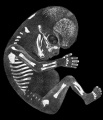
Fig 2 Embryo No. 284
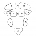
Fig 3 Occipital Ossification
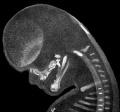
Fig 4 Embryo No. 284 Head
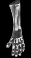
Fig 5 Embryo No. 300 Hand
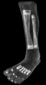
Fig 6 Embryo No. 300 Leg
- Tables
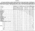
Table 1 Ossification Centers Month 2
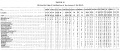
Table 2 Skull Ossification
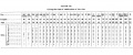
Table 3 Rib Ossification
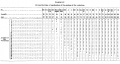
Table 4 Vertebra Ossification
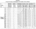
Table 5 Vertebra Body Ossification
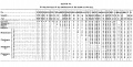
Table 6 Arm Ossification
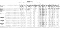
Table 7 Leg Ossification
| Historic Disclaimer - information about historic embryology pages |
|---|
|
- Ossification Centers in Human Embryos: Fig 1 Embryo No. 333 | Fig 2 Embryo No. 284 | Fig 3 Occipital Ossification | Fig 4 Embryo No. 284 Head | Fig 5 Embryo No. 300 Hand | Fig 6 Embryo No. 300 Leg | Table 1 Ossification Centers Month 2 | Table 2 Skull Ossification | Table 3 Rib Ossification | Table 4 Vertebra Ossification | Table 5 Vertebra Body Ossification | Table 6 Arm Ossification | Table 7 Leg Ossification | Franklin Mall | Bone Development Timeline
Reference
Mall FP. On ossification centers in human embryos less than one hundred days old. (1906) Amer. J Anat. 5:433-458.
Cite this page: Hill, M.A. (2026, July 4) Embryology Paper - On Ossification Centers in Human Embryos. Retrieved from https://embryology.med.unsw.edu.au/embryology/index.php/Paper_-_On_Ossification_Centers_in_Human_Embryos
- © Dr Mark Hill 2026, UNSW Embryology ISBN: 978 0 7334 2609 4 - UNSW CRICOS Provider Code No. 00098G