Paper - Human oocyte showing first polar body and metaphase stage in formation of second polar body
| Embryology - 27 Apr 2024 |
|---|
| Google Translate - select your language from the list shown below (this will open a new external page) |
|
العربية | català | 中文 | 中國傳統的 | français | Deutsche | עִברִית | हिंदी | bahasa Indonesia | italiano | 日本語 | 한국어 | မြန်မာ | Pilipino | Polskie | português | ਪੰਜਾਬੀ ਦੇ | Română | русский | Español | Swahili | Svensk | ไทย | Türkçe | اردو | ייִדיש | Tiếng Việt These external translations are automated and may not be accurate. (More? About Translations) |
Dixon AF. Human oocyte showing first polar body and metaphase stage in formation of second polar body. (1927) Irish Jour. Med. Sci., 53: 149-151.
| Historic Disclaimer - information about historic embryology pages |
|---|
| Pages where the terms "Historic" (textbooks, papers, people, recommendations) appear on this site, and sections within pages where this disclaimer appears, indicate that the content and scientific understanding are specific to the time of publication. This means that while some scientific descriptions are still accurate, the terminology and interpretation of the developmental mechanisms reflect the understanding at the time of original publication and those of the preceding periods, these terms, interpretations and recommendations may not reflect our current scientific understanding. (More? Embryology History | Historic Embryology Papers) |
Human Oocyte showing First Polar Body and Metaphase Stage in Formation of Second Polar Body
By A. Francis Dixon
(1929)
Introduction
The section, portions of which are illustrated in this note, shows a ripe human ovarian follicle containing an oöcyte in which the first polar body is present, and in which the chromosomes of the remaining nucleus are in the metaphase of the mitosis which would have given rise to the second polar body.
The specimen was received through the kindness of Doctor Bethel Solomons, and was removed during the course of an operation from the body of a young adult woman. The ovary was placed in formol solution while still warm, and sections obtained from it were stained in Delafield’s haematoxylin followed by orange A G.
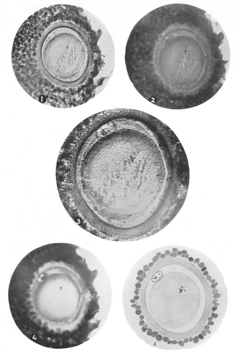
The ovarian follicle which contained the oöcyte described was not spherical, but was. flattened and somewhat compressed between two other larger follicles. The oöcyte, including its oölemma (zona pellucida), measures 0.1 to 0.13 mm. and is not quite spherical, but slightly ovoid in form. (Fig. 1). The otilemma measures .01 mm. in thickness and is coloured orange in the section. It exhibits no radial, orconcentric, markings. The cells of the cumulus oliphoros (discus proligerus), which immediately surround the otilemma and which would have formed the corona radiate, are in ‘many places not actually in contact with the oölemma, but are separated from it by irregularly shaped spaces crossed by fine fibrils. The outer aspect of the a oölemma appears in some places to be pulled out into little angular processes by these fibrils as they pass towards the investing cells of the cumulus , (Figs. 1 and 3). The minute fibrils appear to suspend the entire oöcyte and its oéilemma in a space bounded by the surrounding cells of the cumulus. These spaces have been illustrated by Professor Thomson‘ in his paper on the maturation of the human ovum, and he shows them larger and more extensive than in the specimen here described. The cytoplasm of the oöcyte is nearly spherical and measures .08 to .09. mm. in diameter. Between it and the oölemma there is a narrow crescentic spacethe perivitelline space— in which may be seen, not far from its widest part, the first polar body. The nucleus of this body has a clearly defined wall, and within it is a well-stained nucleolus (Fig. 3).
The chromatin of the nucleus is arranged in a series of communicating strands forming an apparently irregular net. work. This network can easily be traced when the nucleus is carefully focussed in different planes. (Fig 5). The nucleus of the polar body lies with its long axis immediately beneath the overlying oölemma. The perivitelline space, which measures .01 mm. in its widest part, is filled by a finely granular substance which is stained a lighter violet colour and is less dense than the adjacent cytoplasm of the oticyte. At each pole of the nucleus of the polar body the material filling the perivitclline space appears to be slightly denser than elsewhere, and probably represents the cytoplasm of the polar body. At one place only, the material filling the perivitclline space appears to have contracted away from the cytoplasm of the oöcyte; here a very narrow clear slit is visible. This slit is opposite to the place where the cytoplasm is practically in contact with the oölemma. (Fig. 2). There is no indication of a vitelline membrane, and the outer surface of the cytoplasm forms the inner wall of the perivitelline space. The cytoplasm of the oöcyte is for the most part very homogeneous in appearance, and shows very few inclusions. It is stained slightly darker than the contents of the perivitelline space, and its surface where it has been cut by the knife resembles, under the microscope, the cut surface of a mass of gelatine. The section is more than 15 microns in thickness, and when the cytoplasm is carefully focussed a few rod shaped chromosomes may be very distinctly seen. These chromosomes are grouped together in such a way as to show that the section has passed obliquely through the_ equatorial plate of the dividing nucleus in the metaphase of its mitosis. (Figs. 2 and 4). Three chromosomes may be identified in transverse, or slightly oblique section; and in addition at least one characteristically V-shaped dividing chromosome may be seen lying obliquely in the thickness of the section. No trace of achromatic spindle, or of centrosomes, can be recognised. Unfortunately, the next section to the one illustrated is not sufficiently complete to furnish any further details as to the arrangement, or actual total number of the chromosomes of the oticyte.
It is believed that oöcyte II possesses 24 chromosomes, and the first polar body 24 also. In other words, each contains one half of the original 48 chromosomes which existed in oöcyte I, from which both oöcyte II and the first polar body arise by heterotype mitosis (reduction division).
The nucleus of the polar body in the specimen described in this note is not in a stage in which it is possible to recognise, or count, the individual chromosomes.
There does not appear to be any doubt that the oöcyte represents oöcyte II in the metaphase stage of its division to form the second polar body.
The specimen confirms the view put forward by Professor Arthur Thomson, namely, that in the human subject the second polar body is formed before the oöcyte leaves the follicle, and before fertilisation has taken place. In this detail of its maturation the human oöcyte differs then from what is known to occur in other mammals. Further, the appearance of the first polar body nucleus indicates that the division of this body, if it occurs in the human subject, takes place after the formation of the second polar body. Professor Thomson believes that the first polar body divides coincident with the extrusion of the second polar body.
I am pleased to be able to say that Professor James Bronte Gatenby, who very kindly examined the specimen for me, states that he would describe it as a “ ripe oöcyte showing first polar body and second polar body in metaphase of formation ” ; also some other skilled cytologists to whom I have had an opportunity of showing the specimen are satisfied that the condition is substantially as described in this note. The rarity of finding a human oöcyte illustrating a stage in the process of maturation has led me to think the specimen worth recording and illustrating, in spite of the fact that I have failed to secure a series of sections such as could be used to estimate accurately the number of chromosomes in the ripe oöcyte.
The four photographs, 1, 2, 3 and 4, were taken at different depths of focus. In each the oölemma, the nucleus of the first polar body, the perivitelline space and cytoplasm of the oöcyte are shown, also the cells of the cumulus oöphoros.
Fig. 1 shows the ovoid outline of the oölemma and the more circular form of the cytoplasm as seen in section. The minute irregular intervals and the fine fibrils which cross the space between the oölemma and the cells of the cumulus are seen except on the right side of the photograph. [x300 approx.]
Fig. 2 shows in addition some of the chromosomes of the oöcyte; the narrow cleft where the perivitelline substance has contracted away from the cytoplasm is seen on the right side of the photograph. [x300 approx.]
Fig. 3 under higher magnification than the other photographs. The nucleus and nucleolus of the polar body are seen in more detail. In the space surrounding the oölemma the radiating fibrils, some of which are cut transversely, are seen. [x560 approx.]
Fig. 4. The microscope has been focussed to show some of the individual chromosomes of the dividing nucleus of the oöcyte. x300 approx.
Fig. 5. Semi-diagrammatic sketch made by combining various optical sections. [X400 approx.]
Cite this page: Hill, M.A. (2024, April 27) Embryology Paper - Human oocyte showing first polar body and metaphase stage in formation of second polar body. Retrieved from https://embryology.med.unsw.edu.au/embryology/index.php/Paper_-_Human_oocyte_showing_first_polar_body_and_metaphase_stage_in_formation_of_second_polar_body
- © Dr Mark Hill 2024, UNSW Embryology ISBN: 978 0 7334 2609 4 - UNSW CRICOS Provider Code No. 00098G


