File:CNS secondary vesicles.jpg
Original file (987 × 562 pixels, file size: 81 KB, MIME type: image/jpeg)
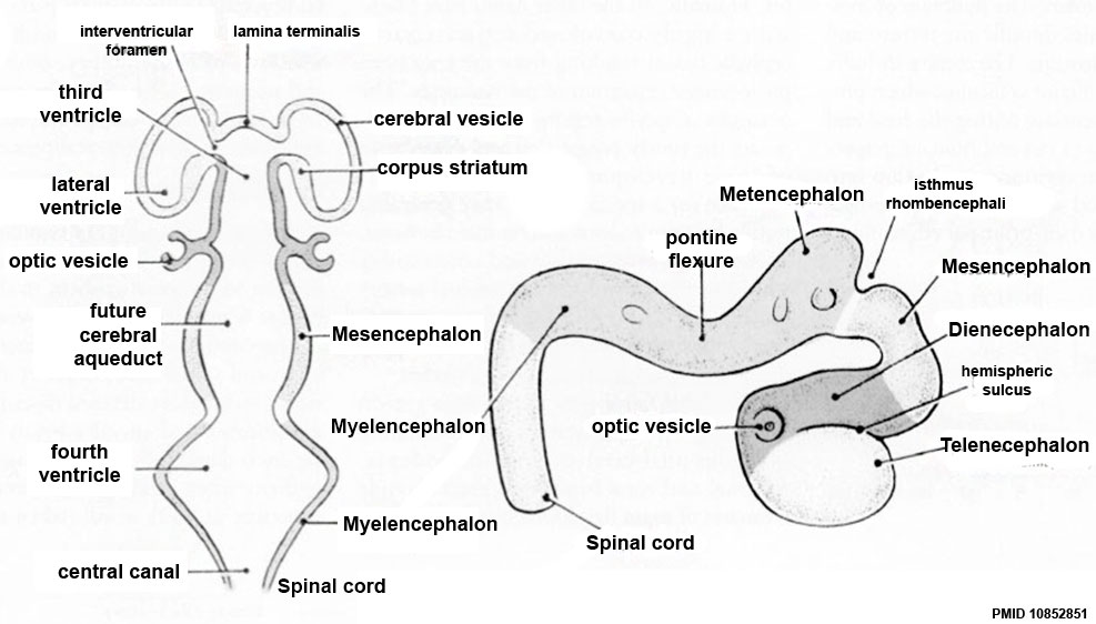
Central Nervous System - Secondary Brain Vesicles
These 5 secondary vesicles developed from the initial 3 primary vesicles of the early neural tube, by division of the forebrain and hindbrain regions.
The more mature brain with five brain vesicles: the horizontal and lateral views correspond to GD 11.5 in rats and 33 ± 1 in humans.
| Neural Tube | Primary Vesicles | Secondary Vesicles | Adult Structures |
|---|---|---|---|
| week 3 | week 4 | week 5 | adult |
| prosencephalon (forebrain) | telencephalon | Rhinencephalon, Amygdala, hippocampus, cerebrum (cortex), hypothalamus, pituitary | Basal Ganglia, lateral ventricles | |
| diencephalon | epithalamus, thalamus, Subthalamus, pineal, posterior commissure, pretectum, third ventricle | ||
| mesencephalon (midbrain) | mesencephalon | tectum, Cerebral peduncle, cerebral aqueduct, pons | |
| rhombencephalon (hindbrain) | metencephalon | cerebellum | |
| myelencephalon | medulla oblongata, isthmus | ||
| spinal cord, pyramidal decussation, central canal | |||
Reference
Rice D & Barone S. (2000). Critical periods of vulnerability for the developing nervous system: evidence from humans and animal models. Environ. Health Perspect. , 108 Suppl 3, 511-33. PMID: 10852851
Copyright: Reproduced with permission from Environmental Health Perspectives (EHP) is a publication of the U.S. Government. Publication of EHP lies in the public domain and is therefore without copyright.
Cite this page: Hill, M.A. (2024, April 29) Embryology CNS secondary vesicles.jpg. Retrieved from https://embryology.med.unsw.edu.au/embryology/index.php/File:CNS_secondary_vesicles.jpg
- © Dr Mark Hill 2024, UNSW Embryology ISBN: 978 0 7334 2609 4 - UNSW CRICOS Provider Code No. 00098G
File history
Click on a date/time to view the file as it appeared at that time.
| Date/Time | Thumbnail | Dimensions | User | Comment | |
|---|---|---|---|---|---|
| current | 16:49, 18 May 2017 | | 987 × 562 (81 KB) | Z8600021 (talk | contribs) | |
| 01:02, 11 August 2009 | 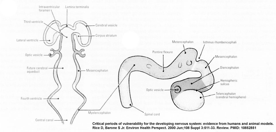 | 961 × 462 (58 KB) | MarkHill (talk | contribs) | CNS primary vesicles cartoon Figure-3CD-PMID10852851.jpg Image Source: Critical periods of vulnerability for the developing nervous system: evidence from humans and animal models. Rice D, Barone S Jr. Environ Health Perspect. 2000 Jun;108 Suppl 3:511-33 |
You cannot overwrite this file.
File usage
The following 27 pages use this file:
- 2009 Lecture 6
- 2010 BGD Lecture - Development of the Embryo/Fetus 2
- 2010 BGD Practical 6 - Week 5
- 2010 Lecture 6
- BGDA Lecture - Development of the Embryo/Fetus 2
- BGDA Lecture - Development of the Nervous System
- BGDA Practical 7 - Week 5
- Lecture - Ectoderm Development
- Lecture - Neural Development
- Neural - Amygdala Development
- Neural - Basal Ganglia Development
- Neural - Cerebellum Development
- Neural - Cerebrum Development
- Neural - Diencephalon Development
- Neural - Hippocampus Development
- Neural - Medulla Oblongata Development
- Neural - Metencephalon Development
- Neural - Myelencephalon Development
- Neural - Pons Development
- Neural - Rhinencephalon Development
- Neural - Spinal Cord Development
- Neural - Tectum Development
- Neural - Telencephalon Development
- Neural - Thalamus Development
- Neural System Development
- S
- Talk:BGDA Lecture - Development of the Nervous System