Fetal Development - 12 Weeks
Introduction
Week 12 or clinical age GA week 14.
Images on this current page show head ossification occurring during the early fetal period (12 weeks approx 92 mm CRL in size). The head undergoes two different forms of ossification (endochondral and intramembranous) in separate regions of the skull. Furthermore the two images (lateral and medial) identify regions of cartilage development. Compare this 12 week fetus with the earlier 10 week Fetal and Carnegie stage embryos: size, head proportions, brain, head, skeletal development.
- 12 Week Images: Sagittal unlabeled | Sagittal labeled | Sagittal medial view | Sagittal lateral view | Pituitary unlabeled | Pituitary labeled | Tongue | Skull Development | Head Development
| Fetal Links: fetal | Week 10 | Week 12 | second trimester | third trimester | fetal neural | Fetal Blood Sampling | fetal growth restriction | birth | birth weight | preterm birth | Developmental Origins of Health and Disease | macrosomia | BGD Practical | Medicine Lecture | Science Lecture | Lecture Movie | Category:Human Fetus | Category:Fetal | |||
|
12 week Head
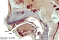
These are 2 views of the same 12 week 92 mm CRL human fetus head, double stained to show both cartilage (blue) and newly-formed bone (red).
Lateral View
In this lateral (external) view, note the distribution of new bone in the plates of the cranial vault, temporal bone, orbit, upper jaw (maxilla) and lower jaw (mandible) regions. Bony regions in the lower jaw (mandible) region also show spaces where tooth formation is occurring.

Medial View
In this medial (internal) view, note the distribution of cartilage from the nasal region through the base of the skull to the atlas/axis (with new bone forming). See also the original Meckel's cartilage within the newly forming bony mandible.

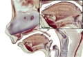
Mandible
Selected extract of above images showing 2 views (lateral/external left, medial/internal right) of the mandible region.
Note: Cartilage in the neck where the hyoid bone will eventually form. Meckel's cartilage visible in the medial view within the newly forming bone.
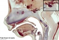
Nose and Mouth
Selected medial head view showing key features of head musculoskeletal and neurological development. Note extensive nasal cartilage, nasal conchae, pituitary, secondary palate, oral cavity, tongue, mandible, hyoid, choana, oropharynx.


Unlabeled
Labeled
Pituitary

Pituitary labeled
Tongue labeled


| Carnegie Embryos - Sensenig (1951) - Spinal Cord Meninges | |||||
|---|---|---|---|---|---|
| Age Group (gestation age) |
Embryo no. |
Crown Rump Length |
Thickness of sections (microns) |
Plane of section |
Illustration |
| 12 weeks | 1455 | 78.5 | 50 | Sagittal | |
| 172 | 80.0 | 100 | Transverse | ||
| Template:CE72181 | 80.0 | 20 | Transverse | Figs. 17, 18, pl. 3 | |
| |||||
References
Sensenig EC. The early development of the meninges of the spinal cord in human embryos. (1951) Contrib. Embryol., Carnegie Inst. Wash. Publ. 611.
Reviews
Articles
{{#pmid:19049908]]
Wilkin H, Tuohy J & Theewis W. (2000). Prenatal diagnosis of fragile X and Turner mosaicism in a 12-week fetus. Prenat. Diagn. , 20, 854-5. PMID: 11038471
Robb A, Forsyth L & Tolmie J. (1987). Partial trisomy 17q and a generalised bone dysplasia in a 12 week fetus. J. Med. Genet. , 24, 502-4. PMID: 3656375
Search PubMed
Note: Week 12 post-fertilization age (used throughout this current website) is Gestational Age GA Week 14 (LMP). Searches for clinical Week 14 gestational age will match this post-fertilization age.
Search PubMed Now: week 12 fetus | week 14 fetus
Terms
choana - the posterior nasal aperture which is the passageway from the back of one side of the nose to the throat. There are two choanae, one on either side of the nose.
mandible
nasal conchae - the thin scroll-shaped bony elements forming three groove-like air passages/chambers in the nasal cavities.
oropharynx - the oral (within the mouth) part of the pharynx which extends from the uvula to the level of the hyoid bone.
Also note the developing tongue musculature and its mandibular attachment site.
Glossary Links
- Glossary: A | B | C | D | E | F | G | H | I | J | K | L | M | N | O | P | Q | R | S | T | U | V | W | X | Y | Z | Numbers | Symbols | Term Link
Cite this page: Hill, M.A. (2026, July 24) Embryology Fetal Development - 12 Weeks. Retrieved from https://embryology.med.unsw.edu.au/embryology/index.php/Fetal_Development_-_12_Weeks
- © Dr Mark Hill 2026, UNSW Embryology ISBN: 978 0 7334 2609 4 - UNSW CRICOS Provider Code No. 00098G