Category:Carnegie Embryo 95
This Embryology category shows pages and images that relate to the Carnegie Collection Embryo No. 95. This embryo would be early fetal development Week 10 based upon the CRL 50 mm.
| Fetal Links: fetal | Week 10 | Week 12 | second trimester | third trimester | fetal neural | Fetal Blood Sampling | fetal growth restriction | birth | birth weight | preterm birth | Developmental Origins of Health and Disease | macrosomia | BGD Practical | Medicine Lecture | Science Lecture | Lecture Movie | Category:Human Fetus | Category:Fetal | |||
|
| Carnegie Collection - Fetal | |||||||||||||||||||||||||||||||||||||||||||||||||||||||||||||||||||||||||||||||||||||||||||||||||||||||||||||||||||||||||||||||||||||||||||||||||||||||||||||||||||||||||||||||||||||||||||||||||||||||||||||||||||||||||
|---|---|---|---|---|---|---|---|---|---|---|---|---|---|---|---|---|---|---|---|---|---|---|---|---|---|---|---|---|---|---|---|---|---|---|---|---|---|---|---|---|---|---|---|---|---|---|---|---|---|---|---|---|---|---|---|---|---|---|---|---|---|---|---|---|---|---|---|---|---|---|---|---|---|---|---|---|---|---|---|---|---|---|---|---|---|---|---|---|---|---|---|---|---|---|---|---|---|---|---|---|---|---|---|---|---|---|---|---|---|---|---|---|---|---|---|---|---|---|---|---|---|---|---|---|---|---|---|---|---|---|---|---|---|---|---|---|---|---|---|---|---|---|---|---|---|---|---|---|---|---|---|---|---|---|---|---|---|---|---|---|---|---|---|---|---|---|---|---|---|---|---|---|---|---|---|---|---|---|---|---|---|---|---|---|---|---|---|---|---|---|---|---|---|---|---|---|---|---|---|---|---|---|---|---|---|---|---|---|---|---|---|---|---|---|---|---|---|
| Serial No. | Size CRL (mm) | Grade | Fixative | Embedding Medium | Plane | Thinness (µm) | Stain | Point Score | Sex | Year | Notes | ||||||||||||||||||||||||||||||||||||||||||||||||||||||||||||||||||||||||||||||||||||||||||||||||||||||||||||||||||||||||||||||||||||||||||||||||||||||||||||||||||||||||||||||||||||||||||||||||||||||||||||||
| 95 | 40 | catalogued as CRL 40 but development suggests 50 stage. Spinal cord - Kunitomo (1920)[1] Colon - Lineback (1920)[2] | |||||||||||||||||||||||||||||||||||||||||||||||||||||||||||||||||||||||||||||||||||||||||||||||||||||||||||||||||||||||||||||||||||||||||||||||||||||||||||||||||||||||||||||||||||||||||||||||||||||||||||||||||||||||
| 96 | 50 | Brain venous sinuses - Streeter (1915)[3] Spinal cord - Kunitomo (1920)[1] Brain vascular - Streeter (1921)[4] Brain weight - Jenkins (1921)[5] | |||||||||||||||||||||||||||||||||||||||||||||||||||||||||||||||||||||||||||||||||||||||||||||||||||||||||||||||||||||||||||||||||||||||||||||||||||||||||||||||||||||||||||||||||||||||||||||||||||||||||||||||||||||||
| 142 | 125 | Spinal cord - Kunitomo (1920)[1] | |||||||||||||||||||||||||||||||||||||||||||||||||||||||||||||||||||||||||||||||||||||||||||||||||||||||||||||||||||||||||||||||||||||||||||||||||||||||||||||||||||||||||||||||||||||||||||||||||||||||||||||||||||||||
| 145 | 33 | Spinal cord - Kunitomo (1920)[1] | |||||||||||||||||||||||||||||||||||||||||||||||||||||||||||||||||||||||||||||||||||||||||||||||||||||||||||||||||||||||||||||||||||||||||||||||||||||||||||||||||||||||||||||||||||||||||||||||||||||||||||||||||||||||
| 184 | 50 | 34 vertebrae, 31 spinal ganglia, Spinal cord - Kunitomo (1920)[1] | |||||||||||||||||||||||||||||||||||||||||||||||||||||||||||||||||||||||||||||||||||||||||||||||||||||||||||||||||||||||||||||||||||||||||||||||||||||||||||||||||||||||||||||||||||||||||||||||||||||||||||||||||||||||
| 211 | 33 | 34 vertebra, 31 spinal ganglia, Spinal cord - Kunitomo (1920)[1] | |||||||||||||||||||||||||||||||||||||||||||||||||||||||||||||||||||||||||||||||||||||||||||||||||||||||||||||||||||||||||||||||||||||||||||||||||||||||||||||||||||||||||||||||||||||||||||||||||||||||||||||||||||||||
| 217 | 45 | Male | Genital - Spaulding (1921)[6] | ||||||||||||||||||||||||||||||||||||||||||||||||||||||||||||||||||||||||||||||||||||||||||||||||||||||||||||||||||||||||||||||||||||||||||||||||||||||||||||||||||||||||||||||||||||||||||||||||||||||||||||||||||||||
| 300 | 73 | 85 days, Bone ossification - Mall (1906)[7] | |||||||||||||||||||||||||||||||||||||||||||||||||||||||||||||||||||||||||||||||||||||||||||||||||||||||||||||||||||||||||||||||||||||||||||||||||||||||||||||||||||||||||||||||||||||||||||||||||||||||||||||||||||||||
| 362 | 30 | Spinal cord - Kunitomo (1920)[1] | |||||||||||||||||||||||||||||||||||||||||||||||||||||||||||||||||||||||||||||||||||||||||||||||||||||||||||||||||||||||||||||||||||||||||||||||||||||||||||||||||||||||||||||||||||||||||||||||||||||||||||||||||||||||
| 448 | 52 | Colon - Lineback (1920)[2] | |||||||||||||||||||||||||||||||||||||||||||||||||||||||||||||||||||||||||||||||||||||||||||||||||||||||||||||||||||||||||||||||||||||||||||||||||||||||||||||||||||||||||||||||||||||||||||||||||||||||||||||||||||||||
| 449 | 36 | Spinal cord - Kunitomo (1920)[1] | |||||||||||||||||||||||||||||||||||||||||||||||||||||||||||||||||||||||||||||||||||||||||||||||||||||||||||||||||||||||||||||||||||||||||||||||||||||||||||||||||||||||||||||||||||||||||||||||||||||||||||||||||||||||
| 538 | |||||||||||||||||||||||||||||||||||||||||||||||||||||||||||||||||||||||||||||||||||||||||||||||||||||||||||||||||||||||||||||||||||||||||||||||||||||||||||||||||||||||||||||||||||||||||||||||||||||||||||||||||||||||||
| 590 | 21 to 23 | Male | Genital - Spaulding (1921)[6] | ||||||||||||||||||||||||||||||||||||||||||||||||||||||||||||||||||||||||||||||||||||||||||||||||||||||||||||||||||||||||||||||||||||||||||||||||||||||||||||||||||||||||||||||||||||||||||||||||||||||||||||||||||||||
| 607 | 37 | Male | Genital - Spaulding (1921)[6] | ||||||||||||||||||||||||||||||||||||||||||||||||||||||||||||||||||||||||||||||||||||||||||||||||||||||||||||||||||||||||||||||||||||||||||||||||||||||||||||||||||||||||||||||||||||||||||||||||||||||||||||||||||||||
| 625 | 220 | Temporomandibular joint - Moffatt (1957)[8] | |||||||||||||||||||||||||||||||||||||||||||||||||||||||||||||||||||||||||||||||||||||||||||||||||||||||||||||||||||||||||||||||||||||||||||||||||||||||||||||||||||||||||||||||||||||||||||||||||||||||||||||||||||||||
| 662 | 80 | Spinal cord - Kunitomo (1920)[1] | |||||||||||||||||||||||||||||||||||||||||||||||||||||||||||||||||||||||||||||||||||||||||||||||||||||||||||||||||||||||||||||||||||||||||||||||||||||||||||||||||||||||||||||||||||||||||||||||||||||||||||||||||||||||
| 693 | 45 | Male | Genital - Spaulding (1921)[6] | ||||||||||||||||||||||||||||||||||||||||||||||||||||||||||||||||||||||||||||||||||||||||||||||||||||||||||||||||||||||||||||||||||||||||||||||||||||||||||||||||||||||||||||||||||||||||||||||||||||||||||||||||||||||
| 847 | 58.8 | Male | Genital - Spaulding (1921)[6] | ||||||||||||||||||||||||||||||||||||||||||||||||||||||||||||||||||||||||||||||||||||||||||||||||||||||||||||||||||||||||||||||||||||||||||||||||||||||||||||||||||||||||||||||||||||||||||||||||||||||||||||||||||||||
| 858 | 57.25 | Temporomandibular joint - Moffatt (1957)[8] | |||||||||||||||||||||||||||||||||||||||||||||||||||||||||||||||||||||||||||||||||||||||||||||||||||||||||||||||||||||||||||||||||||||||||||||||||||||||||||||||||||||||||||||||||||||||||||||||||||||||||||||||||||||||
| 922 | 37 | ||||||||||||||||||||||||||||||||||||||||||||||||||||||||||||||||||||||||||||||||||||||||||||||||||||||||||||||||||||||||||||||||||||||||||||||||||||||||||||||||||||||||||||||||||||||||||||||||||||||||||||||||||||||||
| 928 | 120 | Spinal cord - Kunitomo (1920)[1] | |||||||||||||||||||||||||||||||||||||||||||||||||||||||||||||||||||||||||||||||||||||||||||||||||||||||||||||||||||||||||||||||||||||||||||||||||||||||||||||||||||||||||||||||||||||||||||||||||||||||||||||||||||||||
| 948 | 45 | Male | Genital - Spaulding (1921)[6] | ||||||||||||||||||||||||||||||||||||||||||||||||||||||||||||||||||||||||||||||||||||||||||||||||||||||||||||||||||||||||||||||||||||||||||||||||||||||||||||||||||||||||||||||||||||||||||||||||||||||||||||||||||||||
| 972 | 37 | 34 vertebrae, 30 spinal ganglia, Spinal cord - Kunitomo (1920)[1] | |||||||||||||||||||||||||||||||||||||||||||||||||||||||||||||||||||||||||||||||||||||||||||||||||||||||||||||||||||||||||||||||||||||||||||||||||||||||||||||||||||||||||||||||||||||||||||||||||||||||||||||||||||||||
| 1318 | 37 | Temporomandibular joint - Moffatt (1957)[8] | |||||||||||||||||||||||||||||||||||||||||||||||||||||||||||||||||||||||||||||||||||||||||||||||||||||||||||||||||||||||||||||||||||||||||||||||||||||||||||||||||||||||||||||||||||||||||||||||||||||||||||||||||||||||
| 1388 | 51 | Female | Genital - Spaulding (1921)[6] | ||||||||||||||||||||||||||||||||||||||||||||||||||||||||||||||||||||||||||||||||||||||||||||||||||||||||||||||||||||||||||||||||||||||||||||||||||||||||||||||||||||||||||||||||||||||||||||||||||||||||||||||||||||||
| 1455 | 78.5 | Temporomandibular joint - Moffatt (1957)[8] | |||||||||||||||||||||||||||||||||||||||||||||||||||||||||||||||||||||||||||||||||||||||||||||||||||||||||||||||||||||||||||||||||||||||||||||||||||||||||||||||||||||||||||||||||||||||||||||||||||||||||||||||||||||||
| 1591 | 36 | subcutaneous vascular plexus - Finley (1923)[9] | |||||||||||||||||||||||||||||||||||||||||||||||||||||||||||||||||||||||||||||||||||||||||||||||||||||||||||||||||||||||||||||||||||||||||||||||||||||||||||||||||||||||||||||||||||||||||||||||||||||||||||||||||||||||
| 1656 | 67 | 34 vertebrae, Spinal cord - Kunitomo (1920)[1] | |||||||||||||||||||||||||||||||||||||||||||||||||||||||||||||||||||||||||||||||||||||||||||||||||||||||||||||||||||||||||||||||||||||||||||||||||||||||||||||||||||||||||||||||||||||||||||||||||||||||||||||||||||||||
| 1686 | 40 | Male | Genital - Spaulding (1921)[6] | ||||||||||||||||||||||||||||||||||||||||||||||||||||||||||||||||||||||||||||||||||||||||||||||||||||||||||||||||||||||||||||||||||||||||||||||||||||||||||||||||||||||||||||||||||||||||||||||||||||||||||||||||||||||
| 3990 | 49 | Temporomandibular joint - Moffatt (1957)[8] | |||||||||||||||||||||||||||||||||||||||||||||||||||||||||||||||||||||||||||||||||||||||||||||||||||||||||||||||||||||||||||||||||||||||||||||||||||||||||||||||||||||||||||||||||||||||||||||||||||||||||||||||||||||||
| 4473 | 43 | 20 | Spinal cord meninges - Sensenig (1951)[10] | ||||||||||||||||||||||||||||||||||||||||||||||||||||||||||||||||||||||||||||||||||||||||||||||||||||||||||||||||||||||||||||||||||||||||||||||||||||||||||||||||||||||||||||||||||||||||||||||||||||||||||||||||||||||
| 4475 | 48 | 20 | Spinal cord meninges - Sensenig (1951)[10] | ||||||||||||||||||||||||||||||||||||||||||||||||||||||||||||||||||||||||||||||||||||||||||||||||||||||||||||||||||||||||||||||||||||||||||||||||||||||||||||||||||||||||||||||||||||||||||||||||||||||||||||||||||||||
| 5652 | 49 | Temporomandibular joint - Moffatt (1957)[8] | |||||||||||||||||||||||||||||||||||||||||||||||||||||||||||||||||||||||||||||||||||||||||||||||||||||||||||||||||||||||||||||||||||||||||||||||||||||||||||||||||||||||||||||||||||||||||||||||||||||||||||||||||||||||
| 6581 | 75 | Temporomandibular joint - Moffatt (1957)[8] | |||||||||||||||||||||||||||||||||||||||||||||||||||||||||||||||||||||||||||||||||||||||||||||||||||||||||||||||||||||||||||||||||||||||||||||||||||||||||||||||||||||||||||||||||||||||||||||||||||||||||||||||||||||||
| 7218 | 80 | 20 um | Spinal cord meninges - Sensenig (1951)[10] | ||||||||||||||||||||||||||||||||||||||||||||||||||||||||||||||||||||||||||||||||||||||||||||||||||||||||||||||||||||||||||||||||||||||||||||||||||||||||||||||||||||||||||||||||||||||||||||||||||||||||||||||||||||||
| 1597b | 47 | Female | Genital - Spaulding (1921)[6] | ||||||||||||||||||||||||||||||||||||||||||||||||||||||||||||||||||||||||||||||||||||||||||||||||||||||||||||||||||||||||||||||||||||||||||||||||||||||||||||||||||||||||||||||||||||||||||||||||||||||||||||||||||||||
| 2250a | 40 | Female | Genital - Spaulding (1921)[6] | ||||||||||||||||||||||||||||||||||||||||||||||||||||||||||||||||||||||||||||||||||||||||||||||||||||||||||||||||||||||||||||||||||||||||||||||||||||||||||||||||||||||||||||||||||||||||||||||||||||||||||||||||||||||
| 2250b | 36 | Female | Genital - Spaulding (1921)[6] | ||||||||||||||||||||||||||||||||||||||||||||||||||||||||||||||||||||||||||||||||||||||||||||||||||||||||||||||||||||||||||||||||||||||||||||||||||||||||||||||||||||||||||||||||||||||||||||||||||||||||||||||||||||||
| This table currently contains only has embryo number information.
Abbreviations
| |||||||||||||||||||||||||||||||||||||||||||||||||||||||||||||||||||||||||||||||||||||||||||||||||||||||||||||||||||||||||||||||||||||||||||||||||||||||||||||||||||||||||||||||||||||||||||||||||||||||||||||||||||||||||
References
| |||||||||||||||||||||||||||||||||||||||||||||||||||||||||||||||||||||||||||||||||||||||||||||||||||||||||||||||||||||||||||||||||||||||||||||||||||||||||||||||||||||||||||||||||||||||||||||||||||||||||||||||||||||||||
| |||||||||||||||||||||||||||||||||||||||||||||||||||||||||||||||||||||||||||||||||||||||||||||||||||||||||||||||||||||||||||||||||||||||||||||||||||||||||||||||||||||||||||||||||||||||||||||||||||||||||||||||||||||||||
References
- ↑ Westerway SC, Davison A & Cowell S. (2000). Ultrasonic fetal measurements: new Australian standards for the new millennium. Aust N Z J Obstet Gynaecol , 40, 297-302. PMID: 11065037
- ↑ Kunitomo K. The development and reduction of the tail and of the caudal end of the spinal cord (1920) Contrib. Embryol., Carnegie Inst. Wash. Publ. 272, 9: 163-198.
- ↑ Lineback PE. Studies on the longitudinal muscle of the human colon, with special reference to the development of the taeniae. (1920) Contrib. Embryol., Carnegie Inst. Wash. Publ. 50
Embryo No. 95, 50 mm Crown-Rump Length
Kunitomo K. The development and reduction of the tail and of the caudal end of the spinal cord (1920) Contrib. Embryol., Carnegie Inst. Wash. Publ. 272, 9: 163-198.
Although embryo No. 95 is recorded in the catalogue of the Carnegie Collection as 40 mm crown-rump length, its state of development more nearly corresponds with a 50 mm. embryo, and on this account I have used the latter measurement in the heading. This specimen has 35 vertebrae. The last one is very small and partly fused with the one above it. The column presents a ventral bend at the thirty-first vertebra, giving the typical coccygeal curve. The chorda dorsalis is disappearing in certain areas in the vertebral bodies as far down as the thirtieth vertebra, but in each intervertebral space a fragment remains. Caudal to the thirtieth vertebra the condition of the chorda remains the same as in the younger specimens, and in the thirty-second it gives off a short dorsal branch. The caudal end is more simple in form than in the younger stages, but I am inclined to believe that at an earlier stage it too was winding, as one can see in the thirty-fifth vertebra a few detached globules which probably at an earlier stage were continuous with the chorda and with it formed a terminal loop.
At the caudal end of the spinal cord are two groups of cells connected by a cell-strand. The more caudal one is situated dorsal to the thirty-fourth and thirty-fifth vertebra*; it is somewhat larger than the other, is oblong in form and incloses an oval cavity - a fragment of the central canal of the spinal cord. The other group of cells is situated dorsal to the thirty-second and thirty-third vertebra* and incloses a long, narrow cavity. The ventriculus terminalis extends the length of two vertebrae - the twenty-ninth and thirtieth. At this stage it has acquired its adult form. In none of the earlier specimens have I noted it so perfectly developed, although embryos No. 449, 30 mm., and No. 199, 35 mm., show a cavity at the caudal end of the central canal as the ])rimordium of the ventriculus. In this specimen the structure is cylindrical in shape, has six walls, and measures 0.87 mm long, 0.23 mm. deep, and 0.52 mm. wide. The ventral wall is concave, the dorsal convex, the sides slightly concave. The upper wall or ceiling is irregular and at the front presents a long, narrow diverticulum directed cranio-ventral. Behind this diverticulum is a narrow channel which connects the ventriculus terminalis and the central canal of the spinal cord. The ventriculus terminalis is embedded in the nerve-fibers of the cord. The filum terminale extends from the caudal end of the conus meduUaris, at the level of the thirty-first vertebra, to a point between the thirly-third and thirty-fourth vertebrae close to the column. It is covered by a membrane of the spinal cord and passes through the ventral side of the cell groups at the caudal end of the medullary tube. The pia mater covers closely the whole surface of the spinal cord; it contains blood capillaries, and is visible at the conus meduUaris. The dura mater, which envelops loosely the pia mater, adheres to the wall of the vertebral canal as far as the midlevel of the thirty-first vertebra, at which point it leaves the wall and unites with the caudal end of the conus medullaris. This portion constitutes the primordium of the bursa durge matris. After the dura mater reaches the conus medullaris it envelops the pia mater quite closely, both following a caudal course and forming a sheath for the filum terminale. The point at which these membranes terminate can not be definitely decided. It is probable that the pia mater extends nearly to the end of the filum terminale between the thirty-third and thirty-fourth vertebrae. The fibers of the dura mater appear to enter into the caudal and dorsal portions of the last vertebra.
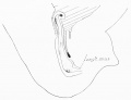
Longitudinal muscle of the human colon
Lineback PE. Studies on the longitudinal muscle of the human colon, with special reference to the development of the taeniae. (1920) Contrib. Embryol., Carnegie Inst. Wash. Publ. 50
A 46-mm fetus, No. 95 C. C. (fig. 2), shows the whole muscle grown a little farther upward and its dorsal fibers extending well into the sigmoid region; no well-defined fibers can be detected in any of the regions higher up. In a 50 mm fetus there is a distinct layer at the mesenteric attachment, the continuation of the dorsal fibers of the 46 mm stage. The layer is well defined and extends throughout the length of the colon. Lewis noted this muscle and stated that in a 75-mm. fetus it was seen along the transverse colon, although he made no mention of it in other parts of the bowel.
Cite this page: Hill, M.A. (2024, April 28) Embryology Carnegie Embryo 95. Retrieved from https://embryology.med.unsw.edu.au/embryology/index.php/Category:Carnegie_Embryo_95
- © Dr Mark Hill 2024, UNSW Embryology ISBN: 978 0 7334 2609 4 - UNSW CRICOS Provider Code No. 00098G
Pages in category 'Carnegie Embryo 95'
The following 2 pages are in this category, out of 2 total.
Media in category 'Carnegie Embryo 95'
The following 2 files are in this category, out of 2 total.
 Lineback1920 fig02.jpg 800 × 612; 34 KB
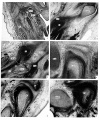 Moffatt1957 plate01.jpg 1,500 × 1,803; 878 KB

